

Biotechnology for Biomedical Engineers - Martin L. Yarmush et al
.pdf13-6 |
Biotechnology for Biomedical Engineers |
FIGURE 13.2 Standard retroviral vector designs.LTRs are shown as black boxes.SD and SA represent splice donor and acceptor sites, respectively.; indicates the packaging signal sequence. Internal promoters are the stippled boxes and transcription start sites are indicated by arrows. (a) A single-gene vector.Transcription is driven from the LTR. (b) A single-gene vector expressed from an internal promoter. (c) A typical two-gene vector. One gene is the therapeutic gene,and the other is a selectable marker gene.Gene A is driven from the LTR,and gene B is expressed from the internal promoter.(d) A two-gene vector in which transcription is driven from the LTR.The efficiency of expression of gene B is a function of the efficiency of splicing.(e) A self-inactivating vector.A deletion in 3’ LTR is copied to the 5’ LTR during reverse transcription. This eliminates any transcription from the LTR. Internal promoters are still active.This vector reduces the chance of insertional activation of a proto-oncogene downstream from the 3’ LTR.
loss of infectivity is difficult [Andreadis et al.,1999].Standard techniques such as centrifugation,column filtration or ultrafiltration have failed [Le Doux et al,1998;McGrath et al.,1978].More recently,hollow fiber [Paul et al., 1993] and tangential flow filtration [Kotani et al., 1994] have been used with some success.A chimeric retrovirus in which the normal envelope proteins were replaced byVSV (Vesicular StomatitisVirus) envelope proteins was readily concentrated by centrifugation [Burns et al., 1993].
Stability and level of expression has also been cited as a significant drawback for retrovirally transduced cells. Although long-term expression has been documented in some systems such as bone marrow, fibroblasts,hepatocytes and muscle cells,there have also been reports of transient expression in retrovirally transduced cells [Roemer and Friedmann, 1992]. This unstable expression has been attributed to several causes including methylation of the DNA and in some cases rearrangement or loss of the proviral sequences [Roemer and Friedmann,1992].
The use of recombinant retroviruses has raised two major safety concerns [Roemer and Friedmann, 1992; Temin, 1990]. Older packaging cell lines occasionally produced replication competent virus by homologous recombination between the retroviral vector and the packaging cell line’s retroviral sequences. New packaging cell lines have made the production of replication competent viruses essentially impossible [Danos and Mulligan, 1988]. The other safety concern was the possibility that the integration of the recombinant retrovirus would activate a proto-oncogene.The probability of this event is very low, and since several mutations are required for a cell to become cancerous, the risk of cellular transformation is extremely low and is typically outweighed by the potential therapeutic benefits [Temin, 1990].
13.3 Recombinant Adenoviruses
Recombinant adenoviruses have a number of properties that make them a useful alternative to recombinant retroviruses for human gene transfer. Recombinant adenoviruses are well-characterized, relatively easy to manipulate, can be grown and concentrated to very high titers (up to 1013 infectious particles/ ml), are stable particles, and can transduce a wide variety of cell types [Crystal, 1995; Hitt et al,1997].Recombinant adenoviruses are also a relatively safe gene transfer system.Replication competent viruses, should they contaminate virus stocks or form by recombination with wild-type adenoviruses
GeneTherapy |
13-7 |
from latent infections, do not pose any significant risk since they elicit only a mild, self-limiting infection [Parks et al., 1996]. Furthermore, genes transferred by recombinant adenoviruses do not integrate, eliminating the risk of insertional mutagenesis of the chromosomal DNA of the target cell [Crystal, 1995].Most important, recombinant adenoviruses efficiently transfer genes to nondividing, as well as dividing,cells that makes possible the in vivo transduction of tissues composed of fully differentiated or slowly dividing cells such as the liver and lung [Mulligan, 1993].
Adenoviruses consist of a large double stranded DNA genome (about 36 kilobase pairs long) packaged within a nonenveloped icosahedral capsid that is primarily composed of three virus-encoded proteins (hexon,penton base,and fiber proteins) [Horwitz,1990].The fiber proteins protrude from the surface of the virus and mediate its attachment to target cells via a high affinity interaction with cell surface receptors.The virus is then internalized into endosomal vesicles via specific interactions between the penton base proteins and av integrins [Wickham et al.,1993].Adenoviruses escape these vesicles by an acid-induced endosomolytic activity and are transported to the nucleus, into which they enter via pores in the nuclear membrane [Greber et al., 1993].
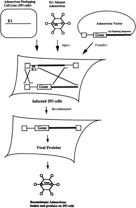
The wild type adenovirus genome consists primarily of five early genes (E1-E5) each of which is expressed from their own promoters [Horwitz, 1990].There are also five late genes (L1-L5), which are expressed from the major late promoter (MLP).The first generation of recombinant adenoviruses were based on a mutant adenovirus in which the El region (and in some cases the E3 region) was deleted. The El region is required for replication. Nevertheless, El minus mutants can be grown on specialized packaging cell lines (293 cells),which express the El gene and therefore provide the necessary functions for virus production [Levine and Friedmann, 1993].
To generate recombinant adenoviruses, a plasmid that contains the gene of interest, flanked by adenovirus sequences, is transfected into 293 cells in which adenoviruses that lack the El region are actively replicating (Fig. 13.3).The virus stock is screened for the rare recombinants in which the gene of interest has correctly recombined with the El minus mutant by homologous recombination.These recombinant virions are purified and grown to high titer on 293 cells [Morgan et al., 1994].
Recombinant adenoviruses have been successfully used for in vivo gene transfer in a number of animal models and in several clinical protocols, including those which delivered a functional copy of the cystic fibrosis transmembrane conductance regulator (CFTR) gene into the nasal epithelium and lungs of cystic fibrosis patients [Boucher and Knowles,1993;Welsh,1993;Wilmott andWhitsett,1993]. Unfortunately,transgene expression was short-lived (5 to 10 days) [Verma and Somia,1997].Short-lived expression is a disadvantage for the treatment of genetic or chronic disorders because, in order to maintain the therapeutic effect of the transgene,recombinant adenoviruses would have to be administered to the patient repeatedly.Repeat administration often fails due to the presence of neutralizing antibodies that form in response to the first administration of the recombinant adenoviruses [Hitt et al., 1997]. Alternatively, short lived high expression from adenovirus vectors might be therapeutically useful for applications where transient expression is preferred. Such applications include the stimulation of angiogenesis by adenoviruses expressing vascular endothelial growth factor (VEGF) [Magovern et al, 1997;Morgan andYarmush,1998].
Short-lived gene expression is currently one of the most significant limitations of recombinant adenoviruses for use in human gene therapy.Although short-lived gene expression could be the result of vector cytotoxicity,promoter shutoff,or loss of the transgene DNA,most investigators believe it is primarily due to the elimination of transduced cells by transgene or adenovirus-specific cytotoxic T-lymphocytes [Dai et al.,1995;Engelhardt et al,1994;Scaria et al,1998;Zsengeller et al.,1995].As a result,most efforts to increase the longevity of gene expression have focused on eliminating the immune rejection of cells transduced by recombinant adenoviruses.Two major strategies have been pursued. One strategy has been to construct recombinant adenoviruses that encode fewer immunogenic adenoviral proteins, such as by deleting portions of the E2 region or the E4 region. Such vectors have the added benefit of being able to accommodate the cloning of much larger inserts of therapeutic DNA (to as much as 36 kilobases) than was possible with the first-generation (El and E3 minus) recombinant adenoviruses which could accommodate only 7 kilobases of foreign DNA [Morsy et al., 1998; Parks et al, 1996].
13-8 |
Biotechnology for Biomedical Engineers |
FIGURE 13.3 Isolation of recombinant adenovirus.A packaging cell line (293 cells),which expresses the El gene, is infected with an El-minus mutant adenovirus.The adenovirus is derived from a plasmid encoding the wild-type adenovirus genome.The El region of the adenovirus genome is replaced with the therapeutic gene,and the resultant plasmid (the adenovirus vector) is transfected into the 293 cell line, which is infected by the mutant adenovirus. Since the therapeutic gene is flanked by adenoviral sequences, the mutant adenovirus genome and the adenovirus vector will occasionally undergo homologous recombination and form an infectious recombinant adenovirus whose genome encodes the therapeutic gene.These rare recombinants are isolated then grown on another 293 cell line.

GeneTherapy |
13-9 |
The second strategy is to block the host immune response. For example, investigators have coinjected recombinant adenoviruses with immunomodulatory compounds (e.g.,deoxyspergualin,IL-12, IFNgamma) that transiently suppressed the immune system, eliminated the humoral response, and made possible repeat administration of the vector [Hitt et al., 1997]. Others have co-injected vectors that encode for immunosuppressive compounds that induce tolerance to recombinant adenoviruses (e.g., CTLA4Ig), or are developing recombinant adenoviruses whose backbones contain genes that encode for immunosuppressive proteins (e.g.,Ad5 E3 19-kDa protein or HSV ICP47) [Nakagawa et al., 1998;Wold and Gooding, 1989;York et al., 1994].
As a result of these and similar efforts, the persistance of gene expression in vivo has been extended to as long as 5 months [Kay et al.,1995;Scaria et al.,1998].It remains to be determined,however,if these methods can eliminate the immune response against recombinant adenoviruses and their products, whether or not elimination of the immune response results in long-term gene expression, and if these methods will be effective in human gene therapy protocols.
13.4 Recombinant Adeno-Associated Viruses
Adeno-associated viruses (AAV) are another virus-based gene transfer system that has significant potential for use in human gene therapy. AAV are small, nonenveloped human parvoviruses that contain a singlestranded DNA genome (4.7 kilobases) and encode two genes required for replication, rep and cap [McCarty and Samulski, 1997]. AAV are stable, have a broad host range, can transduce dividing and nondividing cells,and do not cause any known human disease [Verma and Somia,1997].AAV require the presence of a helper virus (typically adenovirus) to replicate [Muzyczka, 1992].When no helper virus is present,AAV do not replicate but instead tend to establish a latent infection in which they permanently integrate into the chromosomal DNA of the target cells [Inoue and Russell, 1998]. Wild-type AAV preferentially integrates into a specific site in chromosome 19 due to the interaction of the virus-encoded protein Rep with the host cell DNA [McCarty and Samulski, 1997]. Recombinant AAV, however, are Rep negative and as a result integrate randomly into the chromosomal DNA of the target cell.
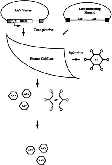
To generate recombinant AAV, human 293 cells are transfected with the AAV vector, a plasmid that contains the therapeutic gene of interest flanked by the 145-bpAAV inverted terminal repeats (ITR) that are necessary for its encapsidation into a virus particle [Ferrari et al., 1997] (Fig. 13.4).The cells are also transfected with a helper plasmid, a plasmid that encodes for the virus proteins necessary for particle formation and replication (i.e.,AAV rep and cap genes).The transfected cells are infected with wild-type adenovirus which supplies helper functions required for amplification of the AAV vector [Morgan and Yarmush, 1998]. Contaminating adenovirus is removed or inactivated by density gradient centrifugation and heat inactivation. Recombinant AAV generated by this and similar methods have been successfully used to achieve long-term expression of therapeutic proteins in a number of cell types and tissues, including in the lung, muscle, liver, central nervous system, retina, and cardiac myocytes [Bartlett et al., 1998; Flannery et al., 1997; Flotte et al., 1993; Maeda et al, 1998; Monahan et al., 1998; Xiao et al., 1998]. Therapeutic effects have been achieved in a number of model systems, including in a dog model of hemophilia and a mouse model for obesity and diabetes [Monahan et al., 1998; Murphy et al., 1997].
Despite these early successes, several technical issues must be addressed before recombinant AAV can be used for human gene therapy on a routine basis. For example, little is known about the conditions or factors which enhance recombinantAAV transduction efficiency in vitro or in vivo [McCarty and Samulski, 1997]. Nor is it well-understood what controls the efficiency with which recombinant AAV integrate into the chromosomal DNA of target cells or why the efficiency of integration is so low (<1%). Perhaps the most significant technical issue,however,is that the current methods for producing recombinant AAV (see above) are tedious, labor intensive, and not well-suited for producing clinical grade virus.
Current methods are not adequate for producing a clinical grade virus for several reasons. First, current methods do not produce enough virus particles to be useful for many gene therapies. For example, it has been estimated that 1014 recombinant viruses will be needed to achieve systemic
13-10 |
Biotechnology for Biomedical Engineers |
FIGURE 13.4 Production of recombinantAAV.Human cells are transfected with a plasmid encoding the therapeutic gene which is driven from a heterologous promoter (cross-hatched box) and flanked with AAV terminal repeats (stippled boxes). The cells are also transfected with a complementing plasmid encoding the AAV rep and cap genes, which cannot be packaged because they are not flanked by AAV terminal repeats.The rep and cap genes, whose products are required for particle formation, are flanked by adenovirus 5 terminal fragments (black boxes) that enhance their expression. The transfected cells are infected with wild-type adenovirus which supplies helper functions required for amplification of the AAV vector.The virus stock contains both AAV recombinant virus and adenovirus.The adenovirus is either separated by density gradient centrifugation or heat inactivated.
production of therapeutic levels of proteins such as factor IX or erythropoietin [Inoue and Russell, 1998].With current production methods,which yield 108 to 109 viruses per ml,about 100 to 1000 liters of virus would have to be produced for each treatment. Second, current methods require the use of helper adenovirus which is a significant disadvantage because their removal is tedious and labor intensive. In addition, there is the risk that residual contaminating adenovirus proteins will be present and stimulate the immune rejection of transduced cells [Xiao et al., 1998]. Finally, the use of transient transfection is not amenable to scale up and significantly increases the likelihood that replication competent AAV will be generated by recombinogenic events [Inoue and Russell, 1998].

GeneTherapy |
13-11 |
To overcome these problems, investigators are working toward the development of stable packaging cell lines that produce recombinant AAV. Substantial increases in the yield of recombinant AAV have been achieved by increasing in the virus producer cell lines the number of copies of the AAV vector or of the helper plasmid. For example, one group developed an AAV packaging cell line that contained integrated helper and vector constructs that were linked to the simian virus 40 replication origin [Inoue and Russell, 1998].These packaging cells could be induced to express SV40 T antigen, which then amplified the helper and vector constructs by 4 to 10 fold, resulting in 5- to 20-fold increases in recombinant AAV production. Progress has also been made toward the elimination of the need for helper adenovirus. For example, adenovirus miniplasmids have been constructed that provide all of the adenovirus helper functions but do not support the production of adenovirus proteins or viruses [Xiao et al., 1998]. Use of adenovirus miniplasmids, in conjunction with an improved helper plasmid, have improved the yield of recombinant AAV by up to 40-fold. Most likely a combination of strategies such as these will be necessary to develop a stable producer cell line that will be free of helper adenovirus and that will generate recombinant AAV at levels that are adequate for use in human gene therapies.
13.5 Direct Injection of Naked DNA
Direct injection of plasmid DNA intramuscularly, intraepidermally, and intravenously is a simple and direct technique for modifying cells in vivo. DNA injected intramuscularly or intraepidermally is internalized by cells proximal to the injection site [Hengge et al.,1995;Wolff et al.,1990].DNA injected intravenously is rapidly degraded in the blood (t1/2 < 10 min.) or retained in various organs in the body, preferentially in the nonparenchymal cells of the liver [Kawabata et al., 1995; Lew et al., 1995]. Gene expression after direct injection has been demonstrated in skeletal muscle cells of rodents and nonhuman primates, cardiac muscle cells of rodents, livers of cats and rats, and in thyroid follicular cells of rabbits [Hengge et al., 1995;Hickman et al., 1994;Levine and Friedmann,1993; Sikes et al., 1994].
The efficiency of gene transfer by direct injection is somewhat inefficient [Doh et al., 1997]. As little as 60 to 100 myocardial cells have been reported to be modified per injection [Acsadi et al.,1991]. Higher efficiencies were observed when plasmids were injected into regenerating muscle cells [Wells, 1993] or co-injected with recombinant adenoviruses [Yoshimura et al., 1993]. Injected DNA does not integrate, yet gene expression can persist for as long as two months [Wolff et al., 1990]. Levels of gene expression are often low but can be increased by improvements in vector design [Hartikka et al, 1996].
Despite the low efficiency of gene transfer and expression, direct injection of plasmid DNA has many promising applications in gene therapy,particularly when low levels of expression are sufficient to achieve the desired biological effect. For example, several patients suffering from critical limb ischemia were successfully treated by injection of DNA encoding human vascular endothelial growth factor (VEGF),a potent angiogenic factor [Isner et al,1996;Isner et al,1996;Melillo et al.,1997;Tsurumi et al, 1996]. Following injection of DNA into the ischemic limbs of ten patients,VEGF expression was detected in their serum, new blood vessels were formed in 7 of the 10 patients, and 3 patients were able to avoid scheduled below-the-knee amputations [Baumgartner et al., 1998].
Direct injection of DNA could also be used to vaccinate patients against pathogens as evidenced by the effects of direct injection of plasmid DNA encoding pathogen proteins or immunomodulatory cytokines [Donnelly et al., 1997; Sato et al., 1996]. Direct injection may also be an effective way to systemically deliver therapeutic proteins [Tripathy et al.,1996].For example,the incidence of autoimmune diabetes in a mouse model of the disease was significantly reduced in mice that were injected intramuscularly with DNA encoding interleukin 10, an immunosuppressive cytokine [Nitta et al., 1998].
In contrast to viral-based delivery systems, there is little restriction on the size of the transgene that can be delivered by direct DNA injection.As a result, direct DNA injection is particularly well-suited for treating disorders that require the delivery of a large transgene. For example, Duchenne’s muscular
13-12 |
Biotechnology for Biomedical Engineers |
dystrophy, a genetic disease of the muscle caused by a defect in the gene for dystrophin (12 kilobases) can potentially be treated by direct DNA injection [Acsadi et al., 1991].
13.6 Particle-Mediated Gene Transfer
Particle-mediated gene transfer is an alternative method used to deliver plasmid DNA to cells. DNA coated gold particles are loaded onto a macro-projectile, which is then accelerated through a vacuum chamber to high velocity by a burst of helium or a voltage discharge until it hits a stopping plate [Yang et al., 1996]. Upon impact, the DNA-coated microprojectiles are released through a hole in the stopping plate, penetrate the target tissue, and the transferred gene is expressed [Morgan andYarmush, 1998;Yang et al., 1996]. Genes have been introduced and expressed in a number of cell types and tissues,including skin,liver,spleen,muscle,intestine,hematopoietic cells,brain,oral mucosa and epidermis, tumor explants,and cells of developing mouse embryos [Jiao et al.,1993;Keller et al, 1996;Mahvi et al, 1996; Morgan and Yarmush, 1998;Verma et al., 1998; Zelenin et al., 1993]. Similar to direct DNA injection, there are few constraints on the size of the DNA that can be delivered.
The efficiency of particle-mediated gene transfer varies with tissue type, but in general it is most efficient in the liver, pancreas, and epidermis of the skin, and least efficient in muscle, vascular, and cardiac tissues [Rakhmilevich andYang, 1997]. For example, 20 percent of bombarded epidermal cells of the skin, but only 1 to 3 percent of bombarded muscle cells, expressed the transferred gene [Williams et al.,1991;Yang et al.,1990].
13.7 Liposome-Mediated Gene Delivery
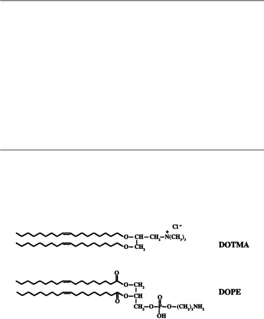
Liposomes made from a mixture of neutral and cationic lipids have also been used to deliver plasmid DNA to cells. Liposomes are relatively easy to make, can be made with well-defined biophysical properties,and can accommodate virtually any size transgene [Kay et al.,1997;Tomlinson and Rolland, 1996]. Small unilamellar liposomes ranging from 20 to 100 nm in diameter are prepared by sonication of a mixture of cationic (e.g., DOTMA, N-{l-(2,3-dioleyloxy)propyl}-N,N,N-triethylammonium) and neutral (e.g.,DOPE,dioleoyl-phosphatidylethanolamine) lipids,followed by extrusion through a porous polycarbonate filter (e.g.,100 nm pore size) (Fig.13.5) [Morgan andYarmush,1998;Radler et al,1997].
FIGURE 13.5 Common constituents of cationic liposomes.A typical cationic liposome is composed of a mixture of cationic lipids and neutral phospholipids. DOTMA {N-[1-(2,3,-dioleyloxy]-N,N,N-trimethylammonium chloride} is a synthetic cationic lipid that attaches to DNA using its positively charged head group. DOPE {1,2- Di[(cis)-9-octadecenoyl]-sn-glycero-3-phosphoethanolamine} is a neutral phospholipid which enhances the activity of these cationic liposomes.The resultant DNA-liposome complex is positively charged and reduces or eliminates the repulsive electrostatic forces between the negatively charged DNA and the negatively charged cell surface.

GeneTherapy |
13-13 |
DNA is added to the cationic liposomes, binds noncovalently to the positively charged cationic lipids, and induces a topological transition from liposomes to multilamellar structures composed of lipid bilayers alternating with DNA monolayers [Felgner and Ringold,1989;Radler et al.,1997].The size of the structures depends on their overall charge, with charged structures (negative or positive) being the smallest (about 100 nm in diameter) due to stabilization by electrostatic repulsion and neutral structures being the largest (greater than 3000 nm in diameter) due to aggregation as a result of van der Waals attractive forces [Radler et al, 1997].The relationship between structure and transfection efficiency is not well-understood, although in general a slight excess of cationic lipid is needed for optimal gene transfer [Kay et al, 1997].
Gene transfer is accomplished by simply mixing or applying the lipid-DNA complexes to the target cells or tissue.Cationic liposome-DNA complexes have been used in a number of applications,including transfer of genes to the arterial wall, lung, skin, and systemically by intravenous injection [Morgan and Yarmush, 1998; Zhu et al, 1993]. Genes delivered by cationic liposomes do not integrate, so gene expression is transient and there is minimal risk of insertional mutagenesis.Liposomes do not carry any viral sequences or proteins, and are relatively nontoxic and nonimmunogenic [Nabel et al., 1993; Zhu et al., 1993].
Liposome-mediated gene transfer is somewhat inefficient, however, in part due to the failure of a large fraction of lipid-complexed DNA to escape degradation in cellular endosomes [Xu and Szoka, 1996; Zabner et al., 1995].Several strategies have been taken to overcome this limitation,including use of acidotropic bases to reduce the rate of degradation by raising the pH of the endosomes, coupling of liposomes to endosomolytic or fusogenic virus proteins, and developing new liposome formulations that use pH sensitive cationic lipids that become fusogenic in the acidic cellular endosomes [Budker et al.,1996;Legendre and Szoka,1992;Yonemitsu et al.,1997].Substantial improvements in the efficiency of liposome-mediated gene transfer will only be achieved when the fundamental mechanisms that govern the interactions of cationic lipid-DNA complexes with cells are better understood.
13.8 Other Gene Transfer Methods
Several other gene transfer technologies have been tested in clinical trials or are in various stages of development.These include recombinant viruses such as vaccinia virus [Qin and Chatterjee, 1996], herpes simplex virus [Glorioso et al.,1997],canarypox virus [Kawakita et al.,1997],and fowlpox [Wang et al.,1995],and nonviral vectors that mimic some of the properties of viruses,such as DNA conjugated to proteins that promote binding to specific cell-surface receptors, fusion, or localization to the nucleus [Wu et al., 1994].These methods are not discussed here in greater detail because even though they are capable of transferring genes to cells they have not yet been well-developed or extensively tested in the clinic.
13.9 Summary and Conclusion
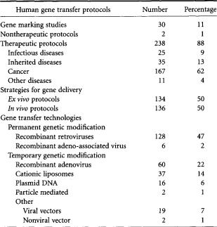
Several gene transfer systems have been developed that have successfully transferred genes to cells, and have elicited various biological effects.To date,nearly 300 clinical trials have been approved to test their safety and efficacy (Table 13.4). Each system has unique features, advantages, and disadvantages that determine if its use in a particular application is appropriate.One principal consideration is whether or not permanent or temporary genetic modification is desired. Other important considerations include what the setting of gene transfer will be (ex vivo or in vivo), what level of gene expression is needed,and whether or not the host has preexisting immunity against the vector. No single gene transfer system is ideal for any particular application and it is unlikely that such a universal gene transfer system will ever be developed. More likely, the current gene transfer systems will be further improved and modified, and new systems developed, that are optimal for the treatment of specific diseases.
13-14 |
Biotechnology for Biomedical Engineers |
TABLE 13.4 Current Clinical Experience Using Gene Transfer
(as of December 1, 1998)
*“Other” viral vectors refers to recombinant canarypox virus, vaccinia virus, fowlpox, and herpes simplex virus. “Other” nonviral vector refers to RNA transfer and antisense delivery.
Defining Terms
Chimeric retrovirus: A recombinant retrovirus whose structural proteins are derived from two or more different viruses.
Ex vivo: Outside the living body, referring to a process or reaction occurring therein.
In vitro: In an artificial environment, referring to a process or reaction occurring therein, as in a test tube or culture dish.
In vivo: In the living body, referring to a process or reaction occurring therein. Liposome: A spherical particle of lipid substance suspended in an aqueous medium.
Packaging cell line: Cells that express all the structural proteins required to form an infectious viral particle.
Plasmid: A small, circular extrachromosomal DNA molecule capable of independent replication in a host cell, typically a bacterial cell.
Recombinant:A virus or vector that has DNA sequences not originally (naturally) present in their DNA. Retrovirus:A virus that possesses RNA-dependent DNA polymerase (reverse transcriptase) that reverse transcribes the virus’ RNA genome into DNA,then integrates that DNA into the host cell’s genome. Transduce:To effect transfer and integration of genetic material to a cell by infection with a recombinant
retrovirus.
Vector:A plasmid or viral DNA molecule into which a DNA sequence (typically encoding a therapeutic protein) is inserted.
References
Acsadi G,Dickson G,Love DR et al.,1991.Human dystrophin expression in mdx mice after intramuscular injection of DNA constructs [see comments]. Nature. 352:815–8.
GeneTherapy |
13-15 |
Acsadi G, Jiao SS, Jani A et al., 1991. Direct gene transfer and expression into rat heart in vivo. New Biol 3:71–81.
AndersonWF.1992.Human gene therapy.Science. 256:808–13.
Andreadis ST, Roth CM, Le Doux JM et al., 1999. Large scale processing of recombinant retroviruses for gene therapy.Biotechnol Prog. Jan-Feb;15(1):1–11.
Bartlett JS, Samulski RJ, and McCown TJ. 1998. Selective and rapid uptake of adeno-associated virus type 2 in brain. Hum. GeneTher. 9:1181–6.
Baumgartner I,PieczekA,Manor O et al.,1998.Constitutive expression of phVEGF165 after intramuscular gene transfer promotes collateral vessel development in patients with critical limb ischemia [see comments]. Circulation. 97:1114–23.
Boucher RC, and Knowles MR. 1993. Gene therapy for cystic fibrosis using El deleted adenovirus:A phase I trial in the nasal cavity. Office of Recombinant DNA Activity, NIH.
Budker V, Gurevich V, Hagstrom JE et al., 1996. pH-sensitive, cationic liposomes: a new synthetic viruslike vector.Nat. Biotechnol. 14:760–4.
Burns JC, Friedmann T, Driever W et al., 1993.Vesicular stomatitis virus G glycoprotein pseudotyped retroviral vectors: concentration to very high titer and efficient gene transfer into mammalian and nonmammalian cells [see comments]. Proc. Natl.Acad. Sci. USA. 90:8033–7.
Capecchi MR. 1980. High efficiency transformation by direct microinjection of DNA into cultured mammalian cells. Cell 22:479–88.
Chen C,and Okayama H.1987. High-efficiency transformation of mammalian cells by plasmid DNA.
Mol Cell Biol 7:2745–52.
Crystal RG. 1995.Transfer of genes to humans: early lessons and obstacles to success. Science. 270:404– 10.
DaiY, Schwarz EM, Gu D et al., 1995. Cellular and humoral immune responses to adenoviral vectors containing factor IX gene: tolerization of factor IX and vector antigens allows for long-term expression. Proc. Natl Acad. Sci. USA. 92:1401–5.
Danos O, and Mulligan RC. 1988. Safe and efficient generation of recombinant retroviruses with amphotropic and ecotropic host ranges. Proc. Natl Acad. Sci. USA. 85:6460–4.
Doh SG,Vahlsing HL, Hartikka J et al., 1997. Spatial-temporal patterns of gene expression in mouse skeletal muscle after injection of lacZ plasmid DNA. Gene Ther. 4:648–63.
Donnelly JJ,Ulmer JB,Shiver JW et al.,1997.DNA vaccines.Ann. Rev. Immunol. 15:617–48. Engelhardt JF,Ye X, Doranz B et al, 1994. Ablation of E2A in recombinant adenoviruses improves
transgene persistence and decreases inflammatory response in mouse liver. Proc. Natl. Acad. Sci. USA. 91:6196–200.
Felgner PL,and Rhodes G. 1991.Gene therapeutics.Nature. 349:351–2.
Felgner PL,and Ringold GM.1989.Cationic liposome-mediated transfection.Nature. 337:387–8. Ferrari FK,Xiao X,McCarty D et al.,1997.New developments in the generation of Ad-free,high-titer
rAAV gene therapy vectors.Nat. Med. 3:1295–7.
Flannery JG, Zolotukhin S,Vaquero MI et al., 1997. Efficient photoreceptor-targeted gene expression in vivo by recombinant adeno-associated virus. Proc. Natl Acad. Sci. USA. 94:6916–21.
FlotteTR,Afione SA,Conrad C et al,1993.Stable in vivo expression of the cystic fibrosis transmembrane conductance regulator with an adeno-associated virus vector.Proc. Natl Acad. Sci. USA. 90:10613–7.
FriedmannT.1989.Progress toward human gene therapy.Science. 244:1275–81.
Glorioso JC, GoinsWF, Schmidt MC et al., 1997. Engineering herpes simplex virus vectors for human gene therapy.Adv.Pharmacol 40:103–36.
Greber UF,Willetts M,Webster P et al., 1993. Stepwise dismantling of adenovirus 2 during entry into cells. Cell. 75:477–86.
Hartikka J, Sawdey M, Cornefert-Jensen F et al., 1996.An improved plasmid DNA expression vector for direct injection into skeletal muscle. Hum. Gene.Ther. 7:1205–17.
Hengge UR, Chan EF, Foster RA et al., 1995. Cytokine gene expression in epidermis with biological effects following injection of naked DNA. Nat. Genet. 10:161–6.