


38E P I T H E L I U M A N D G L A N D S
Intermediate filaments enter and leave the plaques, resembling hairpins.
Embedded into the plaques are transmembrane, calcium-dependent cadherins, desmogleins and desmocollins.
The extracellular moieties of desmogleins and desmocollins contact those of the adjacent cell and in the presence of calcium attach the two cells to each other.
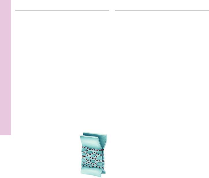
•At gap junctions (communicating junctions, nexus), the two cell membranes are very close to each other, about 2 nm apart.
Interposed within the cell membrane of each cell and meeting each other are connexons composed of six subunits, known as a connexins; these multipass proteins form a cylindrical structure with a central pore.
A connexon of one cell matches the connexon of the other cell and thus forms an aqueous channel, about 2 nm in diameter, between the two cells that permits water, ions, and molecules smaller than 1 kDa in size to traverse the channel and go from one cell into the next.
Each cell has the ability to open or close the channel, and this regulation is calcium as well as pH dependent. In this fashion, a healthy cell can shut off communication with a cell that may be damaged.
Basal Surface Modifications (see Graphic 2-1)
The basal cell membrane of the cell is affixed to the basal lamina by adhering junctions known as the hemidesmosomes.
•A hemidesmosome resembles half of a desmosome, but its biochemical composition and clinical significance demonstrate enough dissimilarity that hemidesmosomes are no longer viewed as being merely a half of a desmosome.
A hemidesmosome has an intracellular plaque, composed mostly of plectin, BP230, and erbin.
Intermediate filaments terminate in the plaque, by interacting with BP230 and plectin.
Hemidesmosomes also possess transmembrane protein components, known as integrin molecules whose cytoplasmic moiety is embedded in the plaque and is attached to it by interacting with BP230 and erbin.
The extracellular region of the integrin molecules contacts laminin and type IV collagen of the basal lamina and binds to them if extracellular calcium is present.
In this manner, hemidesmosomes assist in the anchoring of epithelial sheets to the adjacent basal lamina.
•The three components of the basement membrane, when viewed with the electron microscope, are the lamina lucida, lamina densa (collectively known as the basal lamina), and the lamina reticularis.
The lamina lucida is that region of the basal lamina that houses the extracellular moieties of the transmembrane laminin receptors, integrin and dystroglycans molecules and the glycoproteins laminin, entactin, and perlacans.
The lamina densa is composed of type IV collagen, coated by laminin, entactin, and perlacan on its epithelial surface, and fibronectin on the lamina reticularis surface. Additionally, two other collagen types, XV and XVIII, are also present in the lamina densa. The lamina densa adheres to the lamina reticularis.
The lamina reticularis composed mostly of type III collagen, proteoglycans, glycoproteins, and slender elastic fibers, by anchoring fibers (type VII collagen) and microfibrils (fibrillin).
Basal laminae function as structural supports for the epithelium, as molecular filters (e.g., in the renal glomerulus), in regulating the migration of certain cells across epithelial sheaths (e.g., preventing entry to fibroblasts but permitting access to lymphoid cells), in epithelial regeneration (e.g., in wound healing where it forms a surface along which regenerating epithelial cells migrate), and in cell-to-cell interactions (e.g., formation of myoneural junctions).
GLANDS
Most glands are formed by epithelial downgrowths into the surrounding connective tissue.
•Glands that deliver their secretions onto the epithelial surface do so via ducts and are known as exocrine glands.
•Glands that do not maintain a connection to the outside (ductless) and whose secretions enter the vascular system for delivery are known as endocrine glands.
The secretory cells of a gland are referred to as its parenchyma and are separated from surrounding connective tissue and vascular elements by a basement membrane.
• Exocrine glands |
are |
classified according to vari- |
ous parameters, |
for |
example, morphology of their |

E P I T H E L I U M A N D G L A N D S 39
TABLE 2-2 • Exocrine Gland Characteristics
Cellular Composition |
Example |
|
|
Unicellular (single cell) |
Goblet cell |
|
|
Multicellular (more than one cell) |
Submandibular gland |
|
|
Duct Form |
Example |
|
|
Simple (unbranched) |
Sweat gland |
|
|
Compound (branched) |
Mammary gland |
|
|
Type of Secretion |
Example |
|
|
Serous (watery) |
Parotid gland |
|
|
Mucus (viscous) |
Palatal glands |
|
|
Mixed (serous and mucus) |
Sublingual gland |
|
|
Mode of Secretion |
Example |
|
|
Merocrine (only secretory product released) |
Parotid gland |
|
|
Apocrine (secretory product along with a portion of |
Lactating mammary gland (according to some authors) |
cell cytoplasm) |
|
|
|
Holocrine (cell dies and becomes the secretion) |
Sebaceous gland |
|
|
functional units, branching of their ducts, types of secretory products they manufacture, and the method whereby their component cells release secretory products (Table 2-2).
•The classification of endocrine glands is much more complex, but morphologically, their secretory units either are composed of follicles or are arranged in cords and clumps of cells (see Graphic 2-2).

40 E P I T H E L I U M A N D G L A N D S
CLINICAL CONSIDERATIONS
Bullous Pemphigoid
Bullous pemphigoid, a rare autoimmune disease, is caused by autoantibodies binding to some of the protein components of hemidesmosomes. Individuals afflicted with this disease exhibit skin blistering of the groin and axilla about the flexure areas and often in the oral cavity. Fortunately, it can be controlled by steroids and immunosuppressive drugs.
carcinomas, whereas those developing from glandular epithelium are called adenocarcinomas.
Metaplasia
Epithelial cells are derived from certain germ cell layers, possess a definite morphology and location, and perform specific functions; however, under certain pathological conditions, they may undergo metaplasia, transforming into another epithelial cell type. An example of such metaplasia occurs in the lining epithelium of the oral cavity of individuals who smoke or use chewing tobacco as well as in Barrett’s esophagus, where the long-term gastric reflux causes the epithelium of the lower portion of the esophagus to resemble the cardiac stomach but with the presence of goblet cells rather than surface lining cells.
Bullous pemphigoid. Note that the epidermis is lifted from the dermis, a characteristic of bullous pemphigoid because the hemidesmosomes are attacked by the immune system thus separating the epidermis from the underlying dermis, which displays the presence of an inflammatory infiltrate of neutrophils, lymphocytes, and eosinophils. (Reprinted with permission from Mills SE, Carter D, Greenson JK, Reuter VE, Stoler MH, eds. Sternberger’s Diagnostic Surgical Pathology, 5th ed. Philadelphia: Lippincott Williams & Wilkins. 2010. p. 17.)
Pemphigus Vulgaris
Pemphigus vulgaris is an autoimmune disease, caused by autoantibodies binding to some of the components of desmosomes. This disease causes blistering and is usually found occurring in middle-aged individuals. It is a relatively dangerous disease since the blistering can easily lead to infections. Frequently, this disease also responds to steroid therapy.
Tumor Formation
Under certain pathologic conditions, mechanisms that regulate cell proliferation do not function properly; thus, epithelial proliferation gives rise to tumors that may be benign if they are localized, or malignant if they wander from their original site and metastasize (seed) to another area of the body and continue to proliferate. Malignant tumors that arise from surface epithelium are termed
Metaplasia in a case of Barrett’s esophagus. Note that the normal esophageal epithelium, stratified squamous nonkeratinized, has been replaced by a simple columnar epithelium resembling that of the cardiac stomach but rich in goblet cells. (Reprinted with permission from Mills SE. Histology for Pathologists, 3rd ed. Philadelphia: Lippincott Williams & Wilkins, 2007. p. 580.)
E P I T H E L I U M A N D G L A N D S 41
Cholera
Cholera toxins cause the release of tremendous volumes of fluid from the individual afflicted by that disease. The toxin attacks the zonulae occludentes by disturbing the proteins ZO-1 and ZO-2, thereby disrupting the zonulae occludentes and permitting the paracellular movement of water and electrolytes. The patient has uncontrolled diarrhea and subsequent fluid and electrolyte loss. If the fluids and salts are not replaced in a timely manner, the patient dies.
Psoriasis Vulgaris
Psoriasis affects approximately 2% of the population and may have a familial trait. It usually begins its course between 10 and 40 years of age, and it first appears as patches of dry skin that is raised and is reddish in color on the knees, scalp, elbows, back, or the buttocks. It is believed to be an immune disorder that causes a higher than normal mitotic activity of the cells of the stratified squamous keratinized epithelium, the epidermis, of the skin. In most individuals, this condition has no symptoms other than the unsightly appearance of the skin. In some individuals, however, the condition is accompanied by pain and/or itching, or both.
The normal keratinized stratified squamous epithelium of skin of this patient is greatly modified. Note that the stratum spinosum layer is greatly thickened and the cells of the stratum corneum appear to possess nuclei. Higher magnification of that area, however (not shown), demonstrates that the nuclei belong to neutrophils that invaded the epithelium. Also, note the absence of the strata granulosum and lucidum, which confirms that this specimen is not taken from regions of thick skin, namely, the palm of the hand or the sole of the foot. The large number of nuclei present in the papillary layer of the dermis belong to lymphocytic infiltrate. (Reprinted with permission from Mills SE, Carter D, et al., eds. Sternberg’s Diagnostic Surgical Pathology, 5th ed. Philadelphia: Lippincott, Williams & Wilkins, 2010. p. 6.)

Complex Junctional • 1-2 GRAPHIC
42 E P I T H E L I U M A N D G L A N D S
Zonulae occludentes are occluding junctions where the outer leaflets of the apposing cell membranes fuse with each other, preventing material from taking the paracellular route between the connective tissue and the lumen. They extend along the entire circumference of the cell.
Integrins
(transmembrane receptor proteins)
Hemidesmosomes function in mediating the adherence of epithelial cells to the underlying basal lamina.
Extracellular
space
Strands of transmembrane proteins
Adjacent plasma membranes
Extracellular space
Actin filaments
Zonulae adherentes are located just basal to the zonulae occludentes and are distinguished by the presence of E-cadherins, transmembrane glycoproteins. Intracellularly, actin filaments form a meshwork that is attached to the E-cadherins by the other molecules.
Desmogleins
and E-cadherins
Plaque
Intermediate filaments
Maculae adherentes
are characterized by desmogleins and E-cadherins transmembrane glycoproteins, whose cytoplasmic ends
are associated with a plaque composed of desmoplakins. Intermediate filaments, forming hairpin loops,
enter and exit the plaque.
Adjacent plasma membranes
Connexons
Extracellular
space
Gap junctions are communicating
junctions where ions and small molecules are permitted to pass between adjoining cells. They couple
adjacent cells metabolically and electrically.

E P I T H E L I U M A N D G L A N D S 43
Serous cell
|
|
|
|
|
|
Myoepithelial cell |
|
|
Serous acinus |
|
|
Intercalated duct |
|
|
|
|
|
|||
|
|
|
|
|||
|
|
|
|
cell |
||
|
|
|
|
|||
|
|
|
|
|
|
|
Mucous acinus |
|
|
|
|
||
|
|
|
|
|||
|
Intercalated |
|
duct |
|
Striated duct |
|
Mucous |
Serous demilunes |
cell |
|
Striated duct cell
SIMPLE |
STRATIFIED |
TRANSITIONAL |
|
|
Squamous
Squamous keratinized |
Relaxed |
|
Cuboidal
Squamous nonkeratinized |
Distended |
|
Columnar
PSEUDOSTRATIFIED
Columnar
Columnar
Gland Salivary• 2-2 GRAPHIC
Cuboidal

Epithelium Pseudostratified and Epithelia imple • S1-2 PLATE
44 E P I T H E L I U M A N D G L A N D S
FIGURE 1. Simple squamous epithelium. Kidney. Monkey. Plastic section. ×540.
The lining of the lumen (L) of this small arteriole is composed of a simple squamous epithelium (SE) (known as the endothelium). The cytoplasm of these cells is highly attenuated and can only be approximated in this photomicrograph as a thin line (between the arrowheads). The boundaries of two contiguous epithelial cells cannot be determined with the light microscope. The nuclei (N) of the squamous epithelial cells bulge into the lumen, characteristic of this type of epithelium. Note that some of the nuclei appear more flattened than others. This is due to the degree of agonal contraction of the smooth muscle (M) cells of the vessel wall.
FIGURE 3. Simple columnar epithelium. Monkey. Plastic section. ×540.
The simple columnar epithelium of the duodenum in this photomicrograph displays a very extensive brush border (MV) on the apical aspect of the cells. The terminal web (TW), where microvilli are anchored, appears as a dense line between the brush border and the apical cytoplasm. Distinct dots (arrowheads) are evident, which, although they appear to be part of the terminal web, are actually terminal bars, resolved by the electron microscope to be junctional complexes between contiguous cells. Note that the cells are tall and slender, and their nuclei (N), more or less oval in shape, are arranged rather uniformly at the same level in each cell. The basal aspects of these cells lie on a basal membrane (arrows), separating the epithelium from the connective tissue (CT). The round nuclei (rN) noted within the epithelium actually belong to leukocytes migrating into the lumen (L) of the duodenum. A few goblet cells (GC) are also evident.
FIGURE 2. Simple squamous and simple cuboidal epithelia. x.s. Kidney. Paraffin section. ×270.
The medulla of the kidney provides ideal representation of simple squamous and simple cuboidal epithelia. Simple squamous epithelium, as in the previous figure, is easily recognizable due to flattened but somewhat bulging nuclei (N). Note that the cytoplasm of these cells appears as thin, dark lines (between arrowheads); however, it must be stressed that the dark lines are composed of not only attenuated cells but also the surrounding basal membranes. The simple cuboidal epithelium (CE) is very obvious. The lateral cell membranes (arrow) are clearly evident in some areas; even when they cannot be seen, the relationships of the round nuclei permit an imaginary approximation of the extent of each cell. Note that simple cuboidal cells, in section, appear more or less like small squares with centrally positioned nuclei.
FIGURE 4. Pseudostratified columnar epithelium with cilia. Paraffin section. ×270.
The first impression conveyed by this epithelium from the nasal cavity is that it is stratified, being composed of at least four layers of cells; however, careful observation of the inset (×540) demonstrates that these are closely packed cells of varying heights and girth, each of which is in contact with the basal membrane. Here, unlike in the previous photomicrograph, the nuclei (N) are not uniformly arranged, and they occupy about three-fourths of the epithelial layer. The location and morphology of the nuclei provide an indication of the cell type. The short basal cells (BCs) display small, round to oval nuclei near the basal membrane. The tall, ciliated cells (arrows) possess large, oval nuclei. The terminal web (TW) supports tall, slender cilia (C), which propel mucus along the epithelial surface. The connective tissue is highly vascularized and presents good examples of simple squamous epithelia (arrowheads) that compose the endothelial lining of blood (BV) and lymph vessels (LV).
PSEUDOSTRATIFIED
SIMPLE
Squamous |
Cuboidal |
|
Columnar |
Columnar |
KEY
BC |
basal cell |
GC |
goblet cell |
N |
nucleus |
BV |
blood vessel |
L |
lumen |
rN |
round nucleus |
C |
cilia |
LV |
lymph vessel |
SE |
simple squamous epithelium |
CE |
simple cuboidal epithelium |
M |
smooth muscle |
TW |
terminal web |
CT |
connective tissue |
MV |
brush border |
|
|

M
SE
N
L
M
FIGURE 1
L
MV
TW GC
N
CT
rN
FIGURE 3
|
|
|
|
N |
CE |
|
|
|
S1-2PLATE |
|
|
|
|
|
|
|
|
|
• |
|
|
|
|
Pseudostratified and Epithelia imple |
|
|
|
|
|
N |
|
|
|
Epithelium |
|
|
|
|
FIGURE 2
LV
BV |
c |
|
LV
TW
 TW c
TW c
BC
FIGURE 4

Epithelium Transitional and Epithelia tified Stra• 2-2 PLATE
46 E P I T H E L I U M A N D G L A N D S
FIGURE 1. Stratified cuboidal epithelium. Monkey. Plastic section. ×540.
Stratified cuboidal epithelium is characterized by two or more layers of cuboid-shaped cells, as illustrated in this photomicrograph of a sweat gland duct. The lumen (L) of the duct is surrounded by cells whose cell boundaries are not readily evident, but the layering of the nuclei (N) demonstrates that this epithelium is truly stratified. The epithelium of the duct is surrounded by a basal membrane (BM). The other thick tubular profiles are tangential sections of the secretory (s) portions of the sweat gland, composed of simple cuboidal epithelium. Note the presence of a capillary (Cp), containing a single red blood cell, and the bulging nucleus (arrow) of the epithelial cell constituting the endothelial lining. The large empty space in the lower right-hand corner of this photomicrograph represents the lumen of a lymph vessel (LV) whose endothelial lining presents a flattened nucleus bulging into the lumen. Note that more cytoplasm is evident near the pole of the nucleus (arrowhead) than elsewhere.
FIGURE 2. Stratified squamous nonkeratinized epithelium. Plastic section. ×270.
The lining of the esophagus provides a good example of stratified squamous nonkeratinized epithelium. The lack of vascularity of the epithelium, which is approximately 30 to 35 cell layers thick, is clearly evident. Nourishment must reach the more superficial cells via diffusion from blood vessels of the connective tissue (CT). Note that the deepest cells, which lie on the basal membrane and are known as the basal layer (BL), are actually cuboidal in shape. Due to their mitotic activity, they give rise to the cells of the epithelium, which, as they migrate toward the surface, become increasingly flattened. By the time they reach the surface, to be sloughed off into the esophageal lumen (EL), they are squamous in morphology. The endothelial lining of a vessel is shown as scattered nuclei (N) bulging into the lumen (L), providing an obvious contrast between stratified and simple squamous epithelia.
FIGURE 3. Stratified squamous keratinized epithelium. Skin. Paraffin section. ×132.
The palm of the hand is covered by a thick stratified squamous keratinized epithelium. The definite difference between this and the preceding photomicrograph is the thick layer of nonliving cells containing keratin (K), which functions in protecting the deeper living cells and tissues from abrasion, desiccation, and invasion by bacterial flora. Although the various layers of this epithelium are examined in greater detail in Chapter 11, certain features need to be examined here. Note that the interdigitation between the connective tissue dermal ridges (P) and the epithelial ridges (R) provides a larger surface area for adhesion and providing nutrients than would be offered by a merely flat interface. The basal membrane (BM) is a definite interval between the epithelium and the connective tissue. The basal layer of this epithelium, composed of cuboidal cells, is known as the stratum germinativum, which possesses a high mitotic activity. Cells originating here press toward the surface and, while on their way, change their morphology, manufacture proteins, and acquire different names. Note the duct (D) of a sweat gland piercing the base of an epidermal ridge as it continues toward the outside (arrows).
FIGURE 4. Transitional epithelium. Bladder. Monkey. Plastic section. ×132.
The urinary bladder, as most of the excretory portion of the urinary tract, is lined by a specialized type of stratified epithelium—the transitional epithelium. This particular specimen was taken from an empty, relaxed bladder, as indicated by the large, round, dome-shaped (rC) cells, some of which are occasionally binucleated (arrow), abutting the lumen (L). The epithelial cells lying on the basal membrane (BM) are quite small but increase in size as they migrate superficially and begin to acquire a pear shape. When the bladder is distended, the thickness of the epithelium decreases and the cells become flattened, more squamous-like. The connective tissue-epithelium interface is flat, with very little interdigitation between them. The connective tissue (CT) is very vascular immediately deep to the epithelium, as is evident from the sections of the arterioles (A) and venules (V) in this field. Observe the simple squamous endothelial linings of these vessels, characterized by their bulging nuclei (arrowheads).
STRATIFIED |
STRATIFIED |
STRATIFIED |
TRANSITIONAL |
Cuboidal |
Relaxed |
|
Squamous nonkeratinized |
||
Squamous keratinized |
KEY
A |
arteriole |
EL |
esophageal lumen |
R |
epithelial ridge |
BL |
basal layer |
K |
keratin |
rC |
round-shaped cell |
BM |
basal membrane |
L |
lumen |
S |
secretory portion |
CP |
capillary |
LV |
lymph vessel |
V |
venule |
CT |
connective tissue |
N |
nucleus |
|
|
D |
duct |
P |
dermal ridge |
|
|

EL
s |
|
|
L |
|
|
|
|
|
|
|
|||
CP |
|
|
|
|||
|
|
|||||
|
|
|
|
|||
|
|
N |
|
N |
|
|
|
|
|
|
|
||
|
BM |
LV |
|
|||
|
|
|
CT |
BL |
||
|
|
|
|
|
||
|
|
L |
|
|
|
|
FIGURE 1 |
FIGURE 2 |
L |
rC |
K |
BM |
A
CT
V
V
P
BM
R
D
FIGURE 3 |
FIGURE 4 |
Epithelium Transitional and Epithelia tified Stra• 2-2 PLATE

Microscopy Electron Epithelium, Columnar Ciliated seudostratified • P3-2 PLATE
48 E P I T H E L I U M A N D G L A N D S
FIGURE 1. Pseudostratified ciliated columnar epithelium. Hamster trachea. Electron microscopy. ×6,480.
The pseudostratified ciliated columnar epithelium of the trachea is composed of several types of cells, some of which are presented here. Since this is an oblique section through the epithelium, it is not readily evident here that all of these cells touch the basal lamina (BL). Note that the pale-staining ciliated cells (CC) display rough endoplasmic reticulum (rER), mitochondria (M), Golgi apparatus (G), and numerous cilia (C) interspersed with microvilli (MV). Each cilium, some of which are seen in cross section, displays its plasma membrane and its axoneme (A). The cilia are anchored in the terminal web via their basal bodies (BB). The mitochondria appear to be concentrated in this area of the cell. The second cell types to be noted are the mucous cells (MC),
also known as goblet cells. These cells produce a thick, viscous secretion, which appears as secretory granules (SG) within the apical cytoplasm. The protein moiety of the secretion is synthesized on the rough endoplasmic reticulum (rER), whereas most of the carbohydrate groups are added to the protein in the Golgi apparatus (G). The mucous cells are nonciliated but do present short, stubby microvilli (MV) on their apical surface. When these cells release their secretory product, they change their morphology. They no longer contain secretory granules, and their microvilli become elongated and are known as brush cells. They may be recognized by the filamentous structures within the supranuclear cytoplasm. The lower right-hand corner of this electron micrograph presents a portion of a capillary (Ca) containing a red blood cell (RBC). Observe that the highly attenuated endothelial cell (EC) is outside of but very close to the basal lamina (BL) of the tracheal epithelium. (Courtesy of Dr. E. McDowell.)
Pseudostratified columnar epithelium
KEY
A |
axoneme |
CC |
ciliated cell |
MV |
microvillus |
BB |
basal body |
EC |
endothelial cell |
RBC |
red blood cell |
BL |
basal lamina |
G |
Golgi apparatus |
rER |
rough endoplasmic |
C |
cilium |
M |
mitochondrion |
|
reticulum |
Ca |
capillary |
MC |
mucous cell |
SG |
secretory granule |

PLATE 2-3P • seudostratified Ciliated Columnar Epithelium, Electron Microscopy
FIGURE 1
Microscopy Electron tions, Junc Epithelial• 4-2 PLATE
50 E P I T H E L I U M A N D G L A N D S
FIGURE 1. Epithelial junction. Human. Electron microscopy. ×27,815.
This electron micrograph represents a thin section of an intercellular canaliculus between clear cells of a human eccrine sweat gland stained with ferrocyanide-reduced osmium tetroxide. A tight junction (arrows) separates the lumen of the intercellular canaliculus (IC) from the basolateral intercellular space. Observe the nucleus (N). (From Briggman JV, Bank HL, Bigelow JB, Graves JS, Spicer SS. Structure of the tight junctions of the human eccrine sweat gland. Am J Anat 1981;162:357–368.)
FIGURE 2. Epithelial junction. Zonula occludens. Human. Electron microscopy. ×83,700.
This is a freeze fracture replica of an elaborate tight junction along an intercellular canaliculus between two clear cells. Note the smooth transition from a region of wavy, nonintersecting, densely packed junctional elements to an area of complex anastomoses. At the step fracture (arrows), it can be seen that the pattern of ridges on the E-face corresponds to that of the grooves on the P-face of the plasma membrane of the adjacent clear cell. In certain areas (arrowheads), several of the laterally disposed, densely packed junctional elements are separated from the luminal band. The direction of platinum shadowing is indicated by the circled arrow. (From Briggman JV, Bank HL, Bigelow JB, Graves JS, Spicer SS. Structure of the tight junctions of the human eccrine sweat gland. Am J Anat 1981;162:357–368.)
Zonulae occludentes
Microscopy Electron tions, Junc Epithelial• 4-2 PLATE
FIGURE 1
FIGURE 2

Glands• 5-2 PLATE
52 E P I T H E L I U M A N D G L A N D S |
|
|
FIGURE 1. Goblet cells. Ileum. Monkey. Plastic |
|
FIGURE 2. Goblet cells. Ileum. Monkey. Plastic |
section. ×270. |
|
section. ×540. |
Goblet cells are unicellular exocrine glands that are found interspersed among simple columnar and pseudostratified columnar epithelia. This photomicrograph of an ileal villus displays numerous goblet cells (GC) located among the simple columnar epithelial cells (EC). The brush border (arrowhead) of the columnar cells is only scantly present on the goblet cells. The expanded apical region of the goblet cell is known as the theca (T) and is filled with mucin (m), which, when released into the lumen of the gut, coats and protects the intestinal lining. The lower right-hand corner of the simple columnar epithelium was sectioned somewhat obliquely through the nuclei of the epithelial cells, producing the appearance of a stratified epithelium (asterisk). Looking at the epithelium above the double arrows, however, it is clearly simple columnar. The occasional round nuclei (rN) are those of lymphocytes migrating through the epithelium into the lumen (L). Figure 2 is a higher magnification of the boxed area.
FIGURE 3. Sebaceous gland. Scalp. Paraffin section. ×132.
Sebaceous glands are usually associated with hair follicles. They discharge their sebum into the follicle, although in certain areas of the body they are present independent of hair follicles. These glands, surrounded by slender connective tissue capsules (Ca), are pear-shaped saccules with short ducts. Each saccule is filled with large, amorphous cells with nuclei in various states of degeneration (arrows). The periphery of the saccule is composed of small, cuboidal basal cells (BC), which act in a regenerative capacity. As the cells move away from the periphery of the saccule, they enlarge and increase their cytoplasmic fat (f) content. Near the duct, the entire cell degenerates and becomes the secretion (se). Therefore, sebaceous glands are classified as simple, branched, acinar glands with a holocrine mode of secretion. Smooth muscles (M), arrector pili, are associated with sebaceous glands. Observe the secretory (s) and duct (D) portions of a sweat gland above the sebaceous gland.
This photomicrograph is a higher magnification of the boxed area of the previous figure, demonstrating the light microscopic morphology of the goblet cell. The mucin (m) in the expanded theca
(T) of the goblet cell has been partly precipitated and dissolved during the dehydration procedure. The nucleus (N) of the goblet cell is relatively dense due to the condensed chromatin. Between the nucleus and the theca is the Golgi zone (GZ), where the protein product of the cell is modified and packaged into secretory granules for delivery. The base (b) of the goblet cell is slender, almost as if it were “squeezed in” between neighboring columnar epithelial cells, but it touches the basal membrane (BM). The terminal web and brush border of the goblet cell are greatly reduced but not completely absent (arrowheads). The round nuclei (rN) belong to leukocytes migrating through the epithelium into the lumen (L) of the ileum.
FIGURE 4. Eccrine sweat glands. Skin. Paraffin section. ×270.
Eccrine sweat glands are the most numerous glands in the body, and they are extensively distributed. The glands are simple, unbranched, and coiled tubular, producing a watery solution. The secretory portion (s) of the gland is composed of a simple cuboidal type of epithelium with two cell types, a lightly staining cell that makes up most of the secretory portion and a darker staining cell that usually cannot be distinguished with the light microscope. Surrounding the secretory portion are myoepithelial cells (MC), which, with their numerous branching processes, encircle the secretory tubule and assist in expressing the fluid into the ducts. The ducts (D) of sweat glands are composed of a stratified cuboidal type of epithelium, whose cells are smaller than those of the secretory unit. In histologic sections, therefore, the ducts are always darker than the secretory units. The large, empty-looking spaces are adipose (fat) cells (AC). Note the numerous small blood vessels (arrows) in the vicinity of the sweat gland.
Goblet cell
KEY
AC |
adipose cell |
F |
fat |
rN |
round nucleus |
b |
base |
GC |
goblet cell |
S |
secretory |
BC |
basal cell |
GZ |
Golgi zone |
Se |
secretion |
BM |
basal membrane |
L |
lumen |
T |
theca |
Ca |
capsule |
M |
smooth muscle |
|
|
D |
duct |
m |
mucin |
|
|
EC |
simple columnar epithelial |
MC |
myoepithelial cell |
|
|
|
cell |
N |
nucleus |
|
|

L m
GZ b  T N
T N
BM 
rN
FIGURE 1 FIGURE 2
s D
M D
|
|
BC |
s |
|
|
|
|
|
|
|
MC |
|
Ca |
|
AC |
se |
f |
|
AC |
|
|
||
|
|
|
Glands• 5-2 PLATE
FIGURE 3 |
FIGURE 4 |

Glands• 6-2 PLATE
54 E P I T H E L I U M A N D G L A N D S
FIGURE 1. Compound tubuloacinar (alveolar) serous gland. Pancreas. Monkey. Plastic section. ×540.
This is a photomicrograph of the exocrine portion of the pancreas, a compound tubuloacinar (alveolar) serous gland. The duct system of this gland is studied in Chapter 15 on the Digestive System. Only its secretory cells are considered at this point. Each acinus, when sectioned well, presents a round appearance with a small central lumen (L), with the secretory cells arranged like a pie cut into pieces. The connective tissue (CT) investing each acinus is flimsy in the pancreas. The secretory cells are more or less trapezoid-shaped, with a round, basally situated nucleus (N). The cytoplasm contains numerous zymogen granules (ZG), which are the membrane-bound digestive enzymes packaged by the Golgi apparatus.
FIGURE 3. Compound tubuloacinar (alveolar) mixed gland. Sublingual gland. Monkey. Plastic
section. ×540.
The sublingual gland is a mostly mucous, compound tubuloacinar gland that contains many mucous tubules and acini. These profiles of mucous acini are well represented in this photomicrograph. Note the open lumen (L) bordered by several trapezoidshaped cells whose lateral plasma membranes are clearly evident (double arrows). The nuclei (N) of these mucous cells appear to be flattened against the basal plasma membrane and are easily distinguishable from the round nuclei of the cells of serous acini. The cytoplasm appears to possess numerous vacuole-like structures that impart a frothy appearance to the cell. The serous secretions of this gland are derived from the few serous cells that appear to cap the mucous units, known as serous demilunes (SD). The secretory products of the serous demilunes gain entrance to the lumen of the secretory unit via small intercellular spaces between neighboring mucous cells.
FIGURE 2. Compound tubuloacinar (alveolar) mucous glands. Soft palate. Paraffin section. ×132.
The compound tubuloacinar glands of the palate are purely mucous and secrete a thick, viscous fluid. The secretory acini of this gland are circular in section and are surrounded by fine connective tissue (CT) elements. The lumina (L) of the mucous acini are clearly distinguishable, as are the trapezoid-shaped parenchymal cells (PC), which manufacture the viscous fluid. The nuclei (N) of the trapezoid-shaped cells are dark, dense structures that appear to be flattened against the basal cell membrane. The cytoplasm has an empty, frothy appearance, which stains a light grayish-blue with hematoxylin and eosin.
FIGURE 4. Compound tubuloacinar (alveolar) mixed gland. Submandibular gland. Monkey. Plastic section. ×540.
The submandibular gland is a compound tubuloacinar gland that produces a mixed secretion, as does the sublingual gland of the previous figure. However, this gland contains many purely serous acini (SA) and very few purely mucous ones, namely, because the mucous acini are capped by serous demilunes (SD). Also, this gland possesses an extensive system of ducts (D). Note that the cytoplasm of the serous cells appears to be blue when stained with hematoxylin and eosin. Also notice that the lumina of the acini are so small that they are not apparent, whereas those of mucous units (L) are obvious. Observe the difference in the cytoplasms of serous and mucus-secreting cells as well as the density of the nuclei of individual cells. Finally, note that the lateral cell membranes (arrows) of mucus-producing cells are clearly delineated, whereas those of the serous cells are very difficult to observe.
Salivary gland
KEY
CT |
connective tissue |
N |
nucleus |
SD |
serous demilunes |
D |
duct |
PC |
parenchymal cell |
ZG |
zymogen granules |
L |
lumen |
SA |
serous acini |
|
|

ZG
N
L
CT
FIGURE 1
SD
N
L
FIGURE 3
Glands• 6-2 PLATE
N
 PC
PC
CT
L
FIGURE 2
D
SA
D
SD
L
FIGURE 4