


N E R V O U S T I S S U E 153
TABLE 7-1 • Common Neurotransmitters
Neurotransmitter |
Location |
Function |
|
|
|
Acetylcholine |
Myoneural junctions; all parasympathetic syn- |
Activates skeletal muscle, autonomic nerves, |
|
apses; preganglionic sympathetic synapses |
brain functions |
|
|
|
Norepinephrine |
Postganglionic sympathetic synapses |
Increases cardiac output |
|
|
|
Glutamate |
CNS; presynaptic sensory and cortex |
Most common excitatory neurotransmitter of CNS |
|
|
|
GABA |
CNS |
Most common inhibitory neurotransmitter of CNS |
|
|
|
Dopamine |
CNS |
Inhibitory and excitatory, depending on receptor |
|
|
|
Glycine |
Brainstem and spinal cord |
Inhibitory |
|
|
|
Serotonin |
CNS |
Pain inhibitor; mood control; sleep |
|
|
|
Aspartate |
CNS |
Excitatory |
|
|
|
Enkephalins |
CNS |
Analgesic; inhibits pain transmission |
|
|
|
Endorphins |
CNS |
Analgesic; inhibits pain transmission |
|
|
|
proteoglycans, and ATP, to fuse with the presynaptic membrane and release their contents into the synaptic cleft.
•The process of fusion depends on receptor molecules in both vesicles and the presynaptic membranes.
These receptor molecules are known as vesicular docking proteins and presynaptic membrane docking proteins.
•After the contents of the synaptic vesicle are released, the presynaptic membrane is larger than prior to fusion, and this excess membrane will be recycled via the formation of clathrin-coated vesicles, thus maintaining the morphology and requisite surface area of the presynaptic membrane.
•The released acetylcholine binds to acetylcholine receptors of the sarcolemma, thus opening sodium channels, resulting in sodium influx into the muscle cell, depolarization of the postsynaptic membrane, and the subsequent generation of an action potential and muscle cell contraction.
•Acetylcholinesterase of the basal lamina cleaves acetylcholine into choline and acetate, ensuring that a single release of the neurotransmitter substance will not continue to generate excess action potentials.
The choline is returned to the end-foot via carrier proteins that are powered by a sodium gradient, where it is combined with activated acetate (derived from mitochondria), a reaction catalyzed by acetylcholine transferase, to form acetylcholine.
The newly formed acetylcholine is transported into forming synaptic vesicles by a proton pump-driven, antiport carrier protein.
Neurotransmitter Substances
Neurotransmitter substances are signaling molecules (chemical messengers) that are released at the presynaptic membrane and effect a response by binding to receptor molecules (integral proteins) of the postsynaptic membrane. Neurotransmitter substances are varied in chemical composition and are categorized according to their chemical construction as cholinergic, monoaminergic, peptidergic, nonpeptidergic, GABAergic, glutamatergic, and glycinergic (Table 7-1).
SUPPORTING CELLS
Neuroglial cells function in the metabolism and the support of neurons. To prevent spontaneous or accidental depolarization of the neuron’s cell membrane, specialized neuroglial cells provide a physical covering over its entire surface. In the CNS, these cells are known as astrocytes and oligodendroglia, whereas in the PNS they are capsule and Schwann cells.
•Oligodendroglia and Schwann cells have the capability of forming myelin sheaths around axons (Graphic 7-2), which increases the conduction velocity of the impulse along the axon (Table 7-2). The region where the myelin sheath of one Schwann cell (or oligodendroglion) ends and the next one begins is referred to as the node of Ranvier.
•Additionally, the CNS possesses microglia, which are macrophages derived from monocytes, and ependymal cells, which line brain ventricles and the central canal of the spinal cord.

154 N E R V O U S T I S S U E
TABLE 7-2 • Nerve Fiber Classification and Conduction Velocities
Fiber Group |
Diameter (μm) |
Conduction Velocity (m/s) |
Function |
|
|
|
|
A fibers—highly |
1–20 |
15–120 |
High velocity—motor to skeletal |
myelinated |
|
|
muscles. Most sensory: pain, touch, |
|
|
|
proprioception, temperature |
|
|
|
|
B fibers—less highly |
1–3 |
3–15 |
Moderate velocity—mostly visceral |
myelinated |
|
|
afferents, preganglionic to gangli- |
|
|
|
on soma, nocioceptive, pressure, |
|
|
|
|
C fibers— |
0.5–1.5 |
0.5–2 |
Slow velocity—chronic pain fibers, |
nonmyelinated |
|
|
postganglionic autonomic fibers |
|
|
|
|
PERIPHERAL NERVES
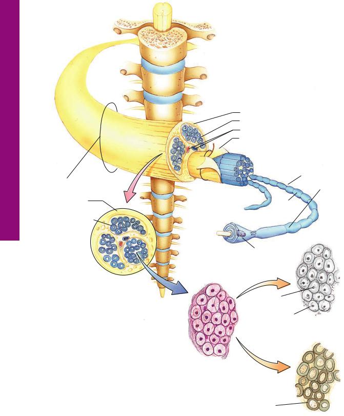
•Peripheral nerves are composed of numerous nerve fibers collected into several fascicles (bundles). These bundles possess a thick connective tissue sheath, the epineurium (see Graphic 7-1).
•Each fascicle within the epineurium is surrounded by a perineurium consisting of an outer connective tissue layer and an inner layer of flattened epithelioid cells.
•Each nerve fiber and associated Schwann cell has its own slender connective tissue sheath, the endoneurium,
whose components include fibroblasts, an occasional macrophage, and collagenous and reticular fibers.
Certain terms must be defined to facilitate understanding of the nervous system. A ganglion is a collection of nerve cell bodies in the PNS, whereas a similar collection of soma in the CNS is called a nucleus. A bundle of axons traveling together in the CNS is known as a tract (or fasciculus or column), whereas a similar bundle in the PNS is known as a peripheral nerve (nerve).
CLINICAL CONSIDERATIONS
Neuroglial Tumors
Almost 50% of the intracranial tumors are due to proliferation of neuroglial cells. Some of the neuroglial tumors, such as oligodendroglioma, are of mild severity, whereas others, such as glioblastoma that are neoplastic cells derived from astrocytes, are highly invasive and usually fatal.
Huntington’s Chorea
Huntington’s chorea is a hereditary condition that becomes evident in the third and fourth decade of life. Initially, this condition affects only the joints but later is responsible for motor dysfunction and dementia. It is thought to be caused by the loss of neurons of the CNS that produce the neurotransmitter GABA (gammaaminobutyric acid). The advent of dementia is thought to be related to the loss of acetylcholine-secreting cells.
Parkinson’s Disease
Parkinson’s disease is related to the loss of the neurotransmitter dopamine in the brain. This crippling
disease causes muscular rigidity, tremor, slow movement, and progressively difficult voluntary movement.
Therapeutic Circumvention of the Blood-Brain Barrier
The selective nature of the blood-brain barrier prevents certain therapeutic drugs and neurotransmitters conveyed by the bloodstream from entering the CNS. For example, the perfusion of mannitol into the blood stream changes the capillary permeability by altering the tight junctions, thus permitting administration of therapeutic drugs. Other therapeutic drugs can be attached to antibodies developed against transferrin receptors located on the luminal aspect of the plasma membranes of these endothelial cells that will permit transport into the CNS.
Guillain-Barré Syndrome
Guillain-Barré syndrome is a form of immune-medi- ated condition resulting in rapidly progressing weakness with possible paralysis of the extremities and,

N E R V O U S T I S S U E 155
occasionally, even of the respiratory and facial muscles. This demyelinating disease is often associated with a recent respiratory or gastrointestinal infection; the muscle weakness reaches its greatest point within 3 weeks of the initial symptoms, and 5% of the afflicted individuals die of the disease. Early recognition of the disease is imperative for complete (or nearly complete) recovery.
Ischemic Injury
Ischemia, the reduction of blood supply to an organ, such as the brain, results in hypoxia and subsequent cell death. The cause of ischemia could be blockage of a blood vessel that serves the particular area, or of another vessel farther away whose responsibility is to supply blood flow to the particular vessels in question. Other causes of diminished blood supply could be lowered blood pressure, cardiac insufficiency, accidental injury to a vessel, as well as a myriad of other factors. Ischemia in the brain is evidenced by the presence of necrotic neurons (different from apoptotic neurons) whose cytoplasm displays a high degree of eosinophilia. These necrotic neurons are known as red neurons.
This devastating condition begins, on the average, around the age of 65 but may affect individuals at a much younger age. The early onset of AD is often masked as symptoms of stress or “senior moments”; however, it progresses to include the incapacity to remember newly acquired information. Additional symptoms develop as the disease continues its progress, namely, personality changes to a more hostile and petulant behavior accompanied by uncertainty and language difficulty. Moreover, the patient experiences an inability to remember previously known personal and general information and the patient eventually becomes unable to take care of bodily functions, resulting in immobility and muscle loss. Individuals diagnosed with AD usually die within 7 to 10 years. Although the cause of the disease is not known, it has been suggested that the intraneuron presence of neurofibrillary tangles, formed by coalescence of modified tau proteins, and the deposition of beta-amyloid-like protein interfere with neuronal function.
This Purkinje cell from the cerebellum of a patient displays a high degree of eosinophilia and is considered to be a red neuron. The presence of such cells indicates that the patient had an ischemic injury of a region of the cerebellum. Note that the cell is reduced in size, its nucleus is pyknotic, and the nucleolus is not evident. If this cell had died because of an apoptotic event, its cytoplasm would be basophilic. (Reprinted with permission from Mills SE, ed. Histology for Pathologists, 3rd ed., Philadelphia: Lippincott, Williams & Wilkins, 2007. p. 287.)
Alzheimer’s Disease
Alzheimer’s disease (AD) is one of the most common forms of dementia that affects approximately 5 million people in the United States and more than 30 million globally.
The neuron depicted in this photomicrographs is from a patient who died as a result of AD. Note the presence of neurofibrillary tangles in its cytoplasm. (Reprinted with permission from Mills SE, Carter D, et al., eds. Sternberg’s Diagnostic Surgical Pathology, 5th ed. Philadelphia: Lippincott, Williams & Wilkins, 2010. p. 441.)
Morphology Nerve Spinal • 1-7 GRAPHIC
156 N E R V O U S T I S S U E
Bundle of nerves
Epineurium
Perineurium
Endoneurium
Nerve trunk (cross section)
Peripheral nerves are composed of bundles of axon and dendrites. Each nerve is enclosed by an epineurium. Bundles (fascicles) of axons and dendrites are surrounded by several layers of flat epithelioid cells, the perineurium, that form occluding junctions with each other. The perineurium is isolated from the connective tissue elements by basal laminae on both its external and internal aspects. Each axon and dendrite is invested by a protective Schwann cell (for insulation and maintenance), which, in turn, is surrounded by its basal lamina and a network of fine reticular fibers, forming the endoneurium.
 Spinal cord
Epineurium
Endoneurium
Blood vessels
Perineurium
 Basal lamina
|
|
Node of |
|
|
|
Ranvier |
|
|
|
Cut end of |
|
|
|
||
Fascicle |
|||
endoneurium |
|||
|
|
||
|
|
Internode |
|
Axon
Schwann cell
Axon of myelinated fiber
Myelin sheath
Nerve fibers (silver stain)
Fascicle detail (H & E stain)
Axon of  myelinated fiber
Myelin sheath
Nerve fibers (osmic stain)
|
|
|
|
|
N E R V O U S T I S S U E |
157 |
|
|
|
|
|
Schwann |
|
|
|
|
|
|
cell |
|
Dendrite |
|
|
|
|
|
|
Unmyelinated |
|
|
|
|
|
|
|
|
|
|
|
|
|
nerve fiber |
|
|
|
|
|
|
Nucleus |
|
|
|
|
|
|
Cell body |
|
|
|
|
|
|
Nissl body |
|
|
|
|
|
|
Axon hillock |
|
|
|
Myelination of |
|
|
|
|
|
|
|
|
|
Node of Ranvier |
|
|
|
nerve fiber |
|
|
Neurilemma sheath |
Axon |
Axon |
|
|||
cell nucleus |
|
|
|
|
||
|
|
|
|
|
||
Myelin sheath |
|
|
|
Schwann |
|
|
Axis cylinder |
|
|
|
|
||
|
|
|
cell |
|
||
|
|
|
|
|
|
|
Schwann |
|
|
|
|
|
|
|
|
|
|
|
||
cell |
|
|
|
|
|
|
Axon |
|
|
|
|
|
|
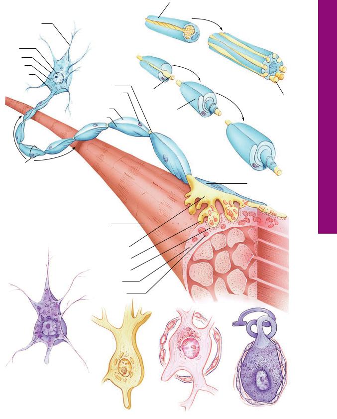
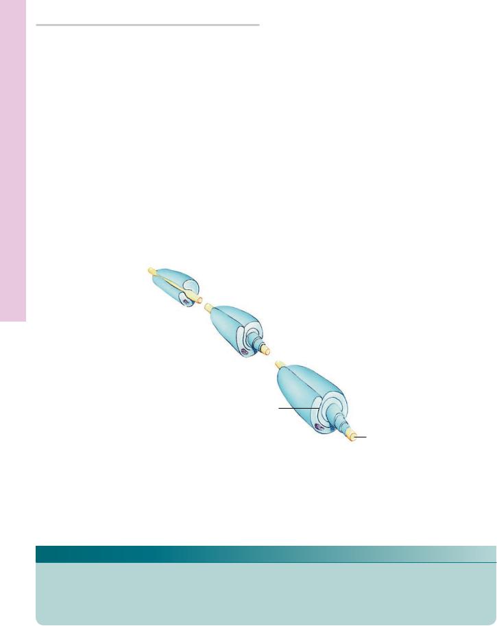
Motor neurons possess numerous |
|
|
dendrites, a large central nucleus, |
|
|
and a long myelinated axon. The |
|
|
RER (Nissl bodies) is segmented by |
|
|
neurotubules and neurofilaments. |
|
|
The axon branches and terminates |
Muscle |
|
as motor end plates. |
||
|
Teloglial cell (shown only in part)
Nerve terminal
Motor end plate
Junctional folds
Sarcoplasm
Mitochondrion
Multipolar cell (spinal cord)
Junctions Myoneural and Neurons• 2-7 GRAPHIC
Multipolar cell |
Multipolar cell |
Unipolar cell |
(cerebellar cortex) |
(autonomic ganglia) |
(cerebrospinal ganglia) |

Cord pinal • S1-7 PLATE
158 N E R V O U S T I S S U E
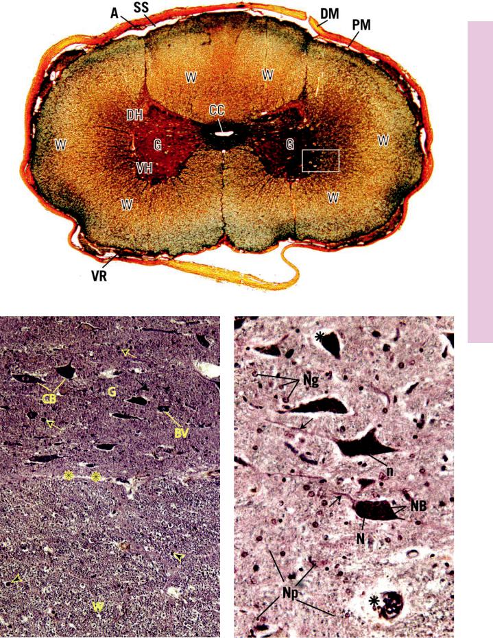
FIGURE 1. Spinal cord. x.s. Cat. Silver stain. Paraffin section. ×21.
The spinal cord is invested by a protective coating, the three-lay- ered meninges. Its outermost fibrous layer, the dura mater (DM), is surrounded by epidural fat, not present in this photomicrograph. Deep to the dura is the arachnoid (A) with its subarachnoid space (SS), which is closely applied to the most intimate layer of the meninges, the vascular pia mater (PM). The spinal cord itself is organized into white matter (W) and gray matter
(G). The former, which is peripherally located and does not contain nerve cell bodies, is composed of nerve fibers, most of which are myelinated, that travel up and down the cord. It is cellular, however, since it houses various types of glial cells. The centrally positioned gray matter contains the cell bodies of the neurons as well as the initial and terminal ends of their processes, many of which are not usually myelinated. These nerve cell processes and those of the numerous glial cells form an intertwined network of fibers that is referred to as the neuropil. The gray matter is subdivided into regions, namely, the dorsal horn (DH), the ventral horn (VH), and the gray commissure (Gc). The central canal (CC) of the spinal cord passes through the gray commissure, dividing it into dorsal and ventral components. Processes of neurons leave and enter the spinal cord as ventral (VR) and dorsal (DR) roots, respectively. A region similar to the boxed area is represented in Figure 2.
FIGURE 2. Spinal cord. x.s. White and gray matter. Human. Paraffin section. ×132.
This photomicrograph represents the boxed region of Figure 1. Observe that the interface between white matter (W) and gray matter (G) is readily evident (asterisks). The numerous nuclei (arrowheads) present in white matter belong to the various neuroglia, which support the axons and dendrites traveling up and down the spinal cord. The large nerve cell bodies (CB) in the ventral horn of the gray matter possess vesicular-appearing nuclei with dense, dark nucleoli. Blood vessels (BV), which penetrate
deep into the gray matter, are surrounded by processes of neuroglial cells, forming the blood-brain barrier, not visible in this photomicrograph. Small nuclei (arrows) in gray matter belong to the neuroglial cells, whose cytoplasm and cellular processes are not evident.
FIGURE 3. Spinal cord. x.s. Ventral horn. Human. Paraffin section. ×270.
The multipolar neurons and their various processes (arrows) are clearly evident in this photomicrograph of the ventral horn. Note the large nucleus (N) and dense nucleolus (n), both of which are characteristic of neurons. Observe the clumps of basophilic material, Nissl bodies (NB), that electron microscopy has demonstrated to be rough endoplasmic reticulum. The small nuclei belong to the various neuroglial cells (Ng), which, along with their processes and processes of the neurons, compose the neuropil (Np), the matted-appearing background substance of gray matter. The white spaces (asterisks) surrounding the soma and blood vessels are due to shrinkage artifacts.
Multipolar cell (spinal cord)
KEY
A |
arachnoid |
G |
gray matter |
PM |
pia mater |
BV |
blood vessel |
Gc |
gray commissure |
SS |
subarachnoid space |
CB |
nerve cell body |
N |
nucleus |
VH |
ventral horn |
CC |
central canal |
N |
nucleolus |
VR |
ventral root |
DH |
dorsal horn |
NB |
Nissl body |
W |
white matter |
DM |
dura mater |
Ng |
neuroglial cell |
|
|
DR |
dorsal root |
Np |
neuropil |
|
|
Cord pinal • S1-7 PLATE
FIGURE 1
FIGURE 2 |
FIGURE 3 |
Microscopy Electron Synapse, erebellum, • C2-7 PLATE
160 N E R V O U S T I S S U E
FIGURE 1. Cerebellum. Human. Paraffin section. ×14.
The cerebellum, in contrast to the spinal cord, consists of a core of white matter (W) and the superficially located gray matter (G). Although it is difficult to tell from this low-magnification photomicrograph, the gray matter is subdivided into three layers: the outer molecular layer (ML), a middle Purkinje cell layer (PL), and the inner granular layer (GL). The less dense appearance of the molecular layer is due to the sparse arrangement of nerve cell bodies, whereas the darker appearance of the granular layer is a function of the great number of darkly staining nuclei packed closely together. A region similar to the boxed area is represented in Figure 2.
FIGURE 3. Purkinje cell. Human cerebellum. Paraffin section. ×540.
This is a higher magnification of the boxed area of Figure 2. The granular layer (GL) of the cerebellum is composed of two cell types, the smaller granule cells (GC) and larger Golgi type II cells (G2). The flask-shaped Purkinje cell (PC) displays its large nucleus
(N) and dendritic tree (D). Nuclei of numerous basket cells (BC) of the molecular layer (ML) as well as the unmyelinated fibers
(UF) of the granule cells are well defined in this photomicrograph. These fibers make synaptic contact (arrows) with the dendritic processes of the Purkinje cells. Inset. Astrocyte. Human cerebellum. Golgi stain. Paraffin section. ×132. Note the numerous processes of this fibrous astrocyte (A) in the white matter of the cerebellum.
FIGURE 2. Cerebellum. Human. Paraffin section. ×132.
This photomicrograph is taken from a region similar to the boxed area in Figure 1. The granular layer (GL) is composed of closely packed granule cells (GC), which, at first glance, resemble lymphocytes due to their dark, round nuclei. Interspersed among these cells are clear spaces called glomeruli or cerebellar islands (CI), where synapses occur between axons entering the cerebellum from outside and dendrites of granule cells. The Purkinje cells (PC) send their axons into the granular layer; their dendrites arborize in the molecular layer (ML). This layer also contains unmyelinated fibers from the granular layer as well as two types of cells, basket cells (BC) and the more superficially located stellate cells (SC). The surface of the cerebellum is invested by pia matter (PM), just barely evident in this photomicrograph. The boxed area is presented at a higher magnification in Figure 3.
FIGURE 4. Synapse. Afferent terminals. Electron microscopy. ×16,200.
The lateral descending nucleus of the fifth cranial nerve displays a primary afferent terminal (AT) that is forming multiple synapses with dendrites (D) and axons (Ax). Observe the presence of synaptic vesicles (SV) in the postsynaptic axon terminals as well as the thickening of the membrane of the primary afferent terminal (arrows). This terminal also houses mitochondria (m) and cisternae (Ci) for the synaptic vesicles. (From Meszler RM. Fine structure and organization of the infrared receptor relays: lateral descending nucleus of V in Boidae and nucleus reticularis caloris in the rattlesnake. J Comp Neurol 1983;220:299–309.)
Multipolar cell (cerebellar cortex)
KEY
A |
fibrous astrocyte |
G |
gray matter |
PC |
Purkinje cell |
AT |
primary afferent terminal |
G2 |
Golgi type II cell |
PL |
Purkinje cell layer |
Ax |
axons |
GC |
granule cell |
PM |
pia mater |
BC |
basket cell |
GL |
granular layer |
SC |
stellate cell |
CI |
cerebellar island |
M |
mitochondrion |
SV |
synaptic vesicle |
Ci |
cistern |
ML |
molecular layer |
UF |
unmyelinated fiber |
D |
dendrite |
N |
nucleus |
W |
white matter |
Microscopy Electron Synapse, erebellum, • C2-7 PLATE
FIGURE 1 |
FIGURE 2 |
FIGURE 3 |
FIGURE 4 |

Cells Neuroglial erebrum, • C3-7 PLATE
162 N E R V O U S T I S S U E
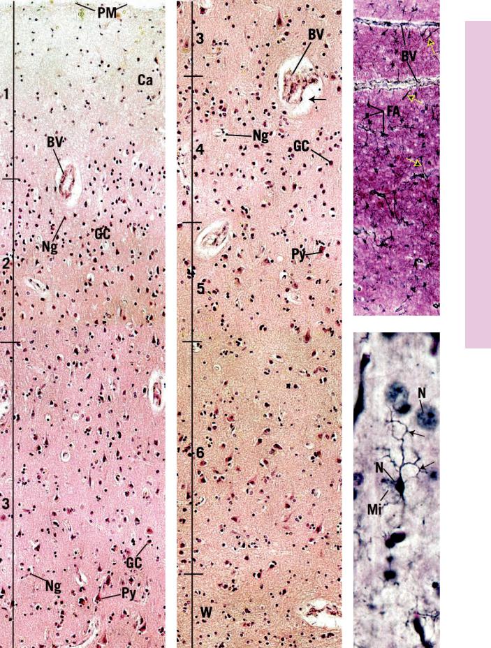
FIGURES 1 and 2. Cerebrum. Human. Paraffin section. ×132.
These figures represent a montage of the entire human cerebral cortex and some of the underlying white matter (W) at a low magnification. Observe that the numerous blood vessels (BV) that penetrate the entire cortex are surrounded by a clear area (arrow), which is due to shrinkage artifact. The six layers of the cortex are not clearly defined but are approximated by brackets. The pia mater (PM), covering the surface of the cortex, is a vascular tissue that provides larger blood vessels as well as capillaries (Ca) that penetrate the brain tissue. Layer one of the cortex is known as the molecular layer (1), which contains numerous fibers and only a few neuron cell bodies. It is difficult to distinguish these somata from the neuroglial cells at this magnification. The second, external granular layer (2) is composed of small granule cells (GC) as well as many neuroglial cells (Ng). The third layer is known as the external pyramidal layer (3), which is the thickest layer in this section of the cerebral cortex. It consists of pyramidal cells (Py) and some granule cells (GC) as well as numerous neuroglia (Ng) interspersed among the soma and fibers. The fourth layer, the internal granular layer (4), is a relatively narrow band whose cell population consists mostly of small and a few large granule cells (GC) and the ever-present neuroglial cells (Ng). The internal pyramidal layer (5) houses medium and large pyramidal cells (Py) as well as the ubiquitous neuroglia (Ng), whose nuclei appear as small dots. Although not evident in this preparation, nerve fibers of the internal band of Baillarger pass horizontally through this layer,
whereas those of the external band of Baillarger traverse the internal granular layer. The deepest layer of the cerebral cortex is the multiform layer (6), which contains cells of various shapes, many of which are fusiform in morphology. Neuroglial cells and Martinotti cells are also present in this layer but cannot be distinguished from each other at this magnification. The white matter (W) appears very cellular, due to the nuclei of the numerous neuroglial cells supporting the cell processes derived from and traveling to the cortex.
FIGURE 3. Astrocytes. Silver stain. Paraffin section. ×132.
This photomicrograph of the white matter of the cerebrum presents a matted appearance due to the interweaving of various nerve cell and glial cell processes. Note also the presence of two blood vessels (BV) passing horizontally across the field. The long processes of the fibrous astrocytes (FA) approach the blood vessels (arrows) and assist in the formation of the blood-brain barrier.
FIGURE 4. Microglia. Silver stain. Paraffin section. ×540.
This photomicrograph is of a section of the cerebral cortex, demonstrating nuclei (N) of nerve cells as well as the presence of microglia (Mi). Note that microglia are very small and possess a dense nucleus (N) as well as numerous cell processes (arrows).
KEY
BV |
blood vessel |
Ng |
neurological cell |
3 |
external pyramidal layer |
Ca |
capillary |
PM |
pia mater |
4 |
internal granular layer |
FA |
fibrous astrocyte |
Py |
pyramidal cell |
5 |
internal pyramidal layer |
GC |
granule cell |
W |
white matter |
6 |
multiform layer |
Mi |
microglia |
1 |
molecular layer |
|
|
N |
nucleus |
2 |
external granular layer |
|
|
Cells Neuroglial erebrum, • C3-7 PLATE
FIGURE 3
FIGURE 1 |
FIGURE 2 |
FIGURE 4 |
Ganglia Sensory Ganglia, ympathetic • S4-7 PLATE
164 N E R V O U S T I S S U E |
|
|
FIGURE 1. Sympathetic ganglion. l.s. Paraffin |
|
FIGURE 2. Sympathetic ganglion. l.s. Paraffin |
section. ×132. |
|
section. ×540. |
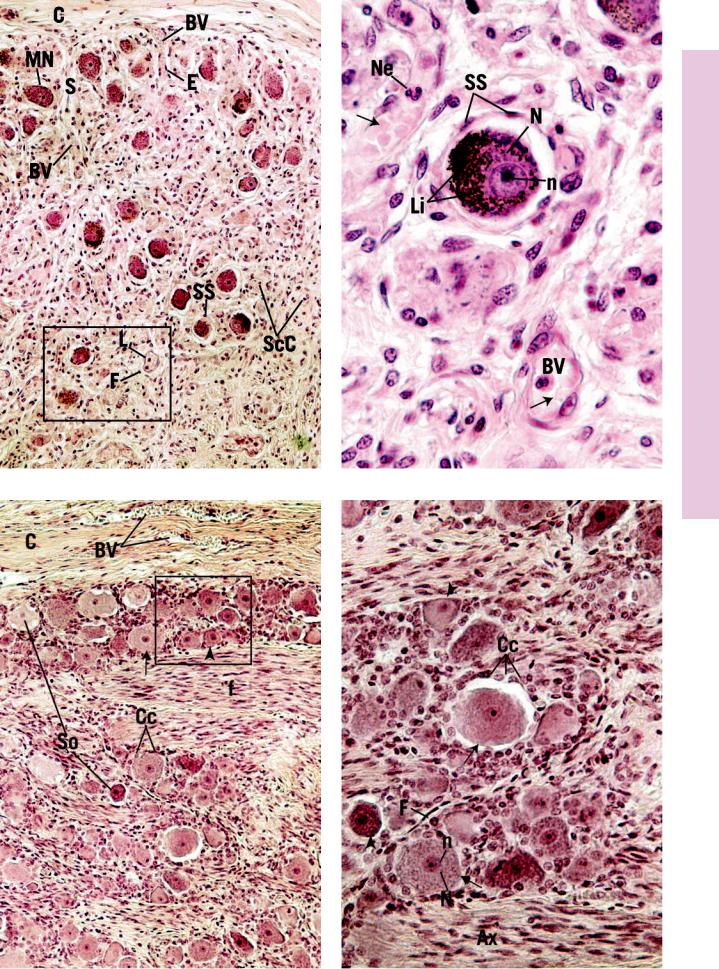
Sympathetic ganglia are structures that receive axons of presynaptic cells, whose soma is within the CNS. Located within the ganglion are somata of postsynaptic neurons upon which the presynaptic cell axons synapse. These ganglia are enveloped by a collagenous connective tissue capsule (C), which sends septa (S) containing blood vessels (BV) within the substance of the ganglion. The arrangement of the cell bodies of the multipolar neurons (MN) within the ganglion appears to be haphazard. This very vascular structure contains numerous nuclei that belong to endothelial cells (E), intravascular leukocytes (L), fibroblasts (F), Schwann cells (ScC), and those of the supporting cells (SS) surrounding the nerve cell bodies. A region similar to the boxed area is presented in Figure 2.
FIGURE 3. Sensory ganglion. l.s. Human. Paraffin section. ×132.
The dorsal root ganglion provides a good representative example of a sensory ganglion. It possesses a vascular (BV) connective tissue capsule (C), which also envelops its sensory root. The neurons of the dorsal root ganglion are pseudounipolar in morphology; therefore, their somata (So) appear spherical in shape. The fibers (f), many of which are myelinated, alternate with rows of cell bodies. Note that some somata are large (arrow), whereas others are small (arrowhead). Each soma is surrounded by neuroectodermally derived capsule cells (Cc). A region similar to the boxed area is presented at a high magnification in Figure 4.
Multipolar cell (autonomic ganglia)
This photomicrograph presents a higher magnification of a region similar to the boxed area of Figure 1. Although neurons of the sympathetic ganglion are multipolar, their processes are not evident in this specimen stained with hematoxylin and eosin. The nucleus (N), with its prominent nucleolus (n), is clearly visible. The cytoplasm contains lipofuscin (Li) a yellowish pigment that is prevalent in neurons of older individuals. The clear space between the soma and the supporting cells (SS) is a shrinkage artifact. Note the numerous blood vessels (BV) containing red blood cells (arrows) and a neutrophil (Ne).
FIGURE 4. Sensory ganglion. l.s. Human. Paraffin section. ×270.
This photomicrograph is a higher magnification of a region similar to the boxed area of Figure 3. The spherical cell bodies display their centrally located nuclei (N) and nucleoli (n). Observe that both small (arrowheads) and large (arrows) somata are present in the field and that the nuclei are not always in the plane of section. Hematoxylin and eosin stain the somata a more or less homogeneous pink, so that organelles such as Nissl substance are not visible. However, the nuclei and cytoplasm of capsule cells (Cc) are clearly evident. Moreover, the small, elongated, densely staining nuclei of fibroblasts (F) are also noted to surround somata, just peripheral to the capsule cells. Axons (Ax) of myelinated nerve fibers belong to the large pseudounipolar neurons.
Unipolar cell (pseudounipolar cell from dorsal root ganglion)
KEY
Ax |
axon |
F |
nerve fiber |
Ne |
neutrophil |
BV |
blood vessel |
L |
leukocyte |
S |
septum |
C |
capsule |
Li |
lipofuscin |
ScC |
Schwann cell |
Cc |
capsule cell |
N |
nucleolus |
So |
soma |
E |
endothelial cell |
MN |
multipolar neuron |
SS |
supporting cell |
F |
fibroblast |
N |
nucleus |
|
|
Ganglia Sensory Ganglia, ympathetic • S4-7 PLATE
FIGURE 1 |
FIGURE 2 |
FIGURE 3 |
FIGURE 4 |

Plexus Choroid Nerve, eripheral • P5-7 PLATE
166 N E R V O U S T I S S U E
FIGURE 1a. Peripheral nerve. l.s. Monkey. Plastic section. ×132.
The longitudinal section of the peripheral nerve fascicle presented in this photomicrograph is enveloped by its perineurium (P), composed of an outer connective tissue layer (CT) and an inner layer of flattened epithelioid cells (E). The perineurium conducts small blood vessels (BV), which are branches of larger vessels traveling in the surrounding epineurium, a structure composed of loose connective tissue with numerous fat cells. The peripheral nerve is composed of numerous nonmyelinated and myelinated nerve fibers, an example of which is presented in Figure 1b. The dense nuclei (arrows) within the nerve fascicle belong to Schwann cells and endoneurial cells. A region similar to the boxed area is presented in Figure 2.
FIGURE 1b. Teased, myelinated nerve fiber. Paraffin section. l.s. ×540.
This longitudinal section of a single myelinated nerve fiber displays its axon (Ax) and the neurokeratin network, the remnants of the dissolved myelin (M). Note the node of Ranvier (NR), a region where two Schwann cells meet. It is here, where the axon is not covered by myelin, that saltatory conduction of impulses occurs. Observe that Schmidt-Lanterman incisures (SL) are clearly evident. These are regions where the cytoplasm of Schwann cells is trapped in the myelin sheath.
FIGURE 3. Peripheral nerve. x.s. Paraffin section. ×132.
This transverse section presents portions of two fascicles, each surrounded by perineurium (P). The intervening loose connective tissue of the epineurium (Ep) with its blood vessels (BV) is clearly evident. The perineurium forms a septum (S), which subdivides this fascicle into two compartments. Note that the axons (Ax) are in the center of the myelin sheath (MS) and occasionally a crescent-shaped nucleus of a Schwann cell (ScC) is evident. The denser, smaller nuclei (arrows) belong to endoneurial cells. Inset.
Peripheral nerve. x.s. Silver stain. Paraffin section. ×540. Silverstained sections of myelinated nerve fibers have the large, clear spaces (arrow) that indicate the dissolved myelin. The axons (Ax) stain well as dark, dense structures, and the delicate endoneurium (En) is also evident.
FIGURE 2. Peripheral nerve. l.s. Paraffin section. ×270.
This is a higher magnification of a region similar to the boxed area of Figure 1a. A distinguishing characteristic of longitudinal sections of peripheral nerves is that they appear to follow a zigzag course, particularly evident in this photomicrograph. The sinuous course of these fibers is accentuated by the presence of nuclei of Schwann cells (ScC), fibroblasts (F), and endothelial cells of capillaries belonging to the endoneurium. Many of these nerve fibers are myelinated (M) as corroborated by the presence of the nodes of Ranvier (NR) and myelin proteins around the axons (Ax).
FIGURE 4. Choroid plexus. Paraffin section. ×270.
The choroid plexus, located within the ventricles of the brain, is responsible for the formation of CSF. This structure is composed of tufts of capillaries (Ca) whose tortuous course is followed by villi (Vi) of the simple cuboidal choroid plexus epithelium (cp). The connective tissue core (CT) of the choroid plexus is contributed by pia-arachnoid, whereas the simple cuboidal epithelium is modified ependymal lining of the ventricle. The clear spaces surrounding the choroid plexus belong to the ventricle of the brain.
Epineurium
 Perineurium
Endoneurium
Nerve trunk (cross section)
KEY
Ax |
axon |
En |
endoneurium |
P |
perineurium |
BV |
blood vessel |
Ep |
epineurium |
S |
septum |
Ca |
capillary |
F |
fibroblast |
ScC |
Schwann cell |
Cp |
choroid plexus epithelium |
M |
myelin |
SL |
Schmidt-Lanterman incisure |
CT |
connective tissue |
MS |
myelin sheath |
Vi |
villus |
E |
epithelioid cell |
NR |
node of Ranvier |
|
|

Plexus Choroid Nerve, eripheral • P5-7 PLATE
FIGURE 1 |
FIGURE 2 |
FIGURE 3 |
FIGURE 4 |
Microscopy Electron Nerve, eripheral • P6-7 PLATE
168 N E R V O U S T I S S U E
FIGURE 1. Peripheral nerve. x.s. Mouse. Electron microscopy. ×33,300.
This electron micrograph presents a cross section of three myelinated and several unmyelinated nerve fibers. Note that the axons (Ax) (although they may be the afferent fibers of pseudounipolar neurons) are surrounded by a thick myelin sheath (MS), peripheral to which is the bulk of the Schwann cell cytoplasm (ScC) housing mitochondria (m), rough endoplasmic reticulum
(rER), and pinocytotic vesicles (PV). The Schwann cell is surrounded by a basal lamina (BL) isolating this cell from the endoneurial connective tissue (CT). The myelin sheath is derived from the plasma membrane of the Schwann cell, which presumably wraps spirally around the axon, resulting in the formation of an external (EM) and internal (IM) mesaxon. The axolemma (Al) is separated from the Schwann cell membrane by a narrow cleft,
the periaxonal space. The axoplasm houses mitochondria (m) as well as neurofilaments (Nf) and neurotubules (Nt). Occasionally, the myelin wrapping is surrounded by Schwann cell cytoplasm on its outer and inner aspects, as in the nerve fiber in the upper right-hand corner. The unmyelinated nerve fibers (f) in the top of this electron micrograph display their relationship to the Schwann cell (ScC). The fibers are positioned in such a fashion that each lies in a complicated membrane-lined groove within the Schwann cell. Some fibers are situated superficially, whereas others are positioned more deeply within the grooves. However, a periaxonal (or peridendritic) space (arrows) is always present.
Mitochondria (m), neurofilaments (Nf), and neurotubules (Nt) are also present. Note that the entire structure is surrounded by a basal lamina (BL), which covers but does not extend into the grooves (arrowheads) housing the nerve fibers. (Courtesy of Dr. J. Strum.)
External mesaxon
Axolemma
Myelination of nerve fiber
KEY
Al |
axolemma |
EM |
external mesaxon |
Nf |
neurofilament |
Ax |
axon |
F |
nerve fiber |
Nt |
neurotubule |
BL |
basal lamina |
IM |
internal mesaxon |
PV |
pinocytotic vesicle |
CT |
endoneurial connective |
M |
mitochondrion |
rER |
rough ER |
|
tissue |
MS |
myelin sheath |
ScC |
Schwann cell cytoplasm |

PLATE 7-6P • eripheral Nerve, Electron Microscopy
FIGURE 1

170 N E R V O U S T I S S U E
Microscopy Electron Body, Cell on Neur• 7-7 PLATE
FIGURE 1
FIGURE 1. Neuron. Lateral descending nucleus. Electron microscopy. ×3,589.
The soma of this neuron presents a typical appearance. Note the large nucleus (N) and nucleolus (n) surrounded by a considerable amount of cytoplasm rich in organelles. Observe the extensive Golgi apparatus (GA), numerous mitochondria (m), and
elements of rough endoplasmic reticulum, which extend into the dendrites (D). Myelinated (M) and nonmyelinated (nM) fibers are also present, as are synapses (arrows) along the cell surface. (From Meszler R, Auker C, Carpenter D. Fine structure and organization of the infrared receptor relay, the lateral descending nucleus of the trigeminal nerve in pit vipers. J Comp Neurol
1981;196:571–584.)