

- •Preface
- •Acknowledgments
- •Reviewers
- •Contents
- •CHAPTER OUTLINE
- •CYTOPLASM
- •Plasmalemma
- •Mitochondria
- •Ribosomes
- •Endoplasmic Reticulum
- •Golgi Apparatus, cis-Golgi Network, and the trans-Golgi Network
- •Endosomes
- •Lysosomes
- •Peroxisomes
- •Proteasomes
- •Cytoskeleton
- •Inclusions
- •NUCLEUS
- •CELL CYCLE
- •CHAPTER OUTLINE
- •EPITHELIUM
- •Epithelial Membranes
- •GLANDS
- •Chapter Summary
- •CHAPTER OUTLINE
- •EXTRACELLULAR MATRIX
- •Fibers
- •Amorphous Ground Substance
- •Extracellular Fluid
- •CELLS
- •CONNECTIVE TISSUE TYPES
- •Chapter Summary
- •CHAPTER OUTLINE
- •CARTILAGE
- •BONE
- •Cells of Bone
- •Osteogenesis
- •Bone Remodeling
- •Chapter Summary
- •CHAPTER OUTLINE
- •FORMED ELEMENTS OF BLOOD
- •Lymphocytes
- •Neutrophils
- •PLASMA
- •COAGULATION
- •HEMOPOIESIS
- •Erythrocytic Series
- •Granulocytic Series
- •Chapter Summary
- •CHAPTER OUTLINE
- •SKELETAL MUSCLE
- •Sliding Filament Model of Muscle Contraction
- •CARDIAC MUSCLE
- •SMOOTH MUSCLE
- •Chapter Summary
- •CHAPTER OUTLINE
- •BLOOD-BRAIN BARRIER
- •NEURONS
- •Membrane Resting Potential
- •Action Potential
- •Myoneural Junctions
- •Neurotransmitter Substances
- •SUPPORTING CELLS
- •PERIPHERAL NERVES
- •Chapter Summary
- •CHAPTER OUTLINE
- •BLOOD VASCULAR SYSTEM
- •HEART
- •ARTERIES
- •Capillary Permeability
- •Endothelial Cell Functions
- •VEINS
- •LYMPH VASCULAR SYSTEM
- •Chapter Summary
- •CHAPTER OUTLINE
- •CELLS OF THE IMMUNE SYSTEM
- •Antigen-Presenting Cells
- •DIFFUSE LYMPHOID TISSUE
- •LYMPH NODES
- •TONSILS
- •SPLEEN
- •THYMUS
- •Chapter Summary
- •CHAPTER OUTLINE
- •PITUITARY GLAND
- •Pars Intermedia
- •Pars Nervosa and Infundibular Stalk
- •Pars Tuberalis
- •THYROID GLAND
- •Parathyroid Glands
- •Suprarenal Glands
- •Cortex
- •Medulla
- •Pineal Body
- •Chapter Summary
- •CHAPTER OUTLINE
- •SKIN
- •Epidermis of Thick Skin
- •Dermis
- •DERIVATIVES OF SKIN
- •Chapter Summary
- •CHAPTER OUTLINE
- •CONDUCTING PORTION OF THE RESPIRATORY SYSTEM
- •Extrapulmonary Region
- •Intrapulmonary Region
- •RESPIRATORY PORTION OF THE RESPIRATORY SYSTEM
- •MECHANISM OF RESPIRATION
- •Chapter Summary
- •CHAPTER OUTLINE
- •ORAL CAVITY AND ORAL MUCOSA
- •Oral Mucosa
- •Tongue
- •Teeth
- •Odontogenesis (See Graphic 13-2)
- •Chapter Summary
- •CHAPTER OUTLINE
- •REGIONS OF THE DIGESTIVE TRACT
- •Esophagus
- •Stomach
- •Small Intestine
- •Large Intestine
- •GUT-ASSOCIATED LYMPHOID TISSUE
- •DIGESTION AND ABSORPTION
- •Carbohydrates
- •Proteins
- •Lipids
- •Water and Ions
- •Chapter Summary
- •CHAPTER OUTLINE
- •MAJOR SALIVARY GLANDS
- •PANCREAS
- •LIVER
- •Exocrine Function of the Liver
- •Endocrine and Other Functions of the Liver
- •GALLBLADDER
- •Chapter Summary
- •CHAPTER OUTLINE
- •KIDNEY
- •Uriniferous Tubule
- •Nephron
- •Collecting Tubules
- •FORMATION OF URINE FROM ULTRAFILTRATE
- •EXTRARENAL EXCRETORY PASSAGES
- •Chapter Summary
- •CHAPTER OUTLINE
- •OVARY
- •Ovarian Follicles
- •Regulation of Follicle Maturation and Ovulation
- •Corpus Luteum and Corpus Albicans
- •GENITAL DUCTS
- •Oviduct
- •Uterus
- •FERTILIZATION, IMPLANTATION, AND THE PLACENTA
- •Fertilization and Implantation
- •Placenta
- •VAGINA
- •EXTERNAL GENITALIA
- •MAMMARY GLANDS
- •Chapter Summary
- •CHAPTER OUTLINE
- •TESTES
- •Spermatogenesis
- •GENITAL DUCTS
- •ACCESSORY GLANDS
- •PENIS
- •Erection and Ejaculation
- •Chapter Summary
- •CHAPTER OUTLINE
- •SENSORY ENDINGS
- •Chapter Summary
- •Terminology of Staining
- •Common Stains Used in Histology
- •Hematoxylin and Eosin
- •Wright Stain
- •Weigert Method for Elastic Fibers and Elastic van Gieson Stain
- •Silver Stain
- •Iron Hematoxylin
- •Bielschowsky Silver Stain
- •Masson Trichrome
- •Periodic Acid-Schiff Reaction (PAS)
- •Alcian Blue
- •von Kossa Stain
- •Sudan Red
- •Mucicarmine Stain
- •Safranin-O
- •Toluidine Blue

resorption and a consequent increase in blood calcium ion concentration.
In the kidneys, PTH prevents urinary calcium loss; thus, ions are returned to the bloodstream.
PTH also controls calcium uptake in the intestines indirectly by modulating kidney production of vitamin D, which is essential for calcium absorption.
Increased levels of PTH cause an elevation in plasma calcium concentration; however, it takes several hours for this level to peak. The concentration of PTH in the blood is also controlled by plasma calcium levels.
•Calcitonin acts as an antagonist to PTH.
•Unlike PTH, calcitonin is fast acting, and since it binds directly to receptors on osteoclasts, it elicits a peak reduction in blood calcium levels within one hour.
•Calcitonin inhibits bone resorption, thus reducing calcium ion levels in the blood. High levels of calcium ions in the blood stimulate calcitonin release.
Absence of parathyroid glands is not compatible with life.
Suprarenal Glands
The suprarenal glands (adrenal glands in some animals) are invested by a connective tissue capsule (see Table 10-2 and Graphics 10-2 and 10-3). The glands are derived from two different embryonic origins, namely, mesodermal epithelium, which gives rise to the cortex, and neuroectoderm, from which the medulla originates. The rich vascular supply of the gland is conveyed to the interior in connective tissue elements derived from the capsule.
E N D O C R I N E S Y S T E M 233
These glucocorticoids regulate carbohydrate metabolism, facilitate the catabolism of fats and proteins, exhibit anti-inflammatory activity, and suppress the immune response.
•The innermost region of the cortex, the zona reticularis, is arranged in anastomosing cords of cells with a rich intervening capillary network.
Zona reticularis cells secrete weak androgens that promote masculine characteristics.
Medulla
Parenchymal cells of the medulla, derived from neural crest material, are disposed in irregularly arranged short cords surrounded by capillary networks. They contain numerous granules that stain intensely when the freshly cut tissue is exposed to chromium salts. This is referred to as the chromaffin reaction, and the cells are called chromaffin cells. There are two populations of chromaffin cells that secrete the two hormones (see Table 10-2) of the suprarenal medulla, mainly
•epinephrine (adrenaline) or
•norepinephrine (noradrenaline).
Secretion of these two catecholamines is directly regulated by preganglionic fibers of the sympathetic nervous system that impinge on the postganglionic sympathetic neuron-like chromaffin cells, which are considered to be related to postganglionic sympathetic neurons (see Graphic 10-3). Catecholamine release occurs in physical and psychological stress. Moreover, scattered, large postganglionic sympathetic ganglion cells in the medulla act on smooth muscle cells of the medullary veins, thus controlling blood flow in the cortex.
Cortex
The cortex is subdivided into three concentric regions or zones that secrete specific hormones (see Table 10-2). Control of these hormonal secretions is mostly regulated by ACTH from the pituitary gland.
•The outermost region, just beneath the capsule, is the zona glomerulosa, where the cells are arranged in arches and spherical clusters with numerous capillaries surrounding them.
Cells of the zona glomerulosa secrete aldosterone, a mineralocorticoid that acts on cells of the distal convoluted tubules of the kidney to modulate water and electrolyte balance.
•The second region, the zona fasciculata, is the most extensive. Its parenchymal cells, usually known as spongiocytes, are arranged in long cords, with numerous capillaries between the cords.
•Zona fasciculata cells secrete cortisol and corticosterone.
Pineal Body
The pineal body (epiphysis) is a projection of the roof of the diencephalon (see Table 10-2 and Graphic 10-2). The connective tissue covering of the pineal body is pia mater, which sends trabeculae and septa into the substance of the pineal body, subdividing it into incomplete lobules. Blood vessels, along with postganglionic sympathetic nerve fibers from the superior cervical ganglia, travel in these connective tissue elements. As the nerve fibers enter the pineal body, they lose their myelin sheath. The parenchyma of the pineal body is composed of pinealocytes and neuroglial cells.
•The pinealocytes form communicating junctions with each other and manufacture melatonin. Interestingly, melatonin is manufactured only at night.
•Neuroglial cells provide physical and nutritional support to pinealocytes.
•The pineal body receives indirect input from the retina, which allows the pineal to differentiate between

234 E N D O C R I N E S Y S T E M
TABLE 10-2 • Hormones of the Thyroid, Parathyroid, Adrenal, and Pineal Glands
Gland |
Hormone |
Stimulating |
Principal Functions |
|
|
Hormone |
|
|
|
|
|
Thyroid gland |
Thyroxine (T4) and |
Thyroid-stimulating |
Promotes gene transcription and stimu- |
|
triiodothyronine (T3) |
hormone |
lates carbohydrate and fat metabolism. |
|
|
|
Increases basal metabolism, growth |
|
|
|
rates, endocrine gland secretion, heart |
|
|
|
rate, and respiration. Decreases cho- |
|
|
|
lesterol, phospholipid, and triglyceride |
|
|
|
levels and lowers body weight |
|
|
|
|
|
Calcitonin |
|
Lowers blood calcium levels by suppress- |
|
(thyrocalcitonin) |
|
ing osteoclastic activity |
|
|
|
|
Parathyroid gland |
Parathyroid hormone |
|
Increases blood calcium levels |
|
|
|
|
Suprarenal |
|
|
|
(adrenal) gland |
|
|
|
|
|
|
|
Cortex |
|
|
|
|
|
|
|
Zona glomerulosa |
Mineralocorticoids |
Angiotensin II and |
Stimulates distal convoluted tubules of |
|
(aldosterone and |
adrenocorticotropic |
the kidney to resorb sodium and excrete |
|
deoxycorticosterone) |
hormone (ACTH) |
potassium |
|
|
|
|
Zona fasciculata |
Glucocorticoids (cortisol |
ACTH |
Controls carbohydrate, lipid, and protein |
|
and corticosterone) |
|
metabolism. Stimulates gluconeogen- |
|
|
|
esis. Reduces inflammation and sup- |
|
|
|
presses the immune system |
|
|
|
|
Zona reticularis |
Androgens (dehydroepian- |
ACTH |
No significant effect in a healthy individual |
|
drosterone and |
|
|
|
androstenedione) |
|
|
|
|
|
|
Medulla |
Catecholamines |
Preganglionic sympathetic |
Epinephrine—increases blood pressure |
|
(epinephrine and |
and splanchnic nerves |
and heart rate, promotes glucose release |
|
norepinephrine) |
|
by the liver |
|
|
|
Norepinephrine—elevates blood pressure |
|
|
|
via vasoconstriction |
|
|
|
|
Pineal body |
Melatonin |
Norepinephrine |
Influences the individual’s diurnal rhythm |
(pineal gland) |
|
|
|
|
|
|
|

day and night, and, in that manner, assists in the establishment of the circadian rhythm.
•The extracellular spaces of the pineal body contain calcified granular material known as brain sand (corpora arenacea), whose significance, if any, is not known.
E N D O C R I N E S Y S T E M 235
It is unclear how the pineal gland functions in humans, but it does exert an affect on the control of the circadian rhythm. Nonetheless, melatonin is used to treat jet lag and in regulating emotional responses related to shortened daylight during winter, a condition called seasonal affective disorder (SAD).
CLINICAL CONSIDERATIONS
Pituitary Gland
Galactorrhea is a condition in which a male produces breast milk or a woman who is not breast-feeding produces breast milk. In men, it is often accompanied by impotence, headache, and loss of peripheral vision and in women by hot flashes, vaginal dryness, and an abnormal menstrual cycle. This rather uncommon condition is usually a result of prolactinoma, a tumor of prolactin-producing cells of the pituitary gland. The condition is usually treated by drug intervention or surgery, or both.
Postpartum pituitary infarct is a condition due the pregnancy-induced enlarging of the pituitary gland and its concomitant increase in its vascularity. The high vascularity of the pituitary increases the chances of a vascular accident, such as hemorrhage, which results in the partial destruction of the pituitary gland. The condition may be severe enough to produce Sheehan’s syndrome, which is recognized by the lack of milk production, the loss of pubic and axillary hair, and fatigue.
Pituitary Somatotrope Adenoma
Pituitary somatotrope adenoma is one of the pituitary adenomas, benign tumors, that are more common in adults than in children. Somatotrope adenomas involve proliferation of acidophils, which produce an excess of growth hormones which, in children, result in gigantism, whereas in adults it results in acromegaly. These acidophils grow slowly and usually do not grow outside the sella turcica. Individuals afflicted with untreated
acromegaly frequently suffer from complications that increase their chance of succumbing to cardiovascular, cerebrovascular, and respiratory problems. These individuals also present with hypertension.
This is a photomicrograph from the pituitary gland of a patient with pituitary somatotrope adenoma. Note that the adenoma cells are arranged in ribbons and cords. (Reprinted with permission from Rubin R, Strayer D, et al., eds. Rubin’s Pathology. Clinicopathologic Foundations of Medicine, 5th ed. Baltimore: Lippincott Williams & Wilkins, 2008, p. 938.)
Thyroid Gland
Graves’ disease is caused by binding of autoimmune IgG antibodies to TSH receptors thus stimulating increased thyroid hormone production (hyperthyroidism). Clinically, the thyroid gland becomes enlarged, and there is evidence of exophthalmic goiter (protrusion of the eyeballs).

236 E N D O C R I N E S Y S T E M
This photomicrograph is from the thyroid gland of a patient with Graves’ disease. Note that the follicular cells are high columnar hyperplastic cells enclosing pinkish colloid that is scalloped along its periphery. (Reprinted with permission from Rubin R, Strayer D, et al., eds. Rubin’s Pathology. Clinicopathologic Foundations of Medicine, 5th ed. Baltimore: Lippincott Williams & Wilkins, 2008, p. 946.)
Parathyroid Gland
Hyperparathyroidism may be due to the presence of a benign tumor causing the excess production of parathyroid hormone (PTH). The high levels of circulating PTH cause increased bone resorption with a resultant greatly elevated blood calcium. The excess calcium may become deposited in arterial walls and in the kidneys, creating kidney stones.
Suprarenal Gland
Addison’s disease is an autoimmune disease, although it may also be the aftermath of tuberculosis. It is
characterized by decreased production of adrenocortical hormones due to the destruction of the suprarenal cortex, and without the administration of steroid treatment, it may have fatal consequences.
This photomicrograph of the adrenal gland of a patient with Addison’s disease displays cortical fibrosis and inflammation, as well as a mass of atrophic cortical cells. (Reprinted with permission from Rubin R, Strayer D, et al., eds. Rubin’s Pathology. Clinicopathologic Foundations of Medicine, 5th ed. Baltimore: Lippincott Williams & Wilkins, 2008, p. 962.)
Type 2 polyglandular syndrome, a hereditary disorder, affects the thyroid and suprarenal glands in such a fashion that they are underactive (although the thyroid may become overactive). Frequently, patients with this disorder also develop diabetes.

|
|
|
|
|
|
|
|
E N D O C R I N E S Y S T E M |
237 |
Neurosecretory cells |
|
Paraventricular |
Supraoptic |
|
|
||||
located in hypothalamus |
|
|
|
|
|
||||
|
|
|
|
|
|||||
|
nuclei |
|
nuclei |
|
|
|
|
||
secrete releasing and |
|
|
|
|
|
|
|||
|
|
|
|
|
|
|
|
||
inhibitory hormones |
|
|
|
|
Hypothalamus |
|
|||
Water absorption |
|
|
|
|
|
|
|
|
|
|
|
|
|
|
|
|
|||
|
|
|
|
|
|
|
|
|
|
|
Median |
|
|
|
|
|
|
|
|
|
eminence |
|
|
|
|
|
|
|
|
Kidney |
Hypophyseal |
|
|
|
|
|
|
|
|
stalk |
|
|
Portal system |
|
|
|
|
||
|
Pars |
|
|
|
Pars distalis |
|
|||
|
|
|
|
|
|
|
|
||
|
nervosa |
|
|
|
|
|
|
|
|
Contraction |
ADH |
|
|
||
|
|
|
|
|
|
Acidophil Basophil
Uterus Oxytocin
Myoepithelial contraction
Mammary gland |
Growth hormone |
FSH |
|
|
|
|
via insulin-like |
|
|
growth factors |
LH |
|
I and II |
|
|
|
|
|
Prolactin |
|
Growth
Bone
Muscle
Hyperglycemia
|
Adipose |
Elevation |
tissue |
|
|
of free |
Mammary |
fatty acids |
|
|
gland |
|
Milk |
|
secretion |
ACTH
TSH
Secretion
Adrenal cortex
Secretion
Thyroid
Spermatogenesis
Androgen secretion
Testis
Follicular development:
Ovary estrogen secretion
Ovulation: progesterone secretion
Hormones Its and Gland Pituitary• 1-10 GRAPHIC

Glands Endocrine • 2-10 GRAPHIC
238 E N D O C R I N E S Y S T E M
Thyroid
Gland
Follicular cell
Parafollicular
cell
Parathyroid Gland
Oxyphil cell
Chief cell
Capsule
Pineal
Body
Pinealocytes
|
Suprarenal |
Cortex |
Gland |
Medulla |
|
Z. reticularis
⎞
Z. fasciculata
Z. glomerulosa
Capsule
 Capsule
Neuroglial cell

E N D O C R I N E S Y S T E M 239
Preganglionic sympathetic neuron and fiber
Postganglionic sympathetic neuron and fiber 
Dorsal root ganglion
Thoracic |
Ventral root |
|
spinal cord |
||
ganglion |
||
|
Sympathetic Collateral chain ganglion ganglion
Stomach, small intestine,
large intestine Medulla of suprarenal gland
Gland Suprarenal the of Medulla the and Viscera the of Innervation Sympathetic• 3-10 GRAPHIC

Gland y Pituitar• 1-10 PLATE
240 |
E N D O C R I N E S Y S T E M |
|
||
FIGURE 1. Pituitary gland. Paraffin section. ×19. |
|
by an extensive capillary network. However, these capillaries are |
||
wide, endothelially lined vessels known as sinusoids (S). The |
||||
This survey photomicrograph of the pituitary gland demonstrates |
||||
parenchymal cells of the anterior pituitary are divided into two |
||||
the relationship of the gland to the hypothalamus (H), from |
groups: chromophils (Ci) and chromophobes (Co). With hema- |
|||
which it is suspended by the infundibulum. The infundibulum is |
toxylin and eosin, the distinction between chromophils and chro- |
|||
composed of a neural portion, the infundibular stem (IS) and the |
mophobes is obvious. The former stain blue or pink, whereas the |
|||
surrounding pars tuberalis (PT). Note that the third ventricle |
latter stain poorly. The boxed area is presented at a higher magni- |
|||
(3V) of the brain is continuous with the infundibular recess (IR). |
fication in Figure 3. |
|||
The largest portion of the pituitary is the pars anterior (PA), which |
|
|||
is glandular and secretes numerous hormones. The neural com- |
FIGURE 3. Pituitary gland. Pars anterior. Paraffin |
|||
ponent of the pituitary gland is the pars nervosa (PN), which does |
||||
section. ×270. |
||||
not manufacture its hormones but stores and releases them. Even |
||||
at this magnification, its resemblance to the brain tissue and to |
This is a higher magnification of the boxed area of Figure 2. |
|||
the substance of the infundibular stalk is readily evident. Between |
Note that the chromophobes (Co) do not take up the stain |
|||
the pars anterior and pars nervosa is the pars intermedia (PI), |
well and only their nuclei (N) are demonstrable. These cells are |
|||
which frequently presents an intraglandular cleft (IC), a remnant |
small; therefore, chromophobes are easily recognizable since |
|||
of Rathke’s pouch. |
their nuclei appear to be clumped together. The chromophils |
|||
|
|
|
may be classified into two categories by their affinity to his- |
|
FIGURE 2. Pituitary gland. Pars anterior. Paraffin |
tologic dyes: blue-staining basophils (B) and pink-colored |
|||
acidophils (A). The distinction between these two cell types in |
||||
section. ×132. |
||||
|
|
|
sections stained with hematoxylin and eosin is not as apparent |
|
The pars anterior is composed of large cords of cells that branch |
||||
as with some other stains. Note also the presence of a large |
||||
and anastomose with each other. These cords are surrounded |
sinusoid (S). |
|||
}Hypothalamus
Pars tuberalis
Infundibular stem
Pars intermedia
Pars |
} |
|
|
nervosa |
Pars anterior |
||
|
|||
|
Acidophil |
Basophil |
|
|
} |
||
|
Chromophobes |
||
Pituitary gland
KEY
A |
acidophils |
IC |
intraglandular cleft |
PI |
pars intermedia |
B |
basophils |
IR |
infundibular recess |
PN |
pars nervosa |
Ci |
chromophils |
IS |
infundibular stem |
PT |
pars tuberalis |
Co |
chromophobes |
N |
nucleus |
S |
sinusoids |
H |
hypothalamus |
PA |
pars anterior |
3V |
third ventricle |
3V |
|
H |
|
|
|
|
|
H |
|
|
|
IR |
PT |
|
|
3V |
IS |
|
|
|
|
||
|
PT |
|
|
|
|
|
|
PA |
|
IC |
PN |
|
|
|
|
|
|
PI |
|
FIGURE 1
FIGURE 2 |
FIGURE 3 |
Gland y Pituitar• 1-10 PLATE

Gland y Pituitar• 2-10 PLATE
242 E N D O C R I N E S Y S T E M
FIGURE 1. Pituitary gland. Paraffin section. ×540.
It is somewhat difficult to discriminate between the acidophils (A) and basophils (B) of the pituitary gland stained with hematoxylin and eosin. Even at high magnification, such as in this photomicrograph, only slight differences are noted. Acidophils stain pinkish and are slightly smaller in size than the basophils, which stain pale blue. In a black and white photomicrograph, basophils appear darker than acidophils. Chromophobes (Co) are readily recognizable, since their cytoplasm is small and does not take up stain. Moreover, cords of chromophobes present clusters of nuclei (N) crowded together.
FIGURE 2. Pituitary gland. Pars intermedia. Human. Paraffin section. ×270.
The pars intermedia of the pituitary gland is situated between the pars anterior (PA) and the pars nervosa (PN). It is characterized by basophils (B), which are smaller than those of the pars anterior. Additionally, the pars intermedia contains colloid (Cl)-filled follicles, lined by pale, small, low cuboidal-shaped cells (arrows). Note that some of the basophils extend into the pars nervosa. Numerous blood vessels (BV) and pituicytes (P) are evident in this area of the pars nervosa.
FIGURE 3. Pituitary gland. Pars nervosa. Paraffin section. ×132.
The pars nervosa of the pituitary gland is composed of elongated cells with long processes known as pituicytes (P), which are thought to be neuroglial in nature. These cells, which possess more or less oval nuclei, appear to support numerous unmyelinated nerve fibers traveling from the hypothalamus via the hypothalamo-hypophyseal tract. These nerve fibers cannot be distinguished from the cytoplasm of pituicytes in a hematoxylin and eosin–stained preparation. Neurosecretory materials pass along these nerve fibers and are stored in expanded regions at the termination of the fibers, which are then referred to as Herring bodies (HB). Note that the pars nervosa resembles neural tissue. The boxed area is presented at a higher magnification in Figure 4.
FIGURE 4. Pituitary gland. Pars nervosa. Paraffin section. ×540.
This photomicrograph is a higher magnification of the boxed area of Figure 3. Note the numerous more or less oval nuclei (N) of the pituicytes, some of whose processes (arrows) are clearly evident at this magnification. The unmyelinated nerve fibers and processes of pituicytes make up the cellular network of the pars nervosa. The expanded terminal regions of the nerve fibers, which house neurosecretions, are known as Herring bodies (HB). Also observe the presence of blood vessels (BV) in the pars nervosa.
Pars nervosa
Pars intermedia
}
Acidophil Basophil
|
|
|
} |
|
|
|
|
|
Chromophobes |
|
|
|
|
|
Pituitary gland |
|
|
KEY |
|
|
|
|
|
A |
acidophils |
Co |
chromophobes |
PA |
pars anterior |
B |
basophils |
HB |
Herring bodies |
PN |
pars nervosa |
BV |
blood vessels |
N |
nucleus |
|
|
CL |
colloid |
P |
pituicytes |
|
|

Gland y Pituitar• 2-10 PLATE
FIGURE 1 |
FIGURE 2 |
FIGURE 3 |
FIGURE 4 |
Gland Parathyroid Gland, hyroid • T3-10 PLATE
244 E N D O C R I N E S Y S T E M |
|
|
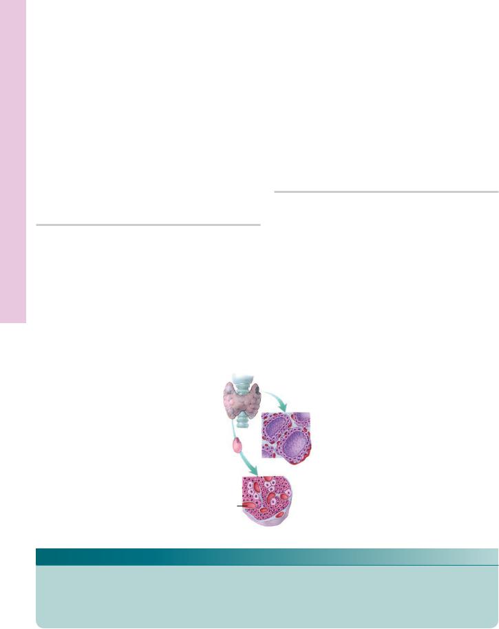
FIGURE 1. Thyroid gland. Monkey. Plastic section. |
|
FIGURE 2. Thyroid gland. Monkey. Plastic section. |
×132. |
|
×540. |
The capsule of the thyroid gland sends septa of connective tissue into the substance of the gland, subdividing it into incomplete lobules. This photomicrograph presents part of a lobule displaying many follicles (F) of varied sizes. Each follicle is surrounded by slender connective tissue (CT), which supports the follicles and brings blood vessels (BV) in close approximation. The follicles are composed of follicular cells (FC), whose low cuboidal morphology indicates that the cells are not producing secretory product. During the active secretory cycle, these cells become taller in morphology. In addition to the follicular cells, another parenchymal cell type is found in the thyroid gland. These cells do not border the colloid, are located on the periphery of the follicles, and are known as parafollicular cells (PF) or C cells. They are large and possess centrally placed round nuclei, and their cytoplasm appears paler.
FIGURE 3. Thyroid and parathyroid glands. Monkey. Plastic section. ×132.
Although the parathyroid (PG) and thyroid glands (TG) are separated by their respective capsules (Ca), they are extremely close to each other. The capsule of the parathyroid gland sends trabeculae (T) of connective tissue carrying blood vessels (BV) into the substance of the gland. The parenchyma of the gland consists of two types of cells, namely, chief cells (CC), also known as principal cells, and oxyphil cells (OC). Chief cells are more numerous and possess darker staining cytoplasm. Oxyphil cells stain lighter and are usually larger than chief cells, and their cell membranes are evident. A region similar to the boxed area is presented at a higher magnification in Figure 4.
The thyroid follicle (F) presented in this photomicrograph is surrounded by several other follicles and intervening connective tissue (CT). Nuclei (N) in the connective tissue may belong either to endothelial cells or to connective tissue cells. Since most capillaries are collapsed in excised thyroid tissue, it is often difficult to identify endothelial cells with any degree of certainty. The follicular cells (FC) are flattened, indicating that these cells are not actively secreting thyroglobulin. Note that the follicles are filled with a colloid (Cl) material. Observe the presence of a parafollicular cell (PF), which may be distinguished from the surrounding cells by its pale cytoplasm (arrow) and larger nucleus.
FIGURE 4. Parathyroid gland. Monkey. Plastic section. ×540.
This photomicrograph is a region similar to the boxed area of Figure 3. The chief cells (CC) of the parathyroid gland form small cords surrounded by slender connective tissue (CT) elements and blood vessels (BV). The nuclei (N) of connective tissue cells may be easily recognized due to their elongated appearance. Oxyphil cells (OC) possess a paler cytoplasm, and frequently, the cell membranes are evident (arrows). The glands of older individuals may become infiltrated by adipocytes.
Thyroid gland
|
|
|
|
|
|
|
|
Blood vessel |
|
|
|
|
|
|
|
|
|
Parathyroid gland |
|
|
|
|
|
|
|
Follicular cell |
|
|
|
|
|
|
|
||
|
|
|
|
|
|
|
|
|
Blood vessel |
|
|
|
|
Oxyphil cell |
|||
|
|
|
|
|||||
|
|
|
|
Chief cell |
||||
|
|
|
|
|||||
|
|
|
|
|
Capsule |
|||
|
|
|
|
|||||
KEY
BV |
blood vessels |
F |
follicle |
PG |
parathyroid gland |
Ca |
capsule |
FC |
follicular cells |
T |
trabeculae |
CC |
chief cells |
N |
nucleus |
TG |
thyroid gland |
CL |
colloid |
OC |
oxyphil cells |
|
|
CT |
connective tissue |
PF |
parafollicular cells |
|
|

Gland Parathyroid Gland, hyroid • T3-10 PLATE
FIGURE 1 |
FIGURE 2 |
FIGURE 3 |
FIGURE 4 |

Gland uprarenal • S4-10 PLATE
246 E N D O C R I N E S Y S T E M
FIGURE 1. Suprarenal gland. Paraffin section. ×14.
The suprarenal gland, usually embedded in adipose tissue (AT), is invested by a collagenous connective tissue capsule (Ca) that provides thin connective tissue elements that carry blood vessels and nerves into the substance of the gland. Since the cortex (Co) of the suprarenal gland completely surrounds the flattened medulla (M), it appears duplicated in any section that completely transects the gland. The cortex is divided into three concentric regions: the outermost zona glomerulosa (ZG), middle zona fasciculata (ZF), and the innermost zona reticularis (ZR). The medulla, which is always bounded by the zona reticularis, possesses several large veins (V), which are always accompanied by a considerable amount of connective tissue.
FIGURE 2. Suprarenal gland. Cortex. Monkey. Plastic section. ×132.
The collagenous connective tissue capsule (Ca) of the suprarenal gland is surrounded by adipose tissue through which blood vessels (BV) and nerves (Ne) reach the gland. The parenchymal cells of the cortex, immediately deep to the capsule, are arranged in an irregular array, forming the more or less oval to round clusters or arch-like cords of the zona glomerulosa (ZG). The cells of the zona fasciculata (ZF) form long, straight columns of cords oriented radially, each being one to two cells in width. These cells are larger than those of the ZG. They present a vacuolated appearance due to the numerous lipid droplets that were extracted during processing and are often referred to as spongiocytes (Sp). The interstitium is richly vascularized by blood vessels (BV).
FIGURE 3. Suprarenal gland. Monkey. Plastic |
FIGURE 4. Suprarenal gland. Monkey. Plastic |
section. ×132. |
|
|
section. ×540. |
|
The columnar arrangement of the cords of the zona fasciculata (ZF) is readily evident by viewing the architecture of the blood vessels indicated by the arrows. The cells in the deeper region of the ZF are smaller and appear denser than the more superficially located spongiocytes (Sp). Cells of the zona reticularis (ZR) are arranged in irregular, anastomosing cords whose interstices contain wide capillaries. The cords of the ZR merge almost imperceptibly with those of the ZF. This is a relatively narrow region of the cortex. The medulla (M) is clearly evident since its cells are much larger than those of the ZR. Moreover, numerous large veins (V) are characteristic of the medulla.
The capsule (Ca) of the suprarenal gland displays its collagen fibers (Cf) and the nuclei (N) of the fibroblasts. The zona glomerulosa (ZG), which occupies the upper part of the photomicrograph, displays relatively small cells with few vacuoles (arrows). The lower part of the photomicrograph demonstrates the zona fasciculata (ZF), whose cells are larger and display a more vacuolated (arrowheads) appearance. Note the presence of connective tissue (CT) elements and blood vessels (BV) in the interstitium between cords of parenchymal cells.
Cortex
Medulla
Z. reticularis
Z. fasciculata
Z. glomerulosa
Capsule
Suprarenal gland
KEY
AT |
adipose tissue |
CT |
connective tissue |
V |
veins |
BV |
blood vessels |
M |
medulla |
ZF |
zona fasciculata |
Ca |
capsule |
N |
nuclei |
ZG |
zona glomerulosa |
Cf |
collagen fibers |
Ne |
nerves |
ZR |
zona reticularis |
Co |
cortex |
Sp |
spongiocytes |
|
|

Gland uprarenal • S4-10 PLATE
FIGURE 1 |
FIGURE 2 |
FIGURE 3 |
FIGURE 4 |

Body Pineal Gland, uprarenal • S5-10 PLATE
248 E N D O C R I N E S Y S T E M |
|
|
FIGURE 1. Suprarenal gland. Cortex. Monkey. Plastic |
|
FIGURE 2. Suprarenal gland. Medulla. Monkey. |
section. ×540. |
|
Plastic section. ×270. |
The upper part of this photomicrograph presents the border between the zona fasciculata (ZF) and the zona reticularis (ZR). Note that the spongiocytes (Sp) of the fasciculata are larger and more vacuolated than the cells of the reticularis. The parenchymal cells of the zona reticularis are arranged in haphazardly anastomosing cords. The interstitium of both regions houses large capillaries containing red blood cells (RBC). Inset. Zona fasciculata. Monkey. Plastic section. ×540. The spongiocytes (Sp) of the zona fasciculata are of two different sizes. Those positioned more superficially in the cortex, as in this inset, are larger and more vacuolated (arrows) than spongiocytes close to the zona reticularis.
FIGURE 3. Pineal body. Human. Paraffin section. ×132.
The pineal body is covered by a capsule of connective tissue derived from the pia mater. From this capsule, connective tissue trabeculae (T) enter the substance of the pineal body, subdividing it into numerous incomplete lobules (Lo). Nerves and blood vessels (BV) travel in the trabeculae to be distributed throughout the pineal, providing it with a rich vascular supply. In addition to endothelial and connective tissue cells, two other types of cells are present in the pineal, namely, the parenchymal cells, known as pinealocytes (Pi), and neuroglial supporting cells (Ng). A characteristic feature of the pineal body is the deposit of calcified material known as corpora arenacea or brain sand (BS). The boxed area is presented at a higher magnification in Figure 4.
The cells of the adrenal medulla, often referred to as chromaffin cells (ChC), are arranged in round to ovoid clusters or in irregularly arranged short cords. The cells are large and more or less round to polyhedral in shape with a pale cytoplasm (Cy) and vesicular appearing nucleus (N), displaying a single, large nucleolus (n). The interstitium presents large veins (V) and an extensive capillary (Cp) network. Large ganglion cells are occasionally noted.
FIGURE 4. Pineal body. Human. Paraffin section. ×540.
This photomicrograph is a higher magnification of the boxed area of Figure 3. With the use of hematoxylin and eosin stain, only the nuclei of the two cell types are clearly evident. The larger, paler, more numerous nuclei belong to the pinealocytes (Pi). The smaller, denser nuclei are those of the neuroglial cells (Ng). The pale background is composed of the long, intertwining processes of these two cell types. The center of the photomicrograph is occupied by brain sand (BS). Observe that these concretions increase in size by apposition of layers on the surface of the calcified material, as may be noted at the arrow.
Cortex
Medulla
|
|
Z. reticularis |
|
|
|
|
Spongiocytes |
Z. fasciculata |
|
|
|
|
|
Z. glomerulosa |
|
Neuroglial cell |
|
|
|
Capsule |
|
|
|
|
|
|
|
|
|
|
Suprarenal gland |
|
Pineal body |
|
|
KEY |
|
|
|
|
|
BS |
brain sand |
N |
nucleus |
T |
trabeculate |
BV |
blood vessels |
n |
nucleolus |
V |
veins |
ChC |
chromaffin cells |
Ng |
neuroglial cells |
ZF |
zona fasciculata |
Cp |
capillaries |
Pi |
pinealocytes |
ZR |
zona reticularis |
Cy |
cytoplasm |
RBC |
red blood cells |
|
|
Lo |
lobules |
Sp |
spongiocytes |
|
|

Body Pineal Gland, uprarenal • S5-10 PLATE
FIGURE 1 |
FIGURE 2 |
FIGURE 3 |
FIGURE 4 |
Microscopy Electron Gland, y Pituitar• 6-10 PLATE
FIGURE 1
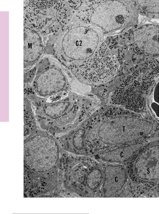
FIGURE 1. Pituitary gland. Pars anterior. Electron microscopy. ×4,950.
Although considerable controversy surrounds the precise fine structural identification of the cells of the pars anterior, it is reasonably certain that the several cell types presented in this electron micrograph are acidophils, basophils, and chromophobes, as
observed by light microscopy. The acidophils are somatotropes (S) and mammotropes (M), whereas only two types of basophils are included in this electron micrograph, namely, type II gonadotropes (G2) and thyrotropes (T). The chromophobes (C) may be recognized by the absence of secretory granules in their cytoplasm. (From Poole M. Cellular distribution within the rat adenohypophysis: a morphometric study. Anat Rec 1982;204:45–53.)
Microscopy Electron Gland, y Pituitar• 7-10 PLATE
FIGURE 1
FIGURE 1. Pituitary gland. Rat. Electron microscopy. ×8,936.
The pars distalis of the rat pituitary houses various cell types, two of which are represented here. The granule-containing gonadotrophs (GN) are surrounded by nongranular folliculostellate cells
(FS), whose processes are demarcated by arrows. The functions of folliculostellate cells are in question, although some believe them to be supportive, phagocytic, regenerative, or secretory in nature. (From Strokreef JC, Reifel CW, Shin SH. A possible phagocytic role for folliculo-stellate cells of anterior pituitary following estrogen withdrawal from primed male rats. Cell Tissue Res 1986;243:255–261.)