

Cartilage and bone form the supporting tissues of the body. In these specialized connective tissues, as in other connective tissues, the extracellular ele-
ments dominate their microscopic appearance.
CARTILAGE
Cartilage forms the supporting framework of certain organs, the articulating surfaces of bones, and the greater part of the fetal skeleton, although most of that will be replaced by bone (see Graphic 4-2).
•There are three types of cartilage in the body, namely, hyaline cartilage, elastic cartilage, and fibrocartilage (see Table 4-1).
Cartilage is a nonvascular, strong, and somewhat pliable structure composed of a firm matrix of proteoglycans whose main glycosaminoglycans are chondroitin-4- sulfate and chondroitin-6-sulfate. The fibrous and cellular components of cartilage are embedded in this matrix. The fibers are either solely collagenous or a combination of elastic and collagenous, depending on the cartilage type.
The cellular components are the
•chondrocytes, which are housed individually in small spaces known as lacunae.
•chondroblasts and chondrogenic cells, both of which are located in the perichondrium.
Most cartilage is surrounded by a dense irregular collagenous connective tissue membrane, the perichondrium, which has an outer fibrous layer and an inner chondrogenic layer.
•The outer fibrous layer, although poor in cells, is composed mostly of fibroblasts and collagen fibers.
•The inner cellular or chondrogenic layer is composed of chondroblasts and chondrogenic cells. The latter give rise to chondroblasts, cells that are responsible for secreting the cartilage matrix. It is from this layer that the cartilage may grow appositionally.
•As the chondroblasts secrete matrix and fibers around themselves, they become incarcerated in their own secretions and are then termed chondrocytes.
These chondrocytes, at least in young cartilage, possess the capacity to undergo cell division, thus contributing to the growth of the cartilage from within (interstitial growth).
When this occurs, each lacuna may house several chondrocytes and is referred to as a cell nest (isogenous group).
In order for these cells to manufacture type II collagen and the other components of the cartilage matrix, these cells need Sox9, a transcription factor.
C A R T I L A G E A N D B O N E 81
•Hyaline cartilage is surrounded by a well-defined perichondrium. The type II collagen fibers of the matrix of this cartilage are mostly very fine and are, therefore, fairly well masked by the surrounding glycosaminoglycans, giving the matrix a smooth, glassy appearance. The acidic nature of the proteoglycans, combined with the enormous size of the proteoglycan-hyaluronic acid complex, results in these molecules possessing huge domains and tremendous capacity for binding cations and water. Additionally, the matrix contains glycoproteins that help the cells maintain contact with the intercellular matrix. Hyaline cartilage is present at the articulating surfaces of most bones, the C rings of the trachea, and the laryngeal, costal, and nasal cartilages, among others.
•Elastic cartilage also possesses a perichondrium. The matrix, in addition to the type II collagen fibers, contains a wealth of coarse elastic fibers that impart to it a characteristic appearance. This cartilage is located in areas like the epiglottis, external ear and ear canal, and some of the smaller laryngeal cartilages.
•Fibrocartilage differs from elastic and hyaline cartilage in that it has no perichondrium. Additionally, the chondrocytes are smaller and are usually oriented in parallel longitudinal rows. The matrix of this cartilage contains a large number of thick type I collagen fiber bundles between the rows of chondrocytes. Fibrocartilage is present in only a few places, namely, in some symphyses, the eustachian tube, intervertebral (and some articular) discs, and certain areas where tendons insert into bone (Table 4-1).
BONE
Bone has many functions, including support, protection, mineral storage, and hemopoiesis. At the specialized cartilage-covered ends, it permits articulation or movement. Bone is a vascular connective tissue consisting of cells and calcified extracellular materials, known as the matrix. The calcified matrix is composed of
•Sixty five percent minerals (mostly calcium hydroxyapatite crystals)
•Thirty five percent organic matter (type I collagen, sulfated glycoproteins, and proteoglycans) including bound water.
The presence of these crystals makes bone the body’s storehouse of calcium, phosphate, and other inorganic ions. Thus, bone is in a dynamic state of flux, continuously gaining and losing inorganic ions to maintain the body’s calcium and phosphate homeostasis.
Bone may be sponge-like (cancellous) or dense (compact).

82 C A R T I L A G E A N D B O N E
TABLE 4-1 • |
Cartilage Types, Characteristics, and Locations |
|
|
Type |
Characteristics |
Perichondrium |
Locations (Major Samples) |
|
|
|
|
Hyaline |
Chondrocytes arranged in |
Usually present except at |
Articular ends of long bones, |
|
groups within a basophilic matrix |
articular surfaces |
ventral rib cartilage, tem- |
|
containing type II collagen |
|
plates for endochondral |
|
|
|
bone formation |
|
|
|
|
Elastic |
Chondrocytes compacted in |
Present |
Pinna of ear, auditory canal, |
|
matrix containing type II |
|
laryngeal cartilages |
|
collagen and elastic fibers |
|
|
|
|
|
|
Fibrocartilage |
Chondrocytes arranged in rows |
Absent |
Intervertebral discs, pubic |
|
in an acidophilic matrix containing |
|
symphysis |
|
type I collagen bundles in rows |
|
|
|
|
|
|
•Cancellous bone, like that present inside the epiphyses (heads) of long bones, is always surrounded by compact bone.
Cancellous bone has large, open spaces surrounded by thin, anastomosing plates of bone.
The large spaces are marrow spaces, and the plates of bones are trabeculae composed of several layers or lamellae.
•Compact bone is much denser than cancellous bone. Its spaces are much reduced in size, and its lamellar organization is much more precise and thicker.
Compact bone is always covered and lined by soft connective tissues.
The marrow cavity is lined by an endosteum composed of osteoprogenitor cells (previously known as osteogenic cells), osteoblasts, and occasional osteoclasts.
The periosteum covering the outer surface of compact bone is composed of an
outer fibrous layer consisting mainly of collagen fibers and populated by fibroblasts.
The inner osteogenic layer consists of some collagen fibers and mostly osteoprogenitor cells and their progeny, the osteoblasts.
The periosteum is affixed to bone via Sharpey’s fibers, collagenous bundles trapped in the calcified bone matrix during ossification.
Cells of Bone
Bone possesses four types of cells: osteoprogenitor cells, osteoblasts, osteocytes, and osteoclasts.
•Osteoprogenitor cells give rise to osteoblasts under the influence of transforming growth factor-β and bone morphogenic protein. However, under hypoxic conditions, osteoprogenitor cells become chondrogenic cells; therefore, these two cells are really the
same cell that expresses different factors under differing oxygen tension.
•Osteoblasts elaborate bone matrix, become surrounded by the matrix they synthesized, and calcify the matrix via matrix vesicles that they release.
When osteoblasts are quiescent, they lose much of their protein synthetic machinery and resemble osteoprogenitor cells.
Osteoblasts function not only in the control of bone matrix mineralization but also in the formation, recruitment, and maintenance of osteoclasts as well as for the initiation of bone resorption.
Osteoblasts express alkaline phosphatase on their cell membranes.
Osteoblasts possess parathyroid receptors on their cell membrane, and in the presence of parathormone, they release macrophage colony–stimulating factor that induces the formation of osteoclast precursors.
Additionally, osteoblasts have expressed on their cell surface RANKL (receptor for activation of nuclear factor kappa B ligand), a molecule that when contacted by the preosteoclast’s surfacebound RANK induces preosteoclasts to differentiate into osteoclasts.
Osteoblasts release osteoclast-stimulating factor which activates osteoclasts to begin resorbing bone.
In order for the osteoclast to attach to bone in a secure fashion, they form a sealing zone on the bone surface, and the formation of this tight adherence is facilitated by another osteoblast-derived factor, osteopontin.
But before the osteoclast can adhere to the bone surface, the osteoblasts must resorb the noncalcified bone matrix that covers the bone surface, and then the osteoblast must leave to provide an available bone surface for the osteoclasts.
•Osteocytes are osteoblasts trapped in the matrix that they have synthesized. Two transcription factors have been implicated in the transformation of osteoblasts to osteocytes, namely, Cbfa1/Runx2 and osterix. Both of these factors are essential for the normal development of mammalian skeleton. As the differentiation occurs, the membrane-bound alkaline phosphatase is no longer expressed.
They occupy lacunae, lenticular-shaped spaces, and possess long osteocytic processes that are housed in tiny canals or tunnels known as canaliculi.
Osteocytes are responsible for the maintenance of bone.
Their cytoplasmic processes contact and form gap junctions with processes of other osteocytes within canaliculi; thus, these cells sustain a communication network.
Large population of osteocytes are able to respond to blood calcium levels as well as to calcitonin and parathormone, released by the thyroid and parathyroid glands, respectively.
Thus, osteocytes are responsible for the short-term calcium and phosphate homeostasis of the body.
•Osteoclasts, large, multinucleated cells derived from monocyte precursors are responsible for the resorption of bone. As they remove bone, they appear to occupy a shallow cavity, Howship’s lacuna (subosteoclastic compartment). Osteoclasts have four regions:
the basal zone, housing nuclei and organelles of the cell;
the ruffled border, composed of finger-like pro-
cesses that are suspended in the subosteoclastic compartment where the resorption of bone is actively proceeding;
The ruffled border possesses many proton pumps that deliver hydrogen ions from the osteoclast into the subosteoclastic compartment.
Additionally, aquapores and chloride channels permit the delivery of water and chloride ions, respectively, forming a concentrated solution of HCl in the subosteoclastic compartment, thus decalcifying bone.
Enzymes are delivered via vesicles into the subosteoclastic compartment to degrade the organic components of bone.
The by-products of degradation are endocytosed by endocytic vesicles and are used by the osteoclast or are exocytosed into the extracellular space where they enter the vascular system for distribution to the rest of the body.
the vesicular zone, housing numerous vesicles that ferry material out of the cell and into the cell from the subosteoclastic compartment; and
C A R T I L A G E A N D B O N E 83
the clear zone, where the osteoclast forms a seal with the bone, isolating the subosteoclastic compartment from the external milieu.
The osteoclast cell membrane also possesses calcitonin receptors;
when calcitonin is bound to the receptors, these cells become inhibited; they stop bone resorption, leave the bone surface, and dissociate into individual cells or disintegrate and are eliminated by macrophages.
Cooperation between osteoclasts and osteoblasts is responsible not only for the formation, remodeling, and repair of bone but also for the long-term maintenance of calcium and phosphate homeostasis of the body.
Since bone, unlike cartilage, is a vascular hard tissue whose blood vessels penetrate and perforate it, canaliculi eventually open into channels known as haversian canals, housing the blood vessels, in order to exchange cellular waste material for nutrients and oxygen and to convey nutrients, hormones, and other necessary substances to and from the osteocytes.
•Each haversian canal with its surrounding lamellae of bone containing canaliculi radiating to it from the osteocytes trapped in the lacunae is known as an osteon or haversian canal system.
•Haversian canals, which more or less parallel the longitudinal axis of long bones, are connected to each other by Volkmann’s canals.
The bony lamellae of compact bone are organized into four lamellar systems: external and internal circumferential lamellae, interstitial lamellae, and the osteons (see Graphic 4-1).
Osteogenesis
Histogenesis of bone occurs via either intramembranous or endochondral ossification.
•Intramembranous ossification arises in a richly vascularized mesenchymal membrane where mesenchymal cells differentiate into osteoblasts (possibly via osteoprogenitor cells), which begin to elaborate bone matrix, thus forming trabeculae of bone.
As more and more trabeculae form in the same vicinity, they will become interconnected.
As they fuse with each other, they form cancellous bone, the peripheral regions of which will be remodeled to form compact bone.
The surfaces of these trabeculae are populated with osteoblasts.
Frequently, an additional cell type, the osteoclast, may be present.
84C A R T I L A G E A N D B O N E
These large, multinucleated cells derived from monocyte precursors are found in shallow depressions on the trabecular surface (Howship’s lacunae) and function to resorb bone.
It is through the integrated interactions of these cells and osteoblasts that bone is remodeled.
The region of the mesenchymal membrane that does not participate in the ossification process will remain the soft tissue component of bone (i.e., periosteum, endosteum).
Newly formed bone is called primary or woven bone, since the arrangement of collagen fibers lacks the precise orientation present in older bone. The integrated interaction between osteoblasts and osteoclasts will act to replace the woven bone with secondary or mature bone.
•Endochondral ossification, responsible for the formation of long and short bones, relies on the presence of a hyaline cartilage model that is used as a template on and within which bone is made (see Graphic 4-2).
Cartilage does not become bone; instead, a bony subperiosteal collar is formed (via intramembranous ossification) around the midriff of the cartilaginous template. This collar increases in width and length.
The chondrocytes in the center of the template hypertrophy and resorb some of their matrix, thus enlarging their lacunae so much that some lacunae become confluent.
The hypertrophied chondrocytes, subsequent to assisting in calcification of the cartilage, degenerate and die.
The newly formed spaces are invaded by the periosteal bud (composed of blood vessels, mesenchymal cells, and osteoprogenitor cells).
Osteoprogenitor cells differentiate into osteoblasts, and these cells elaborate a bony matrix on the surface of the calcified cartilage.
As the subperiosteal bone collar increases in thickness and length, osteoclasts resorb the calcified
cartilage-calcified bone complex, leaving an enlarged space, the future marrow cavity (which will be populated by marrow cells).
The entire process of ossification will spread away from this primary ossification center, and eventually most of the cartilage template will be replaced by bone, forming the diaphysis of a long bone.
The formation of the bony epiphyses (secondary ossification centers) occurs in a modified fashion so that a cartilaginous covering may be maintained at the articular surface.
The growth in length of a long bone is due to the presence of epiphyseal plates of cartilage located between the epiphysis and the diaphysis.
Bone Remodeling
Adult bone is a continuously being remodeled to compensate for changes in the forces being placed on it. As the remodeling of compact bone occurs, haversian canal systems have to be modified by osteoclastic resorption followed by osteoblastic bone formation. Since this progression takes place completely within the substance of compact bone, it is frequently called internal remodeling. The haversian canal system is being remodeled by what is known as a bone remodeling unit, which has two components: resorption cavity (cutting cone) and lamellar formation (closing zone).
•A resorption cavity is formed as osteoclasts enter the haversian canal and begin resorbing bone. Osteoclastic activity is followed by an invasion by capillaries, osteoprogenitor cells, and osteoblasts.
•Once the osteoclastic activity ceases, the osteoprogenitor cells divide, forming osteoblasts, which manufacture lamellae of bone until a new haversian canal system is completed.
•The process of integrated bone resorption and bone replacement is known as coupling.
C A R T I L A G E A N D B O N E 85
CLINICAL CONSIDERATIONS
Cartilage Degeneration
Hyaline cartilage begins to degenerate when the chondrocytes hypertrophy and die, a natural process but one that accelerates with aging. This results in decreasing mobility and joint pain.
Vitamin Deficiency
•Deficiency in Vitamin A inhibits proper bone formation and growth, while an excess accelerates ossification of the epiphyseal plates producing small stature.
•Deficiency in Vitamin D, which is essential for absorption of calcium from the intestine, results in poorly calcified (soft) bone—rickets in children and osteomalacia in adults. When in excess, bone is resorbed.
•Deficiency in Vitamin C, which is necessary for collagen formation, produces scurvy—resulting in poor bone growth and repair.
Hormonal Influences on Bone
Calcitonin inhibits bone matrix resorption by altering osteoclast function, thus preventing calcium release. Parathyroid hormone activates osteoblasts to secrete osteoclast-stimulating factor, thus activating osteoclasts to increase bone resorption resulting in increased blood calcium levels. If in excess, bones become brittle and are susceptible to fracture.
Paget’s Disease of Bone
Paget’s disease of bone is a generalized skeletal disease that usually affects older people. Often, the disease has a familial component, and its results are thickened, but softer, bones of the skull and extremities. It is usually asymptomatic and is frequently discovered after radiographic examination prescribed for other reasons or as a result of blood chemistry showing elevated alkaline phosphatase levels.
Note that the cement lines that surround haversian canal systems are well defined but irregular in morphology. The osteocytes in their lacunae as well as the peripheral osteoblasts, along with the large osteoclasts in their Howship’s lacunae are clearly evident. (Reprinted with permission from Rubin R, Strayer D, et al., eds. Rubin’s Pathology. Clinicopathologic Foundations of Medicine, 5th ed. Baltimore: Lippincott Williams & Wilkins, 2008. p. 1120.)
Osteoporosis
Osteoporosis is a decrease in bone mass arising from lack of bone formation or from increased bone resorption. It occurs commonly in old age because of decreased growth hormone and in postmenopausal women because of decreased estrogen secretion. In the latter, estrogen binding to receptors on osteoblasts stimulate the secretion of bone matrix. Without sufficient estrogen, osteoclastic activity reduces bone mass without the concomitant formation of bone, therefore making the bones more liable to fracture.

86 C A R T I L A G E A N D B O N E
Osteopetrosis
Osteopetrosis is a constellation of heritable disorders that result in denser bones with possible skeletal malformations. The disease may be the early onset type or the delayed onset type. The early onset type may begin in infancy and can result in early death due to anemia, uncontrollable bleeding, and rampant infection. The delayed onset type of osteopetrosis may be quite mild exhibiting no clinical symptoms, but thickening of the bones and slight facial deformities may be evident. As the bones become thicker the diameters of the foramina become smaller and nerves passing through those constricted openings may become compressed and cause considerable pain.
Osteomalacia
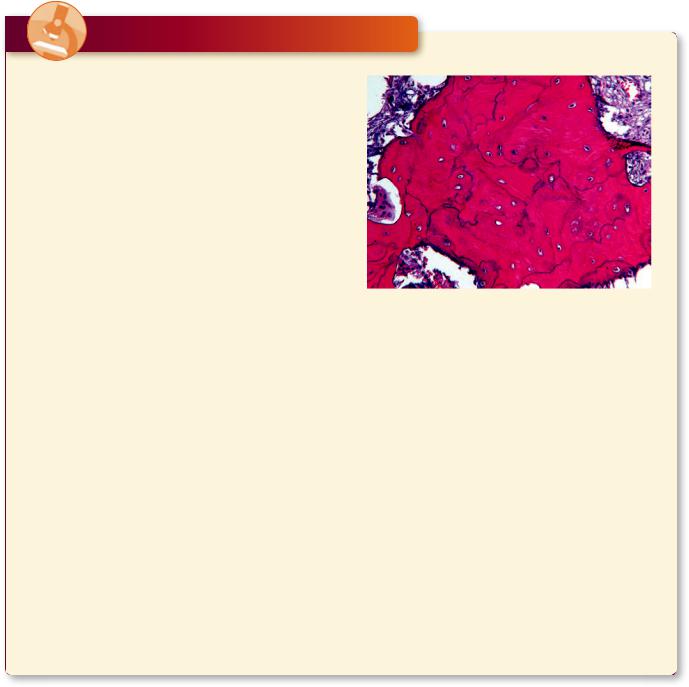
Osteomalacia is a condition in the adult that resembles rickets that occurs in children who have depressed vitamin D levels and, consequently, cannot absorb enough calcium in their gastrointestinal tract. This condition is difficult to diagnose because initially the patient presents with nonspecific symptoms that range from aches and pains to muscle weakness. Once advanced stages of osteomalacia are reached, the symptoms include deep bone pain, difficulty in walking, and bone fractures. Histologic pictures of cancellous bone present overly thin trabeculae of bone with prominent Howship’s lacunae occupied by osteoclasts and the presence of exceptionally thick osteoid over the thin calcified bony trabeculae and spicules.
Observe the large marrow spaces and the thin calcified bone (black) in the histologic image of osteomalacia. Note the very thick osteoid (magenta-colored homogeneous material) covering the calcified bony trabeculae. Osteoclastic activity is apparent in the scalloped indentation on the middle right of the image. (Reprinted with permission from Rubin R, Strayer D, et al., eds., Rubin’s Pathology. Clinicopathologic Foundations of Medicine, 5th ed. Baltimore: Lippincott Williams & Wilkins, 2008, p. 1117.)

C A R T I L A G E A N D B O N E 87
Chondrosarcoma
Chondrosarcoma, a malignant tumor that develops in existing cartilage or bone, is more frequently present in males and is one of the most common cancers of bone. There are three types of chondrosarcoma, depending on their location. The most common type is known as central chondrosarcoma because it develops in the marrow cavity, and patients are usually in their 40s or 50s when the tumor makes its appearance; the next most common is peripheral chondrosarcoma,
because it makes it initial appearance outside and then invades the bone, and patients are usually in their early 20s; the least common form is known as the juxtacortical chondrosarcoma, it begins its development in the region of the metaphysis and invades the bone, and patients suffering from this type of chondrosarcoma are in their mid 40s. The clinical symptom is pain localized to the site of the lesion, and histologic examinations display the presence of malignant chondrocytes in a matrix that resembles that of hyaline cartilage.
Observe the dense population of atypical chondrocytes dispersed within the hyaline cartilage–like matrix in this section from a patient suffering from chondrosarcoma. (Reprinted with permission from Rubin R, Strayer D, et al., eds., Rubin’s Pathology. Clinicopathologic Foundations of Medicine, 5th ed. Baltimore: Lippincott Williams & Wilkins, 2008. p. 1128.)
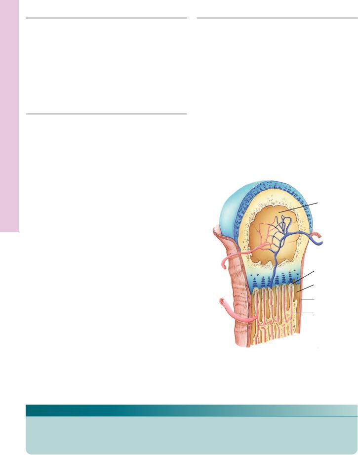
Bone Compact • 1-4 GRAPHIC
88 C A R T I L A G E A N D B O N E
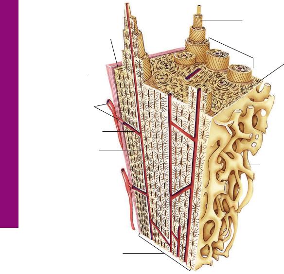
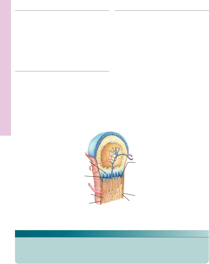
Outer circumferential lamellae
Periosteum Sharpey’s fibers 
Blood vessels
Volkmann’s canal
Haversian canal
Compact bone
Concentric lamellae
 Osteons
Inner circumferential lamellae
Cancellous bone
Marrow cavity
Compact Bone
Compact bone is surrounded by dense irregular collagenous connective tissue, the periosteum, which is attached to the outer circumferential lamellae by Sharpey’s fibers. Blood vessels of the periosteum enter the bone via larger nutrient canals or small Volksmann’s canals, which not only convey blood vessels to the Haversian canals of osteons but also interconnect adjacent Haversian canals. Each osteon is composed of concentric lamellae of bone whose collagen fibers are arranged so that they are perpendicular to those of contiguous lamellae. The inner circumferential lamellae are lined by endosteal lined cancellous bone that protrudes into the marrow cavity.
|
|
C A R T I L A G E A N D B O N E 89 |
|
|
|
D |
|
B |
C |
|
|
(7) |
(9) |
||
|
|||
(2) |
|
(8) |
|
|
|
A
|
(3) |
|
|
|
|
(4) |
|
(1) |
(1) |
|
(10) |
(5) |
|
||
|
|
||
|
|
(1) |
|
|
|
|
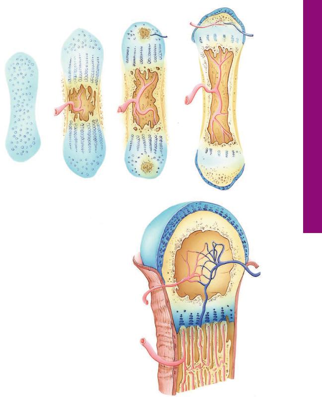
Endochondral Bone Formation
A.Endochondral bone formation requires the presence of a hyaline cartilage model.
B.Vascularization of the diaphysis perichondrium
(2) results in the transformation of chondrogenic cells to osteogenic cells, resulting in the formation of a subperiosteal bone collar (1) (via intramembranous bone formation), which quickly becomes perforated by osteoclastic activity. Chondrocytes in the center of the cartilage hypertrophy (3), and their lacunae become confluent.
C.The subperiosteal bone collar (1) increased in length and width, the confluent lacunae are invaded by the periosteal bud (4), and osteoclastic activity forms a primitive marrow cavity (5) whose walls are composed of calcified cartilage-calcified bone complex. The epiphyses display the beginning of secondary ossification centers (7).
D and E. The subperiosteal bond collar (1) has become sufficiently large to support the developing long bone, so that much of the cartilage has been resorbed, with the exception of the epiphyseal plate (8) and the covering of the epiphyses (9). Ossification in the epiphyses occurs from the center (10), thus the vascular periosteum (11) does not cover the cartilaginous surface. Blood vessels (12) enter the epiphyses, without vascularizing the cartilage, to constitute the vascular network (13) around which spongy bone will be formed.
E
(11)
(13)
(12)
Formation Bone Endochondral• 2-4 GRAPHIC
Cartilages Hyaline and Embryonic • 1-4 PLATE
90 C A R T I L A G E A N D B O N E
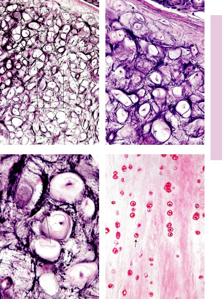
FIGURE 1. Embryonic hyaline cartilage. Pig. Paraffin section. ×132.
The developing hyaline cartilage is surrounded by embryonic connective tissue (ECT). Mesenchymal cells have participated in the formation of this cartilage. Note that the developing perichondrium (P), investing the cartilage, merges both with the embryonic connective tissue and with the cartilage. The chondrocytes in their lacunae are round, small cells packed closely together (arrow), with little intervening homogeneously staining matrix (arrowheads).
FIGURE 3. Hyaline cartilage. Rabbit. Paraffin section. ×270.
The perichondrium is composed of fibrous (F) and chondrogenic (CG) layers. The former is composed of mostly collagenous fibers with a few fibroblasts, whereas the latter is more cellular, consisting of chondroblasts and chondrogenic cells (arrows). As chondroblasts secrete matrix, they become surrounded by the intercellular substance and are consequently known as chondrocytes (C). Note that chondrocytes at the periphery of the cartilage are small and elongated, whereas those at the center are large and ovoid to round (arrowhead). Frequently, they are found in isogenous groups (IG).
FIGURE 2. Hyaline cartilage. Trachea. Monkey. Paraffin section. ×132.
The trachea is lined by a pseudostratified ciliated columnar epithelium (Ep). Deep to the epithelium, observe the large, bloodfilled vein (V). The lower half of the photomicrograph presents hyaline cartilage whose chondrocytes (C) are disposed in isogenous groups (IG) indicative of interstitial growth. Chondrocytes are housed in spaces known as lacunae. Note that the territorial matrix (arrow) in the vicinity of the lacunae stains darker than the interterritorial matrix (asterisk). The entire cartilage is surrounded by a perichondrium (P).
FIGURE 4. Hyaline cartilage. Trachea. Monkey. Plastic section. ×270.
The pseudostratified ciliated columnar epithelium displays numerous goblet cells (arrows). The cilia, appearing at the free border of the epithelium, are clearly evident. Note how the subepithelial connective tissue (CT) merges with the fibrous perichondrium (F). The chondrogenic layer of the perichondrium (Cg) houses chondrogenic cells and chondroblasts. As chondroblasts surround themselves with matrix, they become trapped in lacunae and are referred to as chondrocytes (C). At the periphery of the cartilage, the chondrocytes are flattened, whereas toward the interior they are round to oval. Due to the various histologic procedures, some of the chondrocytes fall out of their lacunae, which then appear as empty spaces. Although the matrix (M) contains many collagen fibrils, they are masked by the glycosaminoglycans; hence, the matrix appears homogeneous and smooth. The proteoglycan-rich lining of the lacunae is responsible for the more intense staining of the territorial matrix, which is particularly evident in Figures 2 and 3.
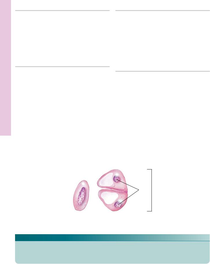
Nuclei
Isogenous group
Nucleus 
Chondroblast Chondrocytes
KEY
C |
chondrocyte |
ECT |
embryonic connective tissue |
IG |
isogenous group |
Cg |
chondrogenic |
Ep |
pseudostratified ciliated |
M |
matrix |
|
perichondrium |
|
columnar epithelium |
P |
perichondrium |
CT |
connective tissue |
F |
fibrous perichondrium |
V |
vein |
ECT
P
FIGURE 1
F
CG
C
IG
EP
V
P P
C
IG
FIGURE 2
CT
F
Cg
 C
C
M
Cartilages Hyaline and Embryonic • 1-4 PLATE
FIGURE 3 |
FIGURE 4 |

ibrocartilages F and Elastic• 2-4 PLATE
92 C A R T I L A G E A N D B O N E
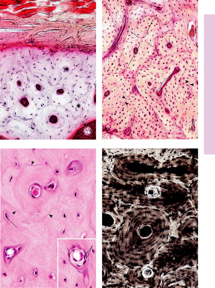
FIGURE 1. Elastic cartilage. Epiglottis. Human. Paraffin section. ×132.
Elastic cartilage, like hyaline cartilage, is enveloped by a perichondrium (P). Chondrocytes (C), which are housed in lacunae (arrow), have shrunk away from the walls, giving the appearance of empty spaces. Occasional lacunae display two chondrocytes (asterisk), indicative of interstitial growth. The matrix has a rich elastic fiber (E) component that gives elastic cartilage its characteristic appearance as well as contributing to its elasticity. The boxed area appears at a higher magnification in Figure 3.
FIGURE 3. Elastic cartilage. Epiglottis. Human. Paraffin section. ×540.
This is a high magnification of the boxed area in Figure 1. The chondrocytes (C) are large, oval to round cells with acentric nuclei (N). The cells accumulate lipids in their cytoplasm, often in the form of lipid droplets, thus imparting to the cell a “vacuolated” appearance. Note that the elastic fibers (E) mask the matrix in some areas and that the fibers are of various thicknesses, especially evident in cross-sections (arrows).
FIGURE 2. Elastic cartilage. Epiglottis. Human. Paraffin section. ×270.
This higher magnification of the perichondrial region of Figure 1 displays the outer fibrous (F) and inner chondrogenic (CG) regions of the perichondrium. Note that the chondrocytes (arrow) immediately deep to the chondrogenic layer are more or less flattened and smaller than those deeper in the cartilage. Additionally, the amount and coarseness of the elastic fibers increase adjacent to the large cells.
FIGURE 4. Fibrocartilage. Intervertebral disc. Human. Paraffin section. ×132.
The chondrocytes (C) of fibrocartilage are aligned in parallel rows, lying singly in individual lacunae. The nuclei of these chondrocytes are easily observed, whereas their cytoplasm is not as evident (arrow). The matrix contains thick bundles of collagen fibers (CF), which are arranged in a more or less regular fashion between the rows of cartilage cells. Unlike elastic and hyaline cartilages, fibrocartilage is not enveloped by a perichondrium.
Nuclei
Nucleus 
Chondroblast Chondrocytes
KEY
C |
chondrocyte |
E |
elastic fiber |
N |
nucleus |
CF |
collagen fiber |
F |
fibrous perichondrium |
P |
perichondrium |
Cg |
chondrogenic |
|
|
|
|
|
perichondrium |
|
|
|
|
P
F
CG
C
E
FIGURE 1 |
FIGURE 2 |
C
E N
C 
CF
ibrocartilages F and Elastic• 2-4 PLATE
FIGURE 3 |
FIGURE 4 |

Bone ompact • C3-4 PLATE
94 C A R T I L A G E A N D B O N E
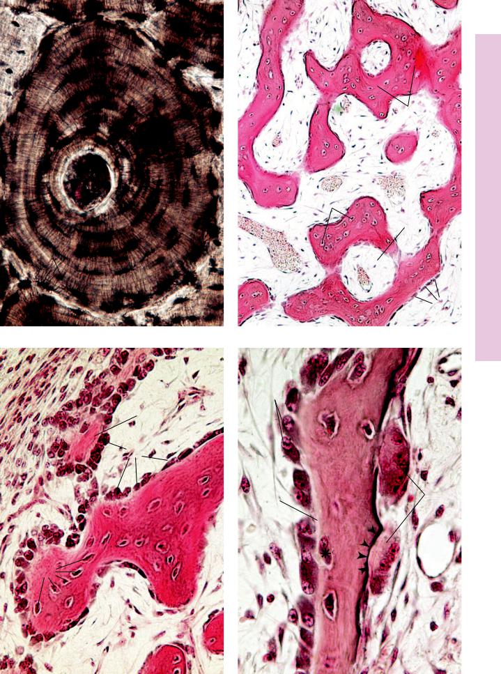
FIGURE 1. Decalcified compact bone. Human. Paraffin section. ×132.
Cross section of decalcified bone, displaying skeletal muscle (SM) fibers that will insert a short distance from this site. The outer
fibrous periosteum (FP) and the inner osteogenic periosteum
(OP) are distinguishable due to the fibrous component of the former and the cellularity of the latter. Note the presence of the inner circumferential (IC) lamellae, osteons (Os), and interstitial lamellae (asterisk). Also observe the marrow (M) occupying the marrow cavity, as well as the endosteal lining (arrow).
FIGURE 2. Decalcified compact bone. Human. Paraffin section. ×132.
This is a cross section of decalcified compact bone, displaying osteons or haversian canal systems (Os) as well as interstitial lamellae (IL). Each osteon possesses a central haversian canal (HC), surrounded by several lamellae (L) of bone. The boundary of each osteon is visible and is referred to as a cementing line (arrowheads). Neighboring haversian canals are connected to each other by Volkmann’s canals (VC), through which blood vessels of osteons are interconnected to each other.
FIGURE 3. Decalcified compact bone. Human. Paraffin section. ×540.
A small osteon is delineated by its surrounding cementing line (arrowheads). The lenticular-shaped osteocytes (Oc) occupy flattened spaces, known as lacunae. The lacunae are lined by uncalcified osteoid matrix. Inset. Decalcified compact bone. Human. Paraffin section. ×540. A haversian canal of an osteon is shown to contain a small blood vessel (BV) supported by slender connective tissue elements. The canal is lined by flattened osteoblasts (Ob) and, perhaps, osteogenic cells (Op).
FIGURE 4. Undecalcified ground compact bone. x.s. Human. Paraffin section. ×132.
This specimen was treated with India ink to accentuate some of the salient features of compact bone. The haversian canals (HC) as well as the lacunae (arrows) appear black in the figure. Note the connection between two osteons at top center, known as Volkmann’s canal (VC). The canaliculi appear as fine, narrow lines leading to the haversian canal as they anastomose with each other and with lacunae of other osteocytes of the same osteon.
 Nucleus
Nucleus
Osteoblast Osteocyte
KEY
BV |
blood vessel |
L |
lamella |
OP |
osteogenic periosteum |
FP |
fibrous periosteum |
M |
marrow |
Os |
osteon |
HC |
haversian canal |
Ob |
osteoblast |
SM |
skeletal muscle fiber |
IC |
inner circumferential lamella |
Oc |
osteocyte |
VC |
Volkmann’s canal |
IL |
interstitial lamella |
Op |
osteogenic cell |
|
|
SM
FP |
HC |
Os
OP
IL
L L |
|
L |
VC |
|
Os
IC
M
FIGURE 1 |
FIGURE 2 |
VC
Oc |
|
HC |
|
|
|
Op |
Ob |
C |
|
||
|
BV |
|
FIGURE 3 |
FIGURE 4 |
Bone ompact • C3-4 PLATE

Ossification Intramembranous and Bone ompact • C4-4 PLATE
96 C A R T I L A G E A N D B O N E
FIGURE 1. Undecalcified ground bone. x.s. Human. Paraffin section. ×270.
This transverse section of an osteon clearly displays the lamellae
(L) of bone surrounding the haversian canal (HC). The cementing line acts to delineate the periphery of the osteon. Note that the canaliculi (C) arising from the peripheral-most lacunae usually do not extend toward other osteons. Instead, they lead toward the haversian canal. Canaliculi, which appear to anastomose with each other and with lacunae, house long osteocytic processes in the living bone.
FIGURE 2. Intramembranous ossification. Pig skull. Paraffin section. ×132.
The anastomosing trabeculae (T) of forming bone appear darkly stained in a background of embryonic connective tissue (ECT). Observe that this connective tissue is highly vascular and that the bony trabeculae are forming primitive osteons (Os) surrounding large, primitive haversian canals (HC), whose center is occupied by blood vessels (BV). Observe that the osteocytes (Oc) are arranged somewhat haphazardly. Every trabecula is covered by osteoblasts (Ob).
FIGURE 3. Intramembranous ossification. Pig skull. Paraffin section. ×270.
This photomicrograph of intramembranous ossification is taken from the periphery of the bone-forming region. Note the developing periosteum (P) in the upper right-hand corner. Just deep to this primitive periosteum, osteoblasts (Ob) are differentiating and are elaborating osteoid (Ot), as yet uncalcified bone matrix. As the osteoblasts surround themselves with bone matrix, they become trapped in their lacunae and are known as osteocytes (Oc). These osteocytes are more numerous, larger, and more ovoid than those of mature bone, and the organization of the collagen fibers of the bony matrix is less precise than that of mature bone. Hence, this bone is referred to as immature (primary) bone, and it will be replaced by mature bone later in life.
FIGURE 4. Intramembranous ossification. Pig skull. Paraffin section. ×540.
This photomicrograph is taken from an area similar to those of Figures 2 and 3. This trabecula demonstrates several points, namely, that osteoblasts (Ob) cover the entire surface and that osteoid (Ot) is interposed between calcified bone and the cells of bone and appears lighter in color. Additionally, note that the osteoblast marked with the asterisk is apparently trapping itself in the matrix it is elaborating. Finally, note the large, multinuclear cells, osteoclasts (Ocl), which are in the process of resorbing bone. The activity of these large cells results in the formation of Howship’s lacunae (arrowheads), which are shallow depressions on the bone surface. The interactions between osteoclasts and osteoblasts are very finely regulated in the normal formation and remodeling of bone.
Outer circumferential |
|
|
|
Concentric lamellae |
||
|
|
|
||||
|
|
|
Osteons |
|||
lamellae |
|
|
|
Inner circumferential |
||
Periosteum |
|
|
|
|
|
|
|
|
|
|
|
lamellae |
|
|
|
|
|
|
||
|
|
|
|
|
|
|
Blood vessels |
|
|
|
|
||
Haversian canal |
|
|
|
|
|
|
|
|
|
|
|
||
Compact bone
KEY
BV |
blood vessel |
L |
lamella |
Os |
osteon |
C |
canaliculus |
Ob |
osteoblast |
Ot |
osteoid |
ECT |
embryonic connective tissue |
Oc |
osteocyte |
P |
periosteum |
HC |
haversian canal |
Ocl |
osteoclast |
T |
trabecula |
C
HC L
L
L
L
L
C 
FIGURE 1
P
Ot
 Ob
Ob
Oc
FIGURE 3
HC
Os
T
ECT
T
Oc
HC
BV
 Ob
Ob
FIGURE 2
Ob
Ot |
Ocl |
|
FIGURE 4
Ossification Intramembranous and Bone Compact • 4-4 PLATE
cation Ossifi Endochondral• 5-4 PLATE
98 C A R T I L A G E A N D B O N E
FIGURE 1. Epiphyseal ossification center. Monkey. Paraffin section. ×14.
Most long bones are formed by the endochondral method of ossification, which involves the replacement of a cartilage model by bone. In this low-power photomicrograph, the diaphysis (D) of the lower phalanx has been replaced by bone, and the medullary cavity is filled with marrow (M). The epiphysis (E) of the same phalanx is undergoing ossification and is the secondary center of ossification (2°), thereby establishing the epiphyseal plate (ED). The trabeculae (T) are clearly evident on the diaphyseal side of the epiphyseal plate.
FIGURE 2. Endochondral ossification. l.s. Monkey. Paraffin section. ×14.
Much of the cartilage has been replaced in the diaphysis of this forming bone. Note the numerous trabeculae (T) and the developing bone marrow (M) of the medullary cavity. Ossification is advancing toward the epiphysis (E), in which the secondary center of ossification has not yet appeared. Observe the periosteum (P), which appears as a definite line between the subperiosteal bone collar and the surrounding connective tissue. The boxed area is represented in Figure 3.
FIGURE 3. Endochondral ossification. Monkey. Paraffin section. ×132.
This montage is a higher magnification of the boxed area of Figure 2. The region where the periosteum and perichondrium meet is evident (arrowheads). Deep to the periosteum is the subperiosteal bone collar (BC), which was formed via intramembranous ossification. Endochondral ossification is evident within the cartilage template. Starting at the top of the montage, note how the chondrocytes are lined up in long columns (arrows), indicative of their intense mitotic activity at the future epiphyseal plate region. In the epiphyseal plate, this will be the zone of cell proliferation (ZP). The chondrocytes increase in size in the zone of cell maturation and hypertrophy (ZH) and resorb some of their lacunar walls, enlarging them to such an extent that some of the lacunae become confluent. The chondrocytes die in the zone of calcifying cartilage (ZC). The presumptive medullary cavity is being populated by bone marrow, osteoclastic and osteogenic cells, and blood vessels. The osteogenic cells are actively differentiating into osteoblasts, which are elaborating bone on the calcified walls of the confluent lacunae. At the bottom of the photomicrograph, observe the bone-covered trabeculae of calcified cartilage (asterisks).
|
|
Diaphysis |
Zone of cell |
|
Calcifying |
proliferation |
|
|
|
cartilage |
|
|
|
|
|
|
Epiphyseal |
|
|
|
Zone of |
|
plate |
|
||
hypertrophy |
|
|
|
|
Epiphysis |
Subperiostal |
|
Periosteum |
bone collar |
|
|
|
|
|
Periosteum |
Subperiosteal |
|
|
bone collar |
|
Endochondral bone formation
KEY
BC |
subperiosteal bone collar |
P |
periosteum |
ZC |
zone of calcifying cartilage |
D |
diaphysis |
2° |
secondary center of |
ZH |
zone of cell maturation and |
E |
epiphysis |
|
ossification |
|
hypertrophy |
ED |
epiphyseal plate |
T |
trabecula |
ZP |
zone of proliferation |
Mmarrow
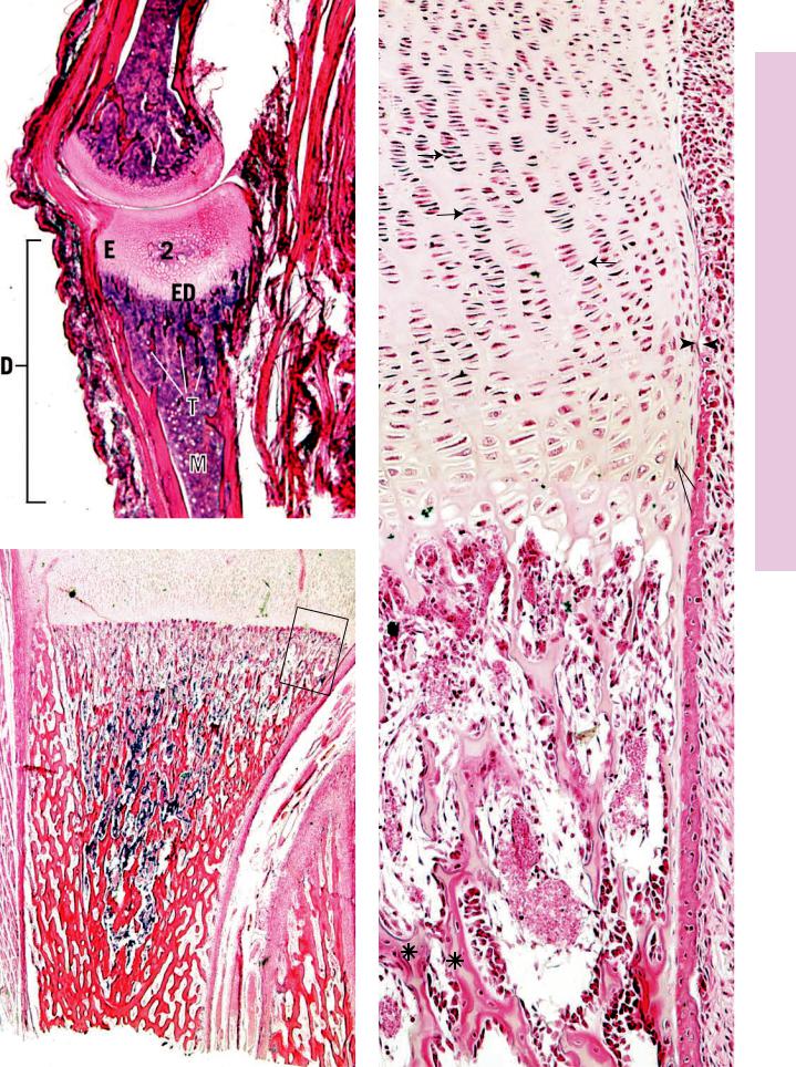
FIGURE 1
E
T
T P
M
FIGURE 2
ZP
BC
ZH
ZC
FIGURE 3
cation Ossifi Endochondral• 5-4 PLATE
cation Ossifi Endochondral• 6-4 PLATE
100 C A R T I L A G E A N D B O N E
FIGURE 1. Endochondral ossification. Monkey. Paraffin section. ×132.
This photomicrograph is a higher magnification of a region of Plate 4-5, Figure 3. Observe the multinucleated osteoclast (arrowheads) resorbing the bone-covered trabeculae of calcified cartilage. The subperiosteal bone collar (BC) and the periosteum (P) are clearly evident, as is the junction between the bone collar and the cartilage (arrows). The medullary cavity is being established and is populated by blood vessels (BV), osteogenic cells, osteoblasts, and hematopoietic cells.
FIGURE 3. Endochondral ossification. x.s. Monkey. Paraffin section. ×196.
A cross section of the region of endochondral ossification presents many round spaces in calcified cartilage that are lined with bone (asterisks). These spaces represent confluent lacunae in the cartilage template, where the chondrocytes have hypertrophied and died. Subsequently, the cartilage-calcified and the invading osteogenic cells have differentiated into osteoblasts (arrowheads) and lined the calcified cartilage with bone. Since neighboring spaces were separated from each other by calcified cartilage walls, bone was elaborated on the sides of the walls. Therefore, these trabeculae, which in longitudinal section appear to be stalactitelike structures of bone with a calcified cartilaginous core, are, in fact, spaces in the cartilage template that are lined with bone. The walls between the spaces are the remnants of cartilage between lacunae that became calcified and form the substructure upon which bone was elaborated. Observe the forming medullary cavity (MC), housing blood vessels (BV), hematopoietic tissue (HT), osteogenic cells, and osteoblasts (arrowheads). The subperiosteal bone collar (BC) is evident and is covered by a periosteum, whose two layers, fibrous (FP) and osteogenic (Og), are clearly discernible.
FIGURE 2. Endochondral ossification. Monkey. Paraffin section. ×270.
This photomicrograph is a higher magnification of the boxed area in Figure 1. Note that the trabeculae of calcified cartilage are covered by a thin layer of bone. The darker staining bone (arrow) contains osteocytes, whereas the lighter staining calcified cartilage (CC) is acellular, since the chondrocytes of this region have died, leaving behind empty lacunae that are confluent with each other. Observe that osteoblasts (Ob) line the trabecular complexes and that they are separated from the calcified bone by thin intervening osteoid (Ot). As the subperiosteal bone collar increases in thickness, the trabeculae of bone-covered calcified cartilage will be resorbed so that the cartilage template will be replaced by bone. The only cartilage that will remain will be the epiphyseal plate and the articular covering of the epiphysis.
Medullary cavity
 Blood
Blood
vessels
Calcified cartilage
Subperiosteal bone collar
Periosteum
Hemopoietic tissue and blood vessel
Endochondral bone formation
KEY
BC |
subperiosteal bone collar |
HT |
hematopoietic tissue |
Og |
osteogenic periosteum |
BV |
blood vessel |
MC |
medullary cavity |
Ot |
osteoid |
CC |
calcified cartilage |
Ob |
osteoblast |
P |
periosteum |
FP |
fibrous periosteum |
|
|
|
|
P
BV
BC
BV
FIGURE 1
MC
HT
BV
BV
FIGURE 3
Ob
Ot 
Ob
CC
FIGURE 2
Og
FP
BC
cation Ossifi Endochondral• 6-4 PLATE
102 C A R T I L A G E A N D B O N E
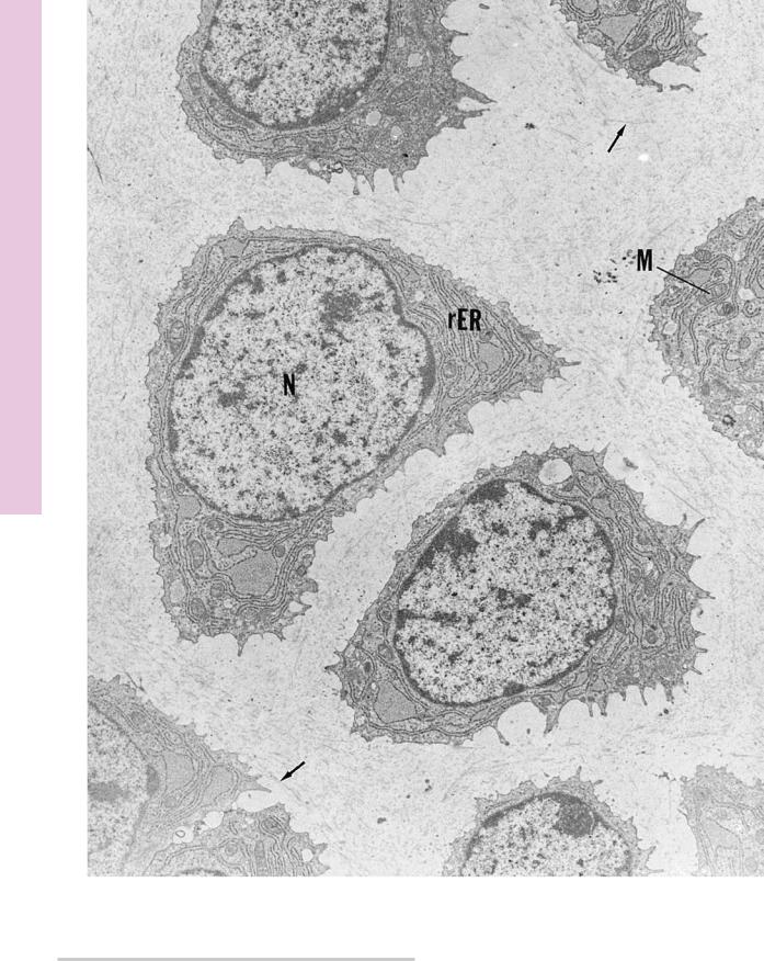
Microscopy Electron Cartilage, aline • Hy7-4 PLATE
FIGURE 1
FIGURE 1. Hyaline cartilage. Mouse. Electron microscopy. ×6,120.
The hyaline cartilage of a neonatal mouse trachea presents chondrocytes, whose centrally positioned nuclei (N) are surrounded
with a rich rough endoplasmic reticulum (rER) and numerous mitochondria (M). The matrix displays fine collagen fibrils (arrows). (From Seegmiller R, Ferguson C, Sheldon H. Studies on cartilage, VI: a genetically determined defect in tracheal cartilage. J Ultrastruct Res 1972;38:288–301.)
C A R T I L A G E A N D B O N E 103
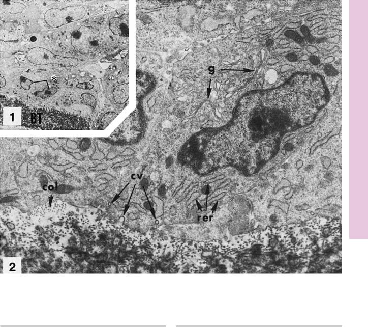
Microscopy Electron eoblasts, •Ost8-4 PLATE
FIGURE 1 and 2
FIGURE 1. Osteoblasts from long bone. Rat. Electron microscopy. ×1,350.
This low-magnification electron micrograph displays numerous fibroblasts and osteoblasts in the vicinity of a bony trabecula (BT). The osteoblasts (asterisk) are presented at a higher magnification in Figure 2. (From Ryder M, Jenkins S, Horton J. The adherence to bone by cytoplasmic elements of osteoclast. J Dent Res 1981;60:1349–1355.)
FIGURE 2. Osteoblasts. Rat. Electron microscopy. ×9,450.
Osteoblasts, at higher magnification, present well-developed Golgi apparatus (g), extensive rough endoplasmic reticulum (rer), and several coated vacuoles (cv) at the basal cell membrane. Observe the cross sections of collagen fibers (col) in the bone matrix. (From Ryder M, Jenkins S, Horton J. The adherence to bone by cytoplasmic elements of osteoclast. J Dent Res 1981;60:1349–1355.)

Microscopy Electron eoclast, •Ost9-4 PLATE
104 C A R T I L A G E A N D B O N E
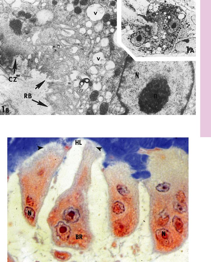
FIGURE 1a. Osteoclast from long bone. Rat. Electron microscopy. ×1,800.
Two nuclei of an osteoclast are evident in this section. Observe that the cell is surrounding a bony surface (asterisk). The region of the nucleus marked by an arrowhead is presented at a higher magnification in Figure 1B.
FIGURE 2. Osteoclasts. Human. Paraffin section. ×600.
The nuclei (N) of these multinuclear cells are located in their basal region (BR), away from Howship’s lacunae (HL). Note that the ruffled border (arrowheads) is in intimate contact with Howship’s lacunae. (Courtesy of Dr. J. Hollinger.)
FIGURE 1b. Osteoclast. Rat. Electron microscopy. ×10,800.
This is a higher magnification of a region of Figure 1A. Note the presence of the nucleus (N) and its nucleolus (n), as well as the ruffled border (RB) and clear zone (CZ) of the osteoclast. Numerous vacuoles (v) of various sizes may be observed throughout the cytoplasm. (From Ryder M, Jenkins S, Horton J. The adherence to bone by cytoplasmic elements of osteoclast. J Dent Res 1981;60:1349–1355.)
C A R T I L A G E A N D B O N E 105
Microscopy Electron eoclast, •Ost9-4 PLATE
FIGURE 1
HL
N
BR |
N |
|
FIGURE 2