

•The O2 is used by neutrophils to form superoxides, hydrogen peroxide, and hypochlorous acid, highly reactive compounds that destroy bacteria within the phagosomes.
•Frequently, the avid response of neutrophils results in the release of some of these highly potent compounds into the surrounding connective tissue precipitating tissue damage.
•Neutrophils also produce leukotrienes from plasmalemma arachidonic acids to aid in the initiation of an inflammatory response.
•Subsequent to the performance of these functions, the neutrophils die and become a major component of pus.
PLASMA
Plasma, the fluid component of blood, comprises approximately 55% of the total blood volume.
•Plasma contains electrolytes and ions, such as calcium, sodium, potassium, and bicarbonate; larger molecules, namely, albumins, globulins, and fibrinogen; and organic compounds as varied as amino acids, lipids, vitamins, hormones, and cofactors.
•Subsequent to clotting, a straw-colored serum is expressed from blood. This fluid is identical to plasma but contains no fibrinogen or other components necessary for the clotting reaction.
COAGULATION
Coagulation is the result of the exquisitely controlled interaction of a number of plasma proteins and coagulation factors. The regulatory mechanisms are in place so that coagulation typically occurs only if the endothelial lining of the vessel becomes injured.
•In the intact blood vessel, the endothelium manufactures inhibitors of platelet aggregation (NO and prostacyclins) as well as display agents, thrombomodulin and heparin-like molecule, on their luminal plasmalemmae that block coagulation.
•If the lining of a blood vessel is damaged, the endothelial cells switch from producing and displaying antiaggregation and anticoagulation agents and release tissue factor (tissue thromboplastin), von Willebrand’s factor and endothelins.
•Tissue factor complexes with Factor VIIa to catalyze the conversion of Factor X to its active form, the protease Factor Xa;
•von Willebrand’s factor activates platelets, facilitating the adhesion of platelets to the exposed laminin and collagens and induces them to release ADP and
B L O O D A N D H E M O P O I E S I S 111
thrombospondin, which encourages their adhesion to each other; and endothelin stimulates the contraction of vascular smooth muscle cells in the region to constrict the damaged blood vessel and, thus, minimize blood loss.
•The process of coagulation ensues in one of two convergent pathways, extrinsic and intrinsic, both of which lead to the final step of converting fibrinogen to fibrin.
The extrinsic pathway has a faster onset and depends on the release of tissue factor.
The intrinsic pathway is initiated slower, is dependent on contact between vessel wall collagen and platelets (or factor XII), and requires the presence of von Willebrand’s factor and factor VIII.
These two factors form a complex that not only binds to exposed collagen but also attaches to receptor sites on the platelet plasmalemma, affecting platelet aggregation and adherence to the vessel wall.
•The two pathways intersect at the conversion of Factor X to Factor Xa and from that point on the remaining steps of the coagulation pathway are referred to as the common pathway.
HEMOPOIESIS
Circulating blood cells have relatively short life spans and must be replaced continuously by newly formed cells. This process of blood cell replacement is known as hemopoiesis (hematopoiesis).
•All blood cells develop from a single pluripotential precursor cell known as the pluripotential hemopoietic stem cell (PHSC).
•These cells undergo mitotic activity, whereby they give rise to two types of multipotential hemopoietic stem cells, CFU-GEMM (Colony-forming unit-gran- ulocyte, erythrocyte, monocyte, megakaryocyte, previously known as CHU-S) and CFU-Ly (colony-forming unit-lymphocyte).
Most PHSCs and other hemopoietic stem cells of adults are located in the red bone marrow of short and flat bones. The marrow of long bones is red in young individuals, but when it becomes infiltrated by fat in the adult, it takes on a yellow appearance and is known as yellow marrow.
•Although it was once believed that adipose cells accumulated the fat, it is now known that the cells actually responsible for storing fat in the marrow are the adventitial reticular cells.
•Stem cells, in response to various hemopoietic growth factors, undergo cell division and maintain the population of circulating erythrocytes, leukocytes, and platelets.
112 B L O O D A N D H E M O P O I E S I S
As stated above, the nomenclature developed for the cells described below is based on their colorations with Wright’s or Giemsa’s modification of the Romanovsky-type stains as applied to blood and marrow smears used in hematology.
Erythrocytic Series
•Erythrocyte development proceeds from CFU-S, which, in response to elevated levels of erythropoietin, gives rise to cells known as BFU-E, which, in response to lower erythropoietin levels, then give rise to CFU-E.
•Later generations of CFU-E are recognizable histologically as proerythroblasts.
These cells give rise to basophilic erythroblasts, which, in turn, undergo cell division to form
polychromatophilic erythroblasts that will divide mitotically to form
orthochromatophilic erythroblasts (normoblasts).
Cells of this stage no longer divide.
will extrude their nuclei, and differentiate into reticulocytes (not to be confused with reticular cells of connective tissue), which, in turn, become mature red blood cells.
Reticulocytes are stained with methylene blue for manual or thiazole orange for automated counting.
Granulocytic Series
The development of the granulocytic series is initiated from the multipotential CFU-S.
•The first histologically distinguishable member of this series is the myeloblast, which gives rise mitotically to
•promyelocytes, which also undergo cell division to yield
•myelocytes. Myelocytes are the first cells of this series to possess specific granules; therefore, neutrophilic, eosinophilic, and basophilic myelocytes may be recognized.
•The next cells in the series are metamyelocytes, which no longer divide, but differentiate into
•band (stab) cells, the juvenile form, which will become mature granulocytes that enter the bloodstream.
Several hemopoietic growth factors activate and promote hemopoiesis. These act by binding to plasma membrane receptors of their target cell, controlling their mitotic rate, as well as the number of mitotic events.Additionally, they stimulate cell differentiation and enhance the survival of the progenitor cell population (Table 5-2). The best known factors are erythropoietin (acts on BFU-E and CFU-E),
•interleukin-3 (acts on PHSC, CFU-S, and myeloid progenitor cells),
•interleukin-7 (acts on CFU-Ly),
•granulocyte-macrophage colony-stimulating factor
(acts on granulocyte and monocyte progenitor cells),
•granulocyte colony-stimulating factor (acts on granulocyte progenitor cells), and
•macrophage colony-stimulating factor (acts on monocyte progenitor cells).

B L O O D A N D H E M O P O I E S I S 113
TABLE 5-2 • Hemopoietic Growth Factors
Factors |
Principal Action of the Factor |
Site of Origin of the Factor |
|
|
|
Stem cell factor |
Facilitates hemopoiesis |
Stromal cells of bone marrow |
|
|
|
GM-CSF |
Facilitates CFU-GM mitosis, differentiation, granulocyte |
T cells, endothelial cells |
|
activity |
|
|
|
|
G-CSF |
Induces mitosis, differentiation of CFU-G; facilitates |
Macrophages, endothelial cells |
|
neutrophil activity |
|
|
|
|
M-CSF |
Facilitates mitosis, differentiation of CFU-M |
Macrophages, endothelial cells |
|
|
|
IL-1 (IL-3, IL-6) |
Facilitates proliferation of PHSC, CFU-S, CFU-Ly; |
Monocytes, macrophages, endothelial |
|
suppresses erythroid precursors |
cells |
|
|
|
IL-2 |
Promotes proliferation of activated T cells, B cells; |
Activated T cells |
|
facilitates NK cell differentiation |
|
|
|
|
IL-3 |
Same as IL-1; also facilitates proliferation of unipotential |
Activated T and B cells |
|
precursors except LyB and LyT |
|
|
|
|
IL-4 |
Promotes activation of T cells, B cells; facilitates |
Activated T cells |
|
development of mast cells, basophils |
|
|
|
|
IL-5 |
Facilitates proliferation of CFU-Eo; activates eosinophils |
T cells |
|
|
|
IL-6 |
Same as IL-1; also promotes differentiation of CTLs and |
Monocyte, fibroblasts |
|
B cells |
|
|
|
|
IL-7 |
Stimulates CFU-LyB and NK cell differentiation |
Adventitial reticular cells |
|
|
|
IL-8 |
Promotes migration and degranulation of neutrophils |
Leukocytes, endothelial cells, smooth |
|
|
muscle cells |
|
|
|
IL-9 |
Promotes activation, proliferation of mast cells, |
T helper cells |
|
modulates IgE synthesis, stimulates proliferation |
|
|
of T helper cells |
|
|
|
|
IL-10 |
Inhibits synthesis of cytokines by NK cells, macrophages, |
Macrophages, T cells |
|
T cells; promotes CTL differentiation and B-cell and |
|
|
mast cell proliferation |
|
|
|
|
IL-12 |
Stimulates NK cells; promotes CTL and NK cell function |
Macrophages |
|
|
|
γ-Interferons |
Activates monocytes, B cells; promotes CTL |
T cells, NK cells |
|
differentiation; enhances expression of class II HLA |
|
|
|
|
Erythropoietin |
Promotes CFU-E differentiation, proliferation of |
Endothelial cells of peritubular capillary |
|
BFU-E |
network of kidney, hepatocytes |
|
|
|
Thrombopoietin |
Enhances mitosis, differentiation of CFU-Meg and |
Not known |
|
megakaryoblasts |
|
CTL, cytotoxic lymphocyte; CFU, colony-forming unit (Eo, eosinophil; G, granulocyte; GM, granulocyte-monocyte; Ly, lymphocyte; S, spleen); CSF, colonystimulating factor (G, granulocyte; GM, granulocyte-monocyte; M, monocyte); HLA, human leukocyte antigen; IL, interleukin.
Modified with permission from Gartner LP, Hiatt JL. Color Textbook of Histology, 2nd ed. Philadelphia: Saunders, 2001.
114 B L O O D A N D H E M O P O I E S I S
CLINICAL CONSIDERATIONS
NADPH Oxidase Deficiency
Certain individuals suffer from persistent bacterial infection due to a hereditary NADPH oxidase deficiency. The neutrophils of these individuals are unable to effect a respiratory burst and, therefore, are incapable of forming the highly reactive compounds, such as hypochlorous acid, hydrogen peroxide, and superoxide, that assist in the killing of bacteria within their phagosomes.
Multiple Myeloma
Multiple myeloma is a relatively uncommon malignant neoplasm with greater incidence in males than females. Its origin is the bone marrow and is characterized by the presence of large numbers of malignant plasma cells that may also be abnormal in morphology. These cells accumulate in the bone marrow of various regions of the skeletal system. Frequently, the cell proliferation is so great in the marrow that the huge number of cells place pressure on the walls of the marrow cavity causing bone pains and even fractures of bones such as the ribs. These cells also produce abnormal proteins such as Bence-Jones proteins that enter the urine where they can be detected to provide a diagnosis for multiple myeloma.
Infectious Mononucleosis
Infection with the Epstein-Barr virus causes infectious mononucleosis, also referred to as the “kissing disease,” because it is common among high school and collegeaged individuals and is frequently spread by saliva. The symptoms of patients suffering from infectious mononucleosis include sore throat, swollen and painful lymph nodes, low energy, and an elevated lymphocyte count. The disease can be life-threatening in immunosuppressed individuals.
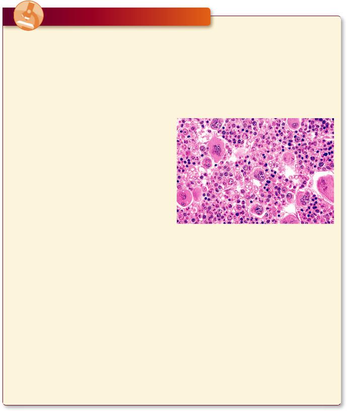
Polycythemia Vera
Polycythemia vera (primary polycythemia) is a rare disorder of the blood that manifests itself by an excess production of red blood cells and, frequently, platelets, resulting in greater blood volume and an increase in the viscosity of blood. It mainly involves individuals who are in their early sixties, although occasionally it occurs in patients who are in their early twenties. Symptoms
may be absent for a number of years after the onset of the condition, but patients suffering from this disorder may exhibit headaches, vertigo, fatigue, shortness of breath, enlarged liver and spleen, burning sensation in the extremities, visual disorders, as well as gingival bleeding, and generalized itching. If left untreated, the patient may die within 2 years, but with proper treatment, the lifespan can be extended by 10 to 20 years.
This is a bone marrow biopsy from a middle-aged woman suffering from polycythemia vera. Observe that the marrow is hypercellular exhibiting an abnormally high number of erythrocyte precursors and megakaryocytes. (Reprinted with permission from Mills SE, Carter D, Greenson JK, Reuter VE, Stoler MH eds. Sternberger’s Diagnostic Surgical Pathology, 5th ed. Philadelphia: Lippincott Williams & Wilkins 2010. p. 635.)
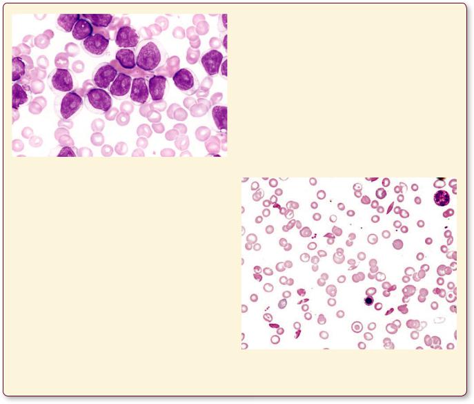
B-Cell Prolymphocytic Leukemia
B-cell prolymphocytic leukemia is a relatively rare form of leukemia that arises relatively late in life, around 60 years of age, and affects males more frequently than females. The histopathologic picture presents bone marrow smears and blood smears with medium to large prolymphocytes. Usually, the disease is accompanied by an enlargement of the spleen. The prognosis is not good because this type of leukemia is quite aggressive and treatment modalities are not very effective; in fact, they are mostly palliative, and usually the patient succumbs in two or three years.
B L O O D A N D H E M O P O I E S I S 115
This blood smear, from a patient suffering from B-cell prolymphocytic leukemia, displays numerous large prolymphocytes whose nucleus presents a coarse chromatin network and large vesicles. (Reprinted with permission from Mills SE, Carter D, Greenson JK, Reuter VE, Stoler MH eds. Sternberger’s Diagnostic Surgical Pathology, 5th ed.., 2010. P. 644.)
Sickle Cell Anemia
Sickle cell anemia, a hereditary disease, is the result of a point mutation in the gene that codes for hemoglobin. A single amino acid substitution of alanine replacing glutamine occurs in some individuals who are descendants of the indigenous population of tropical and subtropical regions of Africa, especially from the sub-Saharan area. Approximately 2 per 1,000 African Americans are afflicted with this disease, and 10% of that population carry one copy of the gene and, therefore, are carriers of the trait but are not afflicted by the disease. The red blood cells of patients with two copies of the gene are
defective and carry a reduced amount of oxygen. These erythrocytes are fragile, do not pass easily through small capillaries, and assume a sickle shape. The abnormally shaped red blood cells have a deleterious effect on the kidneys, brain, bones, and spleen among other organs. Depending on the severity of the condition, the patient’s symptoms may vary from slight to severe, and in the latter case, it may result in death at an early age. Since sickle cell anemia is incurable, it is treated with avoidance of strenuous physical exertions, avoiding high altitudes, and instructing patients to seek treatment for even minor infections.
This blood smear, from a patient suffering from sickle cell anemia, displays numerous red blood cells that are distorted so that they appear spindle-shaped.

116 B L O O D A N D H E M O P O I E S I S
Blood culating • Cir1-5 PLATE
116
FIGURE 1. Red blood cells. Human. ×1,325.
RBCs (arrows) display a central clear region that represents the thinnest area of the biconcave disc. Note that the platelets (arrowheads) possess a central dense region, the granulomere, and a peripheral light region, the hyalomere.
FIGURE 2. Neutrophils. Human. ×1,325.
Neutrophils display a somewhat granular cytoplasm and lobulated (arrowheads) nuclei.
FIGURE 3. Eosinophils. Human. ×1,325.
Eosinophils are recognized by their large, pink granules and their sausage-shaped nucleus. Observe the slender connecting link (arrowhead) between the two lobes of the nucleus.

B L O O D A N D H E M O P O I E S I S 117
FIGURE 4. Basophils. Human. ×1,325.
Basophils are characterized by their dense, dark, large granules.
FIGURE 5. Monocytes. Human. ×1,325.
Monocytes are characterized by their large size, acentric, kidney-shaped nucleus and lack of specific granules.
FIGURE 6. Lymphocytes. Human. ×1,325.
Lymphocytes are small cells that possess a single, large, acentrically located nucleus and a narrow rim of light blue cytoplasm.
1.15 cm = 7.5 µm
Blood culating • Cir1-5 PLATE
118 B L O O D A N D H E M O P O I E S I S
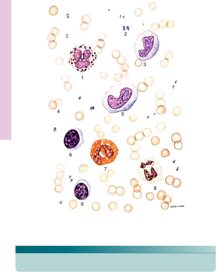
(Drawing) Blood Circulating • 2-5 PLATE
FIGURE 1
KEY
1. |
Basophil |
4. |
Erythrocytes |
7. |
Eosinophil |
2. |
Platelets |
5. |
Monocyte |
8. |
Neutrophil |
3. |
Monocyte |
6. |
Lymphocyte |
9. |
Lymphocyte |

B L O O D A N D H E M O P O I E S I S 119
Hemopoiesis and Blood • 3-5 PLATE
FIGURE 1
KEY
A |
|
5. Neutrophilic stab cell |
D |
|
|
1. |
Basophilic myelocyte |
6. |
Neutrophil |
1. |
Proerythroblast |
2. Basophilic metamyelocyte |
C |
|
2. Basophilic erythroblast |
||
3. |
Basophil stab cell |
1. |
Eosinophilic myelocyte |
3. |
Polychromatophilic erythroblast |
4. |
Basophil |
2. |
Eosinophilic metamyelocyte |
4. |
Orthochromatophilic erythroblast |
B |
|
3. Eosinophil stab cell |
5. Reticulocyte |
||
1. |
Myeloblast |
4. |
Eosinophil |
6. |
Erythrocyte |
2.Promyelocyte
3.Neutrophilic myelocyte
4.Neutrophilic metamyelocyte

Blood Circulating and Marrow one • B4-5 PLATE
120 B L O O D A N D H E M O P O I E S I S
FIGURE 1. Bone marrow. Human. Paraffin section. ×132.
This transverse section of a decalcified human rib displays the presence of haversian canals (H), Volkmann’s canals (V), osteocytes (O) in their lacunae, and the endosteum (E). The marrow presents numerous adventitial reticular cells (A), blood vessels, and sinusoids (S). Moreover, the forming blood elements are also evident as small nuclei (arrows). Note the large megakaryocytes (M), cells that are the precursors of platelets. The boxed area is represented in Figure 2.
FIGURE 3. Blood smear. Human. Wright’s stain. ×270.
This normal blood smear presents erythrocytes (R), neutrophils (N), and platelets (P). The apparent holes in the centers of the erythrocytes represent the thinnest areas of the biconcave discs. Note that the erythrocytes far outnumber the platelets, and they in turn are much more numerous than the white blood cells. Since neutrophils constitute the highest percentage of white blood cells, they are the ones most frequently encountered of the white blood cell population.
FIGURE 2. Bone marrow. Human. Paraffin section. ×270.
This photomicrograph is a higher magnification of the boxed area of Figure 1. Observe the presence of osteocytes (O) in their lacunae as well as the flattened cells of the endosteum (E). The endothelial lining of the sinusoids (arrows) are clearly evident, as are the numerous cells that are in the process of hemopoiesis. Two large megakaryocytes (M) are also discernible.
FIGURE 4. Bone marrow smear. Human. Wright’s stain. ×270.
This normal bone marrow smear presents forming blood cells as well as erythrocytes (R) and platelets (P). In comparison with a normal peripheral blood smear (Figure 3), marrow possesses many more nucleated cells. Some of these are of the erythrocytic series (arrows), whereas others are of the granulocytic series (arrowheads).
KEY
A |
adventitial reticular cell |
M |
megakaryocyte |
R |
erythrocyte |
BV |
blood vessel |
N |
neutrophil |
S |
sinusoid |
E |
endosteum |
O |
osteocyte |
V |
Volkmann’s canal |
H |
haversian canal |
P |
platelet |
|
|
Blood Circulating and Marrow one • B4-5 PLATE
FIGURE 1 |
FIGURE 2 |
FIGURE 3 |
FIGURE 4 |
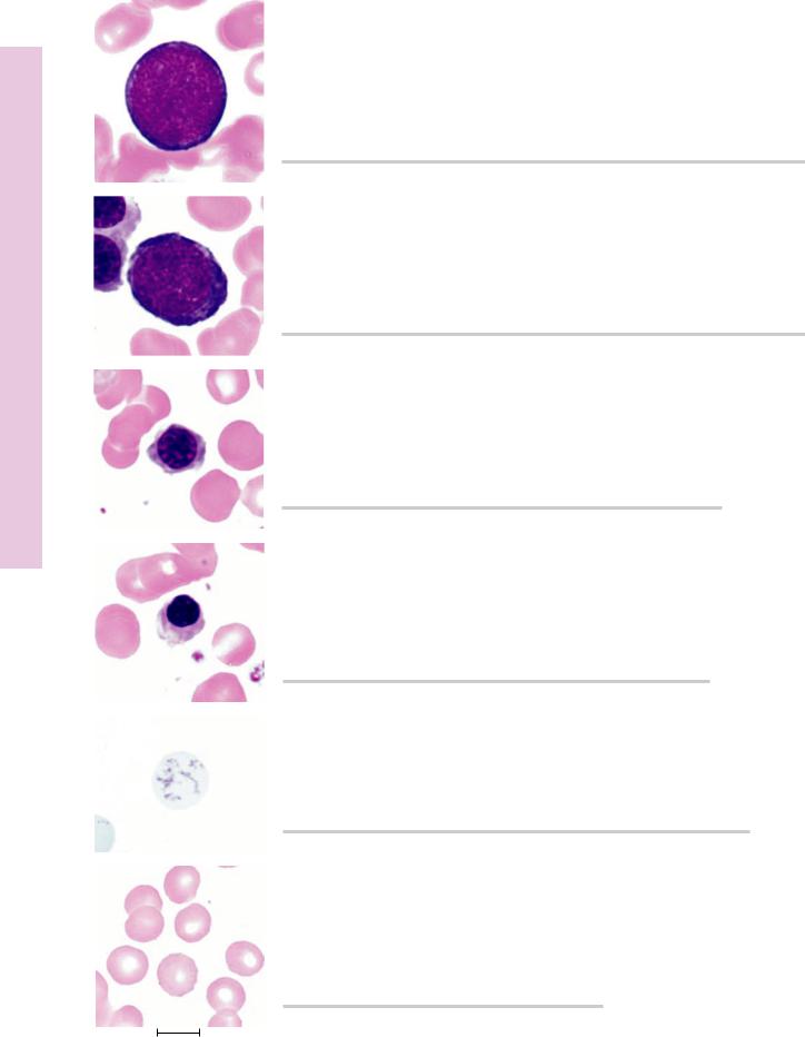
Erythropoiesis • 5-5 PLATE
FIGURE 1. Human marrow smear. ×1,325.
Proerythroblast.
FIGURE 2. Human marrow smear. ×1,325.
Basophilic erythroblast.
FIGURE 3. Human marrow smear. ×1,325.
Polychromatophilic erythroblast.
FIGURE 4. Human marrow smear. ×1,325.
Orthochromatophilic erythroblast.
FIGURE 5. Human marrow smear. Methylene blue stain. ×1,325.
Reticulocyte.
FIGURE 6. Human marrow smear. ×1,325.
Erythrocyte.
1.15 cm = 7.5 µm
122
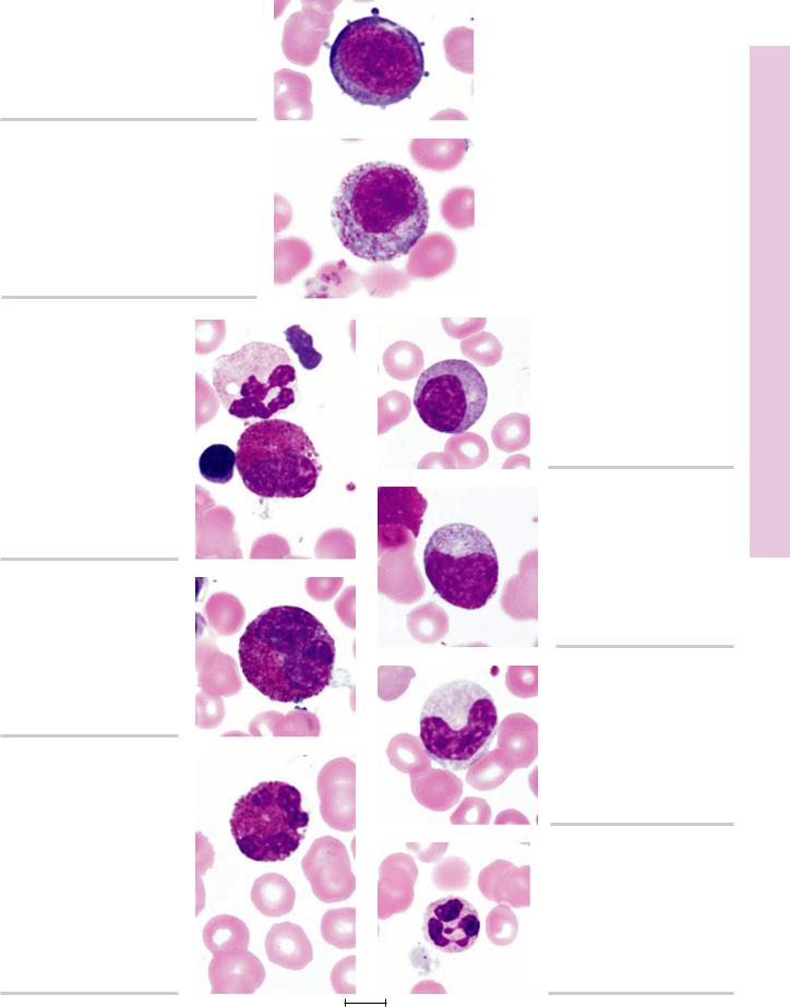
FIGURE 1. Myeloblast. Human bone marrow smear. ×1,325.
FIGURE 2. Promyelocyte. Human bone marrow smear. ×1,325.
ytopoiesis Granuloc• 6-5 PLATE
FIGURE 3a. Eosinophilic myelocyte. Human bone marrow smear. ×1,325.
FIGURE 4a. Eosinophilic metamyelocyte. Human bone marrow smear. ×1,325.
FIGURE 5a. Eosinophilic stab cell. Human bone marrow smear. ×1,325.
FIGURE 3b. Neutrophilic myelocyte. Human bone marrow smear. ×1,325.
FIGURE 4b. Neutrophilic metamyelocyte. Human bone marrow smear.
×1,325.
FIGURE 5b. Neutrophilic stab cell. Human bone marrow smear. ×1,325.
FIGURE 6. Neutrophil. Human bone marrow smear. ×1,325.
1.15 cm = 7.5 µm
123