


Chapter Summary
I. CARTILAGE
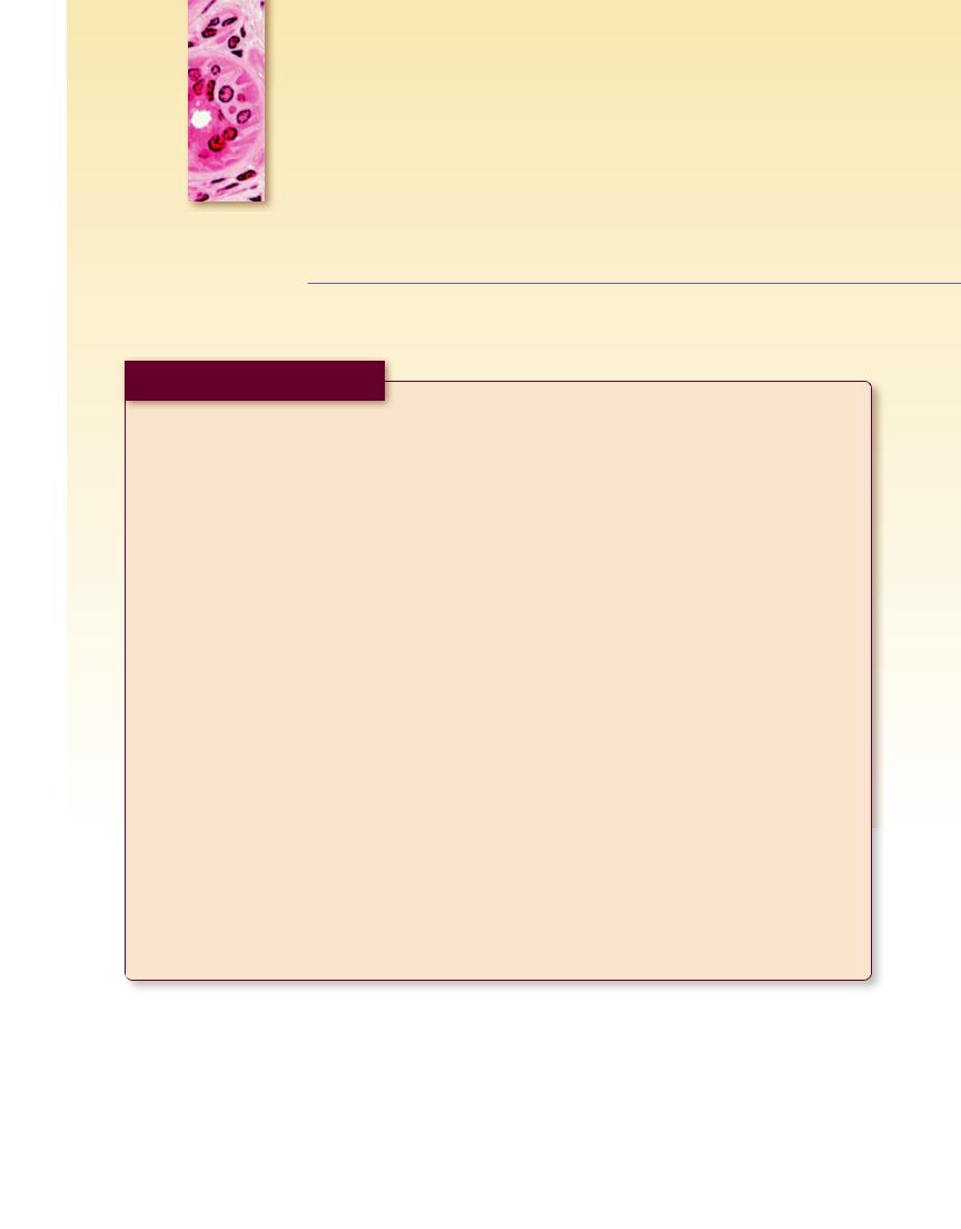
A. Embryonic Cartilage
1. Perichondrium
The perichondrium is very thin and cellular.
2. Matrix
The matrix is scanty and smooth in appearance.
3. Cells
Numerous, small, round chondrocytes are housed in small spaces in the matrix. These spaces are known as lacunae.
B. Hyaline Cartilage
1. Perichondrium
The perichondrium has two layers, an outer fibrous layer, which contains collagen and fibroblasts, and an inner chondrogenic layer, which contains chondrogenic cells and chondroblasts.
2. Matrix
The matrix is smooth and basophilic in appearance. It has two regions, the territorial (capsular) matrix, which is darker and surrounds lacunae, and the interterritorial (intercapsular) matrix, which is lighter in color. The collagen fibrils are masked by the ground substance.
3. Cells
Either chondrocytes are found individually in lacunae or there may be two or more chondrocytes (isogenous group) in a lacuna. The latter case signifies interstitial growth. Appositional growth occurs just deep to the perichondrium and is attributed to chondroblasts.
C. Elastic Cartilage
1. Perichondrium
The perichondrium is the same in elastic cartilage as in hyaline cartilage, but also has elastic fibers.
2. Matrix
The matrix contains numerous dark elastic fibers in addition to the collagen fibrils.
3. Cells
The cells are chondrocytes, chondroblasts, and chondrogenic cells, as in hyaline cartilage.
D. Fibrocartilage
1. Perichondrium
The perichondrium is usually absent.
2. Matrix
The ground substance of matrix is very scanty. Many thick collagen bundles are located between parallel rows of chondrocytes.
3. Cells
The chondrocytes in fibrocartilage are smaller than those in hyaline or elastic cartilage, and they are arranged in parallel longitudinal rows between bundles of thick collagen fibers.
II. BONE
A. Decalcified Compact Bone
1. Periosteum
The periosteum has two layers, an outer fibrous layer, containing collagen fibers and fibroblasts, and an inner osteogenic layer, containing osteoprogenitor cells and osteoblasts. It is anchored to bone by Sharpey’s fibers.
2. Lamellar Systems
Lamellar organization consists of outer and inner circumferential lamellae, osteons (haversian canal systems), and interstitial lamellae.
3. Endosteum
The endosteum is a thin membrane that lines the medullary cavity, which contains yellow or white bone marrow.
4. Cells
Osteocytes are housed in small spaces called lacunae. Osteoblasts and osteoprogenitor cells are found in the osteogenic layer of the periosteum, in the endosteum, and lining haversian canals. Osteoclasts are located in Howship’s lacunae along resorptive surfaces of bone. Osteoid, noncalcified bone matrix, is interposed between the cells of bone and the calcified tissue.
5. Vascular Supply
Blood vessels are found in the periosteum, in the marrow cavity, and in the haversian canals of osteons. Haversian canals are connected to each other by Volkmann’s canals.
106
B. Undecalcified Compact Ground Bone
1. Lamellar Systems
The lamellar organization is clearly evident as wafer-thin layers or lamellae constituting bone. They are then organized as outer and inner circumferential lamellae, osteons, and interstitial lamellae.
Osteons are cylindrical structures composed of concentric lamellae of bone. Their lacunae are empty, but in living bone, they contain osteocytes. Canaliculi radiate from lacunae toward the central haversian canal, which in living bone houses blood vessels, osteoblasts, and osteoprogenitor cells. Cementing lines demarcate the peripheral extent of each osteon. Volkmann’s canals interconnect neighboring haversian canals.
C. Decalcified Cancellous Bone
1. Lamellar Systems
Lamellar organization consists of spicules and trabeculae of bone.
2. Cells
Cells are as before in that osteocytes are housed in lacunae. Osteoblasts line all trabeculae and spicules. Occasionally, multinuclear, large osteoclasts occupy Howship’s lacunae. Osteoid, noncalcified bone matrix, is interposed between the cells of bone and the calcified tissue.
Bone marrow occupies the spaces among and between trabeculae.
D. Intramembranous Ossification
1. Ossification Centers
Centers of ossification are vascularized areas of mesenchymal connective tissue where mesenchymal cells probably differentiate into osteoprogenitor cells, which differentiate into osteoblasts.
2. Lamellar Systems
Lamellar organization begins when spicules and trabeculae form into primitive osteons surrounding blood vessels. The first bone formed is primary bone (woven bone), whose cells are larger and whose fibrillar arrangement is haphazard compared with secondary (mature) bone.
3. Cells
The cellular elements of intramembranous ossification are osteoprogenitor cells, osteoblasts, osteocytes, and osteoclasts. Additionally, mesenchymal and hemopoietic cells are also present.
C A R T I L A G E A N D B O N E 107
E. Endochondral Ossification
1. Primary Ossification Center
The perichondrium of the diaphysis of the cartilage template becomes vascularized, followed by hypertrophy of the centrally located chondrocytes, confluence of contiguous lacunae, calcification of the cartilage remnants, and subsequent chondrocytic death. Concomitant with these events, the chondrogenic cells of the perichondrium become osteoprogenitor cells, which, in turn, differentiate into osteoblasts. The osteoblasts form the subperiosteal bone collar, thus converting the overlying perichondrium into a periosteum. A periosteal bud invades the diaphysis, entering the confluent lacunae left empty by the death of chondrocytes. Osteogenic cells give rise to osteoblasts, which elaborate bone on the trabeculae of calcified cartilage. Hemopoiesis begins in the primitive medullary cavity; osteoclasts (and, according to some, chondroclasts) develop, which resorb the bone-covered trabeculae of calcified cartilage as the subperiosteal bone collar becomes thicker and elongated.
2. Secondary Ossification Center
The epiphyseal (secondary) center of ossification is initiated somewhat after birth. It begins in the center of the epiphysis and proceeds radially from that point, leaving cartilage only at the articular surface and at the interface between the epiphysis and the diaphysis, the future epiphyseal plate.
3. Epiphyseal Plate
The epiphyseal plate is responsible for the future lengthening of a long bone. It is divided into five zones: (1) zone of reserve cartilage, a region of haphazardly arranged chondrocytes; (2) zone of cell proliferation, where chondrocytes are arranged in rows whose longitudinal axis parallels that of the growing bone; (3) zone of cell maturation and hypertrophy, where cells enlarge and the matrix between adjoining cells becomes very thin;
(4) zone of calcifying cartilage, where lacunae become confluent and the matrix between adjacent rows of chondrocytes becomes calcified, causing subsequent chondrocytic death; and (5) zone of provisional ossification, where osteoblasts deposit bone on the calcified cartilage remnants between the adjacent rows. Osteoclasts (and, according to some, chondroclasts) resorb the calcified complex.
5 BLOOD AND HEMOPOIESIS
CHAPTER OUTLINE
Tables
Table 5-1 Formed Elements of Blood Table 5-2 Hemopoietic Growth Factors
Plates
Plate 5-1 |
Circulating Blood p. 116 |
Fig. 1 |
Red blood cells. Human |
Fig. 2 |
Neutrophils. Human |
Fig. 3 |
Eosinophils. Human |
Fig. 4 |
Basophils. Human |
Fig. 5 |
Monocytes. Human |
Fig. 6 |
Lymphocytes. Human |
Plate 5-2 |
Circulating Blood (Drawing) p. 118 |
Plate 5-3 |
Blood and Hemopoiesis p. 119 |
Plate 5-4 |
Bone Marrow and Circulating Blood |
|
p. 120 |
Fig. 1 |
Bone marrow. Human |
Fig. 2 |
Bone marrow. Human |
Fig. 3 |
Blood smear. Human. Wright’s stain |
Fig. 4 |
Bone marrow smear. Human. Wright’s |
|
stain |
Plate 5-5 |
Erythropoiesis p. 122 |
Fig. 1 |
Human marrow smear. Proerythroblast |
Fig. 2 |
Human marrow smear. Basophilic |
|
erythroblast |
Fig. 3 |
Human marrow smear. |
|
Polychromatophilic |
|
erythroblast |
Fig. 4 |
Human marrow smear. |
|
Orthochromatophilic erythroblast |
Fig. 5 |
Human marrow smear. Reticulocyte |
Fig. 6 |
Human marrow smear. Erythrocyte |
Plate 5-6 |
Granulocytopoiesis p. 123 |
Fig. 1 |
Human bone marrow smear. Myeloblast |
Fig. 2 |
Human bone marrow smear. |
|
Promyelocyte |
Fig. 3a |
Human bone marrow smear. |
|
Eosinophilic myelocyte |
Fig. 3b |
Human bone marrow smear. |
|
Neutrophilic myelocyte |
Fig. 4a |
Human bone marrow smear. |
|
Eosinophilic metamyelocyte |
Fig. 4b |
Human bone marrow smear. |
|
Neutrophilic metamyelocyte |
Fig. 5a |
Human bone marrow smear. |
|
Eosinophilic stab cell |
Fig. 5b |
Human bone marrow smear. |
|
Neutrophilic stab cell |
Fig. 6 |
Human bone marrow smear. Neutrophil |
108