

- •Preface
- •Acknowledgments
- •Reviewers
- •Contents
- •CHAPTER OUTLINE
- •CYTOPLASM
- •Plasmalemma
- •Mitochondria
- •Ribosomes
- •Endoplasmic Reticulum
- •Golgi Apparatus, cis-Golgi Network, and the trans-Golgi Network
- •Endosomes
- •Lysosomes
- •Peroxisomes
- •Proteasomes
- •Cytoskeleton
- •Inclusions
- •NUCLEUS
- •CELL CYCLE
- •CHAPTER OUTLINE
- •EPITHELIUM
- •Epithelial Membranes
- •GLANDS
- •Chapter Summary
- •CHAPTER OUTLINE
- •EXTRACELLULAR MATRIX
- •Fibers
- •Amorphous Ground Substance
- •Extracellular Fluid
- •CELLS
- •CONNECTIVE TISSUE TYPES
- •Chapter Summary
- •CHAPTER OUTLINE
- •CARTILAGE
- •BONE
- •Cells of Bone
- •Osteogenesis
- •Bone Remodeling
- •Chapter Summary
- •CHAPTER OUTLINE
- •FORMED ELEMENTS OF BLOOD
- •Lymphocytes
- •Neutrophils
- •PLASMA
- •COAGULATION
- •HEMOPOIESIS
- •Erythrocytic Series
- •Granulocytic Series
- •Chapter Summary
- •CHAPTER OUTLINE
- •SKELETAL MUSCLE
- •Sliding Filament Model of Muscle Contraction
- •CARDIAC MUSCLE
- •SMOOTH MUSCLE
- •Chapter Summary
- •CHAPTER OUTLINE
- •BLOOD-BRAIN BARRIER
- •NEURONS
- •Membrane Resting Potential
- •Action Potential
- •Myoneural Junctions
- •Neurotransmitter Substances
- •SUPPORTING CELLS
- •PERIPHERAL NERVES
- •Chapter Summary
- •CHAPTER OUTLINE
- •BLOOD VASCULAR SYSTEM
- •HEART
- •ARTERIES
- •Capillary Permeability
- •Endothelial Cell Functions
- •VEINS
- •LYMPH VASCULAR SYSTEM
- •Chapter Summary
- •CHAPTER OUTLINE
- •CELLS OF THE IMMUNE SYSTEM
- •Antigen-Presenting Cells
- •DIFFUSE LYMPHOID TISSUE
- •LYMPH NODES
- •TONSILS
- •SPLEEN
- •THYMUS
- •Chapter Summary
- •CHAPTER OUTLINE
- •PITUITARY GLAND
- •Pars Intermedia
- •Pars Nervosa and Infundibular Stalk
- •Pars Tuberalis
- •THYROID GLAND
- •Parathyroid Glands
- •Suprarenal Glands
- •Cortex
- •Medulla
- •Pineal Body
- •Chapter Summary
- •CHAPTER OUTLINE
- •SKIN
- •Epidermis of Thick Skin
- •Dermis
- •DERIVATIVES OF SKIN
- •Chapter Summary
- •CHAPTER OUTLINE
- •CONDUCTING PORTION OF THE RESPIRATORY SYSTEM
- •Extrapulmonary Region
- •Intrapulmonary Region
- •RESPIRATORY PORTION OF THE RESPIRATORY SYSTEM
- •MECHANISM OF RESPIRATION
- •Chapter Summary
- •CHAPTER OUTLINE
- •ORAL CAVITY AND ORAL MUCOSA
- •Oral Mucosa
- •Tongue
- •Teeth
- •Odontogenesis (See Graphic 13-2)
- •Chapter Summary
- •CHAPTER OUTLINE
- •REGIONS OF THE DIGESTIVE TRACT
- •Esophagus
- •Stomach
- •Small Intestine
- •Large Intestine
- •GUT-ASSOCIATED LYMPHOID TISSUE
- •DIGESTION AND ABSORPTION
- •Carbohydrates
- •Proteins
- •Lipids
- •Water and Ions
- •Chapter Summary
- •CHAPTER OUTLINE
- •MAJOR SALIVARY GLANDS
- •PANCREAS
- •LIVER
- •Exocrine Function of the Liver
- •Endocrine and Other Functions of the Liver
- •GALLBLADDER
- •Chapter Summary
- •CHAPTER OUTLINE
- •KIDNEY
- •Uriniferous Tubule
- •Nephron
- •Collecting Tubules
- •FORMATION OF URINE FROM ULTRAFILTRATE
- •EXTRARENAL EXCRETORY PASSAGES
- •Chapter Summary
- •CHAPTER OUTLINE
- •OVARY
- •Ovarian Follicles
- •Regulation of Follicle Maturation and Ovulation
- •Corpus Luteum and Corpus Albicans
- •GENITAL DUCTS
- •Oviduct
- •Uterus
- •FERTILIZATION, IMPLANTATION, AND THE PLACENTA
- •Fertilization and Implantation
- •Placenta
- •VAGINA
- •EXTERNAL GENITALIA
- •MAMMARY GLANDS
- •Chapter Summary
- •CHAPTER OUTLINE
- •TESTES
- •Spermatogenesis
- •GENITAL DUCTS
- •ACCESSORY GLANDS
- •PENIS
- •Erection and Ejaculation
- •Chapter Summary
- •CHAPTER OUTLINE
- •SENSORY ENDINGS
- •Chapter Summary
- •Terminology of Staining
- •Common Stains Used in Histology
- •Hematoxylin and Eosin
- •Wright Stain
- •Weigert Method for Elastic Fibers and Elastic van Gieson Stain
- •Silver Stain
- •Iron Hematoxylin
- •Bielschowsky Silver Stain
- •Masson Trichrome
- •Periodic Acid-Schiff Reaction (PAS)
- •Alcian Blue
- •von Kossa Stain
- •Sudan Red
- •Mucicarmine Stain
- •Safranin-O
- •Toluidine Blue

The organs of special senses include the gustatory, olfactory, visual, auditory, and vestibular systems. The gustatory apparatus, consisting of taste buds,
is discussed in Chapter 13, and the olfactory epithelium is treated in Chapter 12. This chapter details sensory endings: the microscopic morphology of the eye, involved with visual sensations, and the ear, involved with auditory and vestibular sensations.
SENSORY ENDINGS
Sensory endings are located at the terminus of dendrites. These specialized receptors (see Table 19-1) are members of the general somatic or general visceral afferent pathways and are:
•specialized to respond to stimuli, such as pressure, touch, temperature, and pain, on the external surface of the body (exteroceptors); additionally, there are special senses of sight and hearing as well as smell and taste
•incorporated into muscles and tendon to perceive the localization of the body in three-dimensional space (proprioceptors), and
•distributed within organs to monitor the activity of these organs as components of the general visceral afferent pathways.
EYE
The eye is a sensory organ whose lens focuses rays of light originating in the external environment onto photosensitive cells of the retina (see Graphic 19-1). The intensity, location, and wavelengths of the transmitted light are partially processed by the retina, and the assembled information is transmitted, via the optic nerve, for further processing and interpretation by the visual cortex of the brain as three-dimensional color images of the external milieu.
•Because the eyes are set apart and because their visual fields overlap, such three-dimensional imaging becomes possible.
•Each eyeball (bulb, globe), protected by the eyelids, is movable by means of a group of extrinsic skeletal muscles that insert into its fibrous outer tunic, thus assisting in suspending it in its bony orbit and directing the pupil to the most advantageous position for perceiving the image viewed.
Three coats constitute the wall of the bulb: the outer fibrous tunic, the middle vascular tunic (uvea), and the inner retinal tunic.
S P E C I A L S E N S E S |
455 |
•The fibrous tunic (corneoscleral layer) is composed of the opaque, white sclera that covers the posterior aspect of the bulb and the transparent cornea that covers the anterior one-sixth of the eyeball.
The junction between the sclera and the cornea is known as the limbus.
•The vascular tunic consists of several regions: the anteriorly positioned iris and ciliary body and the posteriorly located, highly vascular and pigmented choroid.
Melanocytes located in the epithelium and stroma of the iris block light from passing through the iris, except at the pupil.
Additionally, eye color is related to the abundance of melanin produced by these melanocytes:
A large amount of melanin imparts dark eyes, whereas less melanin renders the eyes light in color.
Intrinsic smooth muscles, represented by the sphincter pupillae and dilatator pupillae muscles, adjust the aperture of the iris.
The ciliary smooth muscles alter tension of the suspensory (zonular) fibers anchored in the lens and thus alter the shape of the lens (accommodation) for near and far vision.
•The innermost retinal tunic is composed of 10 layers responsible for photoreception and impulse generation (see Table 19-2). The two deepest layers, the retinal pigment epithelium and the layer of rods and cones, bear the major responsibility for photoreception. Retinal pigment epithelium functions in esterifying vitamin A and transporting it to the rods and cones, phagocytosing the shed tips of rods and cones, and synthesizing melanin, which absorbs light after rods and cones have been stimulated.
Rods are sensitive to low light intensity and possess many flattened discs containing rhodopsin (an integral membrane protein, opsin, bound to retinal, the aldehyde form of vitamin A) in their outer segment.
When light is absorbed by rhodopsin, it dissociates into retinal and opsin (bleaching),permitting diffusion of bound Ca2+ into the outer segment.
Excess levels of Ca2+ hyperpolarize the cell by closing Na+ channels, thus preventing the entry of Na+ into the cell.
The electrical potential thus generated is relayed to other rods via gap junctions and then along the pathway to the optic nerve.
Dissociated retinal and opsin reassemble, and the Ca2+ ions are recaptured, establishing a normal resting potential.
Cones, sensitive to light of high intensity, producing greater visual acuity, are much more numerous

456 S P E C I A L S E N S E S
TABLE 19-1 • Specialized Receptors, Their Function and Location
Receptor |
Type |
Function and Location |
|
|
|
Peritrichial nerve |
Nonencapsulated |
Are nonmyelinated and have no associated Schwann’s cells. Most are |
endings |
|
coupled with hair follicles and react to the hair’s motion. The sensation is |
|
|
interpreted as touch or being tickled. |
|
|
|
Merkel’s discs |
Nonencapsulated |
Mechanoreceptors located in the stratum basale of the epidermis. |
|
|
|
Meissner’s corpuscles |
Encapsulated |
Located in the dermal papillae of the dermis and respond to touch sensations |
|
|
|
Pacinian corpuscles |
Encapsulated |
Resemble an onion since epithelioid cells form concentric layers around |
|
|
a naked nerve ending. These corpuscles, located in the hypodermis, |
|
|
mesocolon, and mesentery, respond to vibration, pressure, and deep touch. |
|
|
|
Ruffini’s endings |
Encapsulated |
Are composed of highly branched nerve termini surrounded by fibroblast- |
|
|
like cells. They respond to pressure and stretch and are located in nail |
|
|
beds, periodontal ligament, dermis of the skin, and capsules of joints. |
|
|
|
Krause’s end bulbs |
Encapsulated |
These spherical capsules containing a naked nerve ending are located in the |
|
|
connective tissues just deep to the epithelium, capsules of joints, perito- |
|
|
neum, and in the dermis of skin. Their function is not known. |
|
|
|
Muscle spindles |
Encapsulated |
Described in the chapter on Muscle. They function in proprioception. They |
|
|
respond to alteration in the length and rate of change in muscle and thus |
|
|
function in proprioception. |
|
|
|
Golgi tendon organs |
Encapsulated |
Described in the chapter on Muscle. Respond to changes in the tension and |
|
|
the rate of tension change around a joint, thus function in proprioception. |
|
|
|
Thermoreceptors |
Nonencapsulated |
They are assumed to be naked nerve endings located in the epidermis that |
|
|
respond to temperature. Their morphology is not known. |
|
|
|
Nociceptors |
Nonencapsulated |
Branched naked nerve endings located in the epidermis. They are stimulat- |
|
|
ed by extremes in temperature, by damage to the epidermis and underly- |
|
|
ing structures, as well as by certain chemicals as pain sensation. |
|
|
|

S P E C I A L S E N S E S |
457 |
TABLE 19-2 • Layers of the Retina
Layer |
Description |
|
|
Pigment epithelium |
Synthesizes melanin that absorbs light that activated rods and cones; phagocytoses the shed |
|
tips of rods and cones; estrifies vitamin A |
|
|
Layer of rods and |
Photosensitivity; rods are sensitive to low light intensity, and cones are sensitive to bright light |
cones |
and perceive color. |
|
|
External limiting |
Zonulae adherentes formed between the photoreceptor cells and Müller cells (therefore, it is |
membrane |
not a true membrane) |
|
|
Outer nuclear layer |
Houses the nuclear regions of rods and cones |
|
|
Outer plexiform layer |
Region of synapse between axons, photoreceptor cells, and dendrites of horizontal and bipolar cells |
|
|
Inner nuclear layer |
Houses the nuclear regions of Müller, bipolar, amacrine, and horizontal cells |
|
|
Inner plexiform layer |
Region where synapses occur among axons and dendrites of amacrine, bipolar, and ganglion cells |
|
|
Ganglion cell layer |
Region of the cell bodies of multipolar neurons as well as neuroglial cells |
|
|
Optic nerve fiber layer |
Region where the unmyelinated axons of ganglion cells join to form the optic nerve. Once the |
|
fibers pierce the sclera, they become myelinated. |
|
|
Inner limiting membrane |
Composed of the expanded terminal processes of Müller cells and their basal lamina |
|
|

458 S P E C I A L S E N S E S
than rods, and they produce iodopsin, the photopigment responsible for distinguishing color. Three different moieties of opsin are sensitive to either red, green, or blue light.
The mechanism of transducing photoenergy into electrical energy for transmission to the brain via the optic nerve is similar to that described in the rods.
Rods and cones are either stimulated (on) or inhibited (off) by light, that is, they indicate the location of a light pixel and, in the case of cones, its color.
Dendrites of 10 different types of bipolar cells receive information from the rods and cones, and then this information is conveyed by the axons of the bipolar cells into specific strata of the inner plexiform layer of the retina.
The further transmission of the impulses is monitored and modulated by one or more of the 27 types of amacrine cells, whose axons can span several millimeters or just a few micrometers of the retinal expanse.
The outer layer of the retina contains 12 types of ganglion cells whose interactions with bipolar cells and amacrine cells result in the
transmission of 12 different moving images (a continuous moving stream that resembles but is not created frame by frame) of the same scene via the optic nerve to the visual cortex of the brain for further analysis, assembly, and interpretation.
These moving images are very different from each other, in that some consist of highlights, others consist of line drawings of outlines, and still others generate shadows.
It is the function of the visual cortex to assemble these movies into the world that we recognize.
It must be stressed that this is a simplified description of some of the current concepts of vision that will certainly be modified as more information is gained from research in this field.
An additional type of ganglion cells, which make up less than 3% of the ganglion cell population, appear to function in the establishment of the circadian rhythm.
These ganglion cells possess the light-sensitive pigment melanopsin that responds to blue light even in individuals who are blind.
The axons of these ganglion cells project to the suprachiasmatic nucleus, the region of the brain responsible for the regulation of circadian rhythm.
It appears, then, that the suprachiasmatic nucleus receives information from these specialized ganglion cells of the retina as to when there is daylight.
The optic disc, the region where the optic nerve exits the eyeball, contains neither cones nor rods; consequently, it represents and is called a blind spot. Just lateral to the blind spot is the fovea centralis, a depression in the retina. The fovea contains mostly cones that are packed so tightly that not all layers of the retina are present. Visual acuity is the greatest in the fovea centralis.
•Accessory structures of the eye include the conjunctiva, eyelids, and lacrimal gland.
The conjunctiva is a transparent mucous membrane that lines the eyelids and reflects onto the eyeball.
The eyelids contain modified sebaceous glands, the meibomian glands, which are responsible for altering the surface tension of the watery tears, thus slowing evaporation.
The lacrimal glands secrete tears, a complex fluid composed of water, proteins, salts, peptides, and other organic molecules which keep the conjunctiva and cornea moist.
Tears also contain lysozyme, an antibacterial enzyme.
The additional components of the interior of the eyeball are the aqueous humor, a fluid; the vitreous body, a gel; and the lens, all of which serve as parts of the refractive media.
The aqueous humor, located in the posterior and anterior chambers of the eye, and the vitreous body, located behind the lens of the eye, are also important in providing nutrients to the avascular lens and cornea.
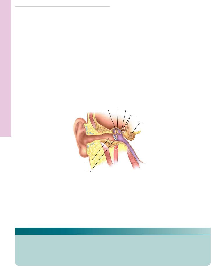
EAR
The ear functions in the reception of sound as well as in the perception of the orientation of the head and therefore the body in relation to the directional forces of gravity (see Graphic 19-2). To perform both functions of hearing and equilibrium (balance), the ear is subdivided into the external, middle, and inner ears.
•The external ear is composed of a cartilaginous, skin-covered auricle (pinna) and the external auditory meatus, with its cartilaginous outer and bony inner aspects, whose internal extent is separated from the middle ear by the thin tympanic membrane.
•The tympanic cavity of the middle ear houses the three auditory ossicles, connected in series to each other: the outermost malleus (hammer), the middle

incus (anvil), and the innermost stapes (stirrup). This cavity is connected to the nasopharynx via the cartilaginous auditory (eustachian) tube, which permits equalization of atmospheric pressures on either side of the tympanic membrane. Sound waves are funneled by the auricle to the tympanic membrane, whose vibrations are amplified and transmitted by the movements of the three bony ossicles to the oval window of the inner ear’s cochlea.
•The bony labyrinth of the inner ear, subdivided into the semicircular canals, vestibule, and cochlea, is filled with perilymph. Loosely contained within it and all of its subdivisions is the endolymph-containing membranous labyrinth.
Movements of the fluid environment within this system are perceived by apical “hairs” of specialized sensory cells contained within the membranous labyrinth and ultimately transduced to electrical impulses for transmission to the brain.
The saccule and utricle, specialized structures of the membranous labyrinth in the vestibule, contain type I and type II hair cells (neuroepithelial cells containing many stereocilia and a single kinocilium), whose free ends are embedded in the otolithic membrane containing calcium carbonate crystals known as otoliths (otoconia).
Static equilibrium and linear acceleration are determined by movements (or the lack of movements) in the stereocilia or kinocilia of these hair cells.
Threshold bending of the stereocilia or kinocilia will depolarize the hair cells, which, in turn, relay (via neurotransmission) information to the processes of primary vestibular neurons located in Scarpa’s ganglion.
Semicircular ducts, specializations of the membranous labyrinth in the semicircular canals,
contain neuroepithelial hair cells located in the cristae ampullares (sensory regions) of the ampullae.
Free ends of these hair cells have stereocilia that are embedded in a viscous glycoprotein known as the cupula.
Movement of the endolymph bathing the cupula depolarize the hair cells, which, in turn, alter the activity in the synaptic endings associated with the base of the hair cells.
This process is sensitive to rotational acceleration in any of the three directions of orientation of the semicircular canals.
Thus, these structures are responsible for the vestibular sensations of balance and orientation.
S P E C I A L S E N S E S |
459 |
TABLE 19-3 • Cells of the Spiral Organ of Corti
Cells |
Function |
|
|
Border cells |
Support the inner aspect of the |
|
organ of Corti |
|
|
Cells of Böttcher |
Unclear |
|
|
Cells of Claudius |
Unclear |
|
|
Cells of Hensen |
Support the outer aspect of the |
|
organ of Corti |
|
|
Inner hair cells |
Transduction of impulses to bipo- |
|
lar cells of the spiral ganglion |
|
|
Inner pillar cells |
Form the medial wall of the |
|
inner tunnel and support the |
|
hair cells |
|
|
Inner |
Surround and support the inner |
phalangeal cells |
hair cells |
|
|
Outer hair cells |
Transduction of impulses to bipo- |
|
lar cells of the spiral ganglion |
|
|
Outer |
Support the outer hair cells and |
phalangeal cells |
their associated nerve fibers |
|
|
Outer pillar cells |
Form the lateral wall of the inner |
|
tunnel and support the hair cells |
|
|
The endolymphatic sac (terminal end of the endolymphatic duct) contains phagocytic cells in its lumen and may function in resorption of endolymph.
The cochlear duct (scala media) contains the spiral organ of Corti, which is bordered by the scala vestibuli and the scala tympani (both scalae contain perilymph and communicate at the helicotrema).
The vestibular membrane located between the scala vestibuli and the cochlear duct functions to maintain the high ion gradient between the perilymph and endolymph.
The spiral organ of Corti (see Table 19-3), sitting on the basilar membrane, contains, among other supporting cells, neuroepithelial inner and outer hair cells whose free ends are embedded in the gel-like tectorial membrane.
-Sound transmission/conduction via the tympanic membrane and ossicles to the oval window sets waves translated to the oval window and sets the perilymph of the scala tympani in motion, which displaces the basilar membrane, thus moving the hair cells but not the tectorial membrane.

460S P E C I A L S E N S E S
-Bending of the hair cells causes them to release neurotransmitter substance, exciting the bipolar cells of the spiral ganglion, resulting in transmission of the impulse to higher centers of the brain.
-Although the basilar membrane vibrates at many frequencies, certain regions vibrate optimally at specific frequencies.
-For example, low-frequency sound waves are detected farther away from the oval window.
-It should be noted that loud sounds, such as those at rock concerts, create a great deal of energy within the hearing mechanism, such that it may take 2 or 3 days for the energy to be completely dissipated and the buzzing to stop.
CLINICAL CONSIDERATIONS
Blue Eye Color
Until approximately 6,000 to 10,000 years ago, every human being had brown eyes; then, a small mutation in the switch that turned off OCA2 gene resulted in the inability of that individual to manufacture P protein in the iris. Protein P is involved in melanin formation; thus, the person with this particular mutation was able to synthesize melanin normally except in the iris, and instead of having brown eyes, that person’s eyes were blue. Thus, it is believed that all blue-eyed individuals are descendants of that one person born in that era.
Myopia and Hyperopia
As an individual ages, the longitudinal axis of the eye changes, as may the curvature of the cornea, and the lens, instead of focusing the image on the retina, focuses it either in front of the retina (myopic vision) or behind the retina (hyperopic vision). The condition may be corrected with lenses (eyeglasses or contact lenses) or by refractive surgery, assisting the lens in focusing on the retina.
Glaucoma
Glaucoma is a condition of high intraocular pressure caused by an obstruction that prevents the aqueous humor from exiting the anterior chamber of the eye. If left untreated, the pressure damages the optic nerve to such an extent that blindness may result.
Cataract
Cataract, a common condition of aging, is caused by excessive UV radiation and by pigments and other substances accumulating in the lens, making it opaque and thus impairing vision. This condition may be corrected by excising the lens and replacing it with a plastic lens.
This photomicrograph is from the lens of an older patient who presented with age-related cataract. Observe the presence of cortical extracellular clefts and globules. (Reprinted with permission from Mills SE, Carter, D, et al., eds. Sternberg’s Diagnostic Surgical Pathology, 5th ed., Philadelphia: Lippincott Williams & Wilkins, 2010, p. 981.)
Detached Retina
Detached retina may result from a trauma in which the neural and pigmented layers of the retina become separated, resulting to ischemic damage to the neurons. This condition may cause partial blindness, but it may be corrected by surgical intervention.
Retinoblastoma
Retinoblastoma is a malignancy of the very young child, usually detected at about 2 years of age, although at the time of diagnosis the child may be 5 or 6 years old. Approximately a third of the cases have familial components, but at least 60% occur without a familial incidence. The tumor appears white with regions of calcification
S P E C I A L S E N S E S |
461 |
and yellow foci of necrosis. The tumor may spread by individual cells invading the optic nerve as well as the choroid. The patient has to lose the eyeball to prevent metastasis.
This photomicrograph is from the eyeball of a child with retinoblastoma. Observe the relatively normal retina on the left-hand side of the image. The arrows indicate regions of necrosis in a field of perivascular tumor cells. (Reprinted with permission from Mills SE, Carter, D, et al., eds. Sternberg’s Diagnostic Surgical Pathology, 5th ed., Philadelphia: Lippincott Williams & Wilkins, 2010, p. 982.)
Conductive Hearing Loss
Conductive hearing loss may arise from a middle ear infection (otitis media), an obstruction, or otosclerosis of the middle ear.
Nerve Deafness
Nerve deafness results from a lesion in the cochlear portion of the vestibulocochlear nerve (cranial nerve VIII). This condition may be the result of disease, prolonged exposure to loud sounds, and/or drugs.
Observe that the footplate of the stapes is fixed to the densely sclerotic bone forming the perimeter of the oval window. (Reprinted with permission from Mills SE, Carter D, et al., eds. Sternberg’s Diagnostic Surgical Pathology, 5th ed., Philadelphia: Lippincott Williams & Wilkins, 2010, p. 944.)
Ménière’s Disease
Ménière’s disease is an inner ear disorder characterized by symptoms such as hearing loss due to excess fluid accumulation in the endolymphatic duct, vertigo, tinnitus, nausea, and vomiting. In severe cases, surgical treatment may be required.
Acoustic Neuroma
The condition known as acoustic neuroma is a benign tumor whose cells of origin are the Schwann cells of the vestibulocochlear nerve (cranial nerve VIII). It is manifested by loss of hearing, loss of balance, vertigo, and tinnitus. If the tumor is not treated early enough, it may involve other cranial nerves in its vicinity. Recent studies suggest the possibility that long-term exposures to the electromagnetic radiation emitted by cell phones may be a causative factor in the development of acoustic neuroma in susceptible individuals.

Eye • 1-19 GRAPHIC
462 S P E C I A L S E N S E S
Retina
Choroid
Sclera
Optic nerve
Lens
Iris
Cornea
Vitreous body
Retina
Choroid
Sclera
Photosensitive region
Metabolic region
Cone
Section of Retina
Rod
Müller cell
Ciliary muscles
Iris
Lens
External plexiform region
Ganglion cell axon to optic nerve
Amacrine cell
Cone
Rod |
Bipolar cell |
|
Horizontal cell
Pigment layer

Stapes
Incus
Malleus
External auditory meatus Tympanum
Outer Hair
Cell
Inner
Hair Cell
Outer hair cell
Cells of Hensen
Outer spiral sulcus
Outer phalangeal cells
Outer pillar cell
Cochlear nerve
Inner pillar cell
S P E C I A L S E N S E S |
463 |
Acoustic
nerve
Semicircular canals
Cochlea
 Auditory tube
Scala vestibuli
Tectorial membrane
Vestibular membrane
Spiral ganglion
Organ of Corti
Basilar membrane
Scala tympani
Organ of Corti
Tectorial membrane
Inner spiral cells
Inner hair cell
Inner phalangeal cells
Cochlear nerve fibers
Ear• 2-19 GRAPHIC

Body Ciliary and Iris, Sclera, Cornea, ye, • E1-19 PLATE
464 S P E C I A L S E N S E S
FIGURE 1. Cornea. Monkey. Paraffin section. ×132.
The cornea is a multilayered, transparent structure. Its anterior surface is covered by a stratified squamous nonkeratinized epithelium (Ep) on the right-hand side of the image, deep to which is a thin, acellular Bowman’s membrane. The bulk of the cornea, the stroma (St), is composed of regularly arranged collagen fibers (CF) and intervening fibroblasts, whose nuclei (N) are readily evident. The posterior surface of the cornea is lined by a simple squamous-to-cuboidal epithelium (Ep) on the left-hand side of the image. A thin, acellular Descemet’s membrane lies between the simple epithelium and the stroma. Inset. Cornea. Monkey. Paraffin section. ×270. A higher magnification of the anterior surface displays the stratified squamous epithelium (Ep) as well as the acellular Bowman’s membrane (BM). Note the regularly arranged bundles of collagen fibers (CF) and intervening fibroblasts (F), whose nucleus is labeled by the lead line.
FIGURE 3. Iris. Monkey. Paraffin section. ×132.
The iris (I) is a pigmented diaphragm that delineates the pupil (P) of the eye. It separates the anterior chamber (AC) from the posterior chamber (PC). The iris is composed of three layers: an outer discontinuous layer of melanocytes and fibroblasts; the intermediate fibrous layer (FL), housing pigment cells (Pc) and fibroblasts; and the posterior double-layered pigmented epithelium (PEp). The sphincter (sM) and dilatator muscles are composed of smooth muscle and smooth muscle–like myoepithelial cells, respectively. The pupillary region of the iris contacts the capsule (Ca) of the lens (L) in living individuals.
FIGURE 2. Sclera. Monkey. Paraffin section. ×132.
The sclera is similar to and continuous with the cornea, but it is not transparent. Note that the epithelium (Ep) of the conjunctiva covers the anterior surface of the sclera. Deep to the epithelium is the loose episcleral tissue (ET), whose small blood vessels (BV) are evident. The stroma (St) is composed of thick collagen fiber (CF) bundles, between which numerous fibroblasts (F) can be seen. The deepest layer of the sclera is the suprachoroid lamina (SL), whose melanocytes (M) containing dark melanin pigment characterize this layer.
FIGURE 4. Ciliary body. Monkey. Paraffin section. ×132.
The ciliary body is composed of ciliary processes (CP), projecting into the posterior chamber (PC), from which suspensory ligaments (zonular fibers) extend to the lens. The bulk of the ciliary body is composed of smooth muscle (SM) disposed more or less in three layers, not evident in this photomicrograph. Numerous pigment cells (Pc) are present in this region. Note that the epithelium of the ciliary body is composed of two layers: an outer pigmented (OP) and an inner nonpigmented (IN) epithelium. The narrow vascular layer (VL) intervenes between the epithelium and ciliary muscles. The base, or root, of the iris is anchored to the ciliary body.
|
Retina |
|
|
|
|
|
Choroid |
|
|
|
|
Optic nerve |
Sclera |
|
|
|
|
Iris |
|
|
|
||
|
Cornea |
Ciliary muscles |
|||
Vitreous body |
|
|
|
|
Iris |
|
|
|
|
||
Retina |
Lens |
|
|
|
|
Choroid |
|
|
|
||
|
|
|
|
|
|
Sclera |
|
|
|
Lens |
|
|
|
|
|||
Eye, ciliary muscles, iris, and lens
KEY
AC |
anterior chamber |
FL |
fibrous layer |
Pc |
pigment cells |
BM |
Bowman’s membrane |
I |
iris |
PEp |
pigmented epithelium |
BV |
blood vessel |
IN |
inner nonpigmented layer |
SEp |
squamous epithelium |
Ca |
capsule |
L |
lens |
SL |
suprachoroid lamina |
CF |
collagen fibers |
M |
melanocytes |
SM |
smooth muscle |
CP |
ciliary process |
N |
nucleus |
sM |
sphincter muscle |
Ep |
epithelium |
OP |
outer pigmented layer |
St |
stroma |
ET |
episcleral tissue |
P |
pupil |
VL |
vascular layer |
F |
fibroblasts |
PC |
posterior chamber |
|
|

Body Ciliary and Iris, Sclera, Cornea, ye, • E1-19 PLATE
FIGURE 1 |
FIGURE 2 |
FIGURE 3 |
FIGURE 4 |

Microscopy Electron Scanning and Light Retina,• 2-19 PLATE
466 S P E C I A L S E N S E S
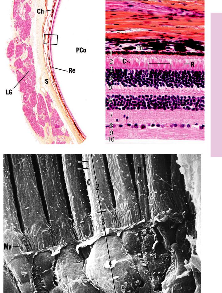
FIGURE 1. Tunics of the eye. Monkey. Paraffin section. ×14.
This survey photomicrograph is of an anterolateral section of the globe of the eye, as evidenced by the presence of the lacrimal gland (LG). Note that the three layers of the globe of the eye are extremely thin in relation to its diameter. The sclera (S) is the outermost layer. The pigment choroid (Ch) and multilayered retina (Re) are easily distinguishable even at this low magnification. The posterior compartment (PCo) lies behind the lens and houses the vitreous body. A region similar to the boxed area is presented at a higher magnification in Figure 2.
FIGURE 3. Rods and cones. Monkey. Scanning electron microscopy. ×6,300.
This scanning electron micrograph of the monkey retina displays regions of several cones (C) that display their thicker morphology and wider nuclear zone and of a few rods (R) whose diameter is narrower, with a thinner nuclear zone. The inner segments of the lamina of rods and cones (2), external limiting membrane (3), and outer nuclear layer (4) are clearly recognizable. The microvilli (Mv) noted in the vicinity of the external limiting membrane belong to Müller’s cells, which were removed during specimen preparation. Observe the longitudinal ridges (arrows) along the surface of the inner segments. (From Borwein B, Borwein D, Medeiros J, McGowan J. The ultrastructure of monkey foveal photoreceptors, with special reference to the structure, shape, size, and spacing of the foveal cones. Am J Anat 1980;159:125–146.)
FIGURE 2. Retina. Pars optica. Monkey. Paraffin section. ×270.
The pars optica of the retina is composed of 10 distinct layers. The pigment epithelium (1), the outermost layer, is closely apposed to the vascular and pigmented choroid (Ch). Various regions of the rods (R) and cones (C) characterize the next four layers. These are the lamina of rods and cones (2), external limiting membrane (3), outer nuclear layer (4), and outer plexiform layer (5). The inner nuclear layer (6) houses the cell bodies of various associative glial (Müller’s) and neural cells. The inner plexiform layer
(7) is a region of synapse formation, whereas the ganglion cell layer (8) houses the cell bodies of multipolar neurons and associated neuroglia. The centrally directed (toward the central nervous system) fibers of these ganglion cells form the optic nerve fiber layer (9), whereas the inner limiting membrane (10) is composed of the expanded processes of Müller’s cells along the inner surface of the eye. A region similar to the boxed area is presented in Figure 3, a scanning electron micrograph of the rods and cones.
Cone
Rod
Pigment layer
Photosensitive region
Metabolic region
|
External plexiform region |
Müller cell |
Cone |
|
|
|
Rod |
|
Ganglion cell axon to optic nerve |
|
Amacrine cell |
|
Bipolar cell |
|
Horizontal cell |
Section of retina
KEY
1 |
pigment epithelium |
7 |
inner plexiform layer |
LG |
lacrimal gland |
2 |
lamina of rods and cones |
8 |
ganglion cell layer |
Mv |
microvilli |
3 |
external limiting membrane |
9 |
optic nerve fiber layer |
PCo |
posterior compartment |
4 |
outer nuclear layer |
10 |
inner limiting membrane |
R |
rods |
5 |
outer plexiform layer |
C |
cones |
Re |
retina |
6 |
inner nuclear layer |
Ch |
choroid |
S |
sclera |
Microscopy Electron Scanning and Light Retina,• 2-19 PLATE
FIGURE 1 |
FIGURE 2 |
FIGURE 3

Glands Lacrimal and Eyelid, Lens, ovea, • F3-19 PLATE
468 S P E C I A L S E N S E S
FIGURE 1. Fovea centralis. Monkey. Paraffin section. ×132.
The retina is greatly reduced in thickness at the fovea centralis (FC) of the macula lutea. This is the region of greatest visual acuity, and cones (C) are the only photoreceptor cells in this area. Note that the retinal layers present are the pigmented epithelium
(1), lamina of cones (2), external limiting membrane (3), outer nuclear layer (4), outer plexiform layer (5), ganglion cell layers
(8), and inner limiting membrane (10). Due to the presence of numerous melanocytes, the vascular choroid (Ch) appears dark.
FIGURE 2a. Lens. Monkey. Paraffin section. ×132.
The lens is a biconvex, flexible, transparent disc covered by a homogenous capsule (Ca), deep to which lies the simple cuboidal lens epithelium (Ep). The fibers (arrows), constituting the bulk of the lens, are composed of closely packed, hexagon-shaped cells whose longitudinal axes are oriented parallel to the surface. The lens is avascular, hence the absence of blood vessels. Inset.
Lens. Monkey. Paraffin section. ×270. Note the presence of the homogeneous capsule (Ca) overlying the simple cuboidal lens epithelium (Ep).
FIGURE 3. Eyelid. Paraffin section. ×14.
The external aspect of the eyelid is covered by thin skin (Sk). The deep surface of the eyelid is lined by a stratified columnar epithelium, the palpebral conjunctiva (pC). The substance of the eyelid is formed by the thick connective tissue tarsal plate (TP) and tarsal glands (TG). Two skeletal muscles are associated with the upper eyelid, the circularly disposed orbicularis oculi (OO) and the longitudinally oriented levator palpebrae superioris. Although the latter muscle is not present in this photomicrograph, its connective tissue aponeurosis is evident (arrow). Eyelashes and the sebaceous ciliary glands (CG) are present at the free end of the lid.
FIGURE 2b. Lens. Monkey. Paraffin section. ×132.
The equator of the lens displays the presence of younger cells that still possess their nuclei (N) and organelles but lose them as these cells mature. Note the suspensory ligaments (SL), capsule (Ca), and the lens epithelium (Ep).
FIGURE 4. Lacrimal gland. Monkey. Paraffin section. ×132.
Lacrimal glands are compound tubuloalveolar glands, separated into lobes and lobules (Lo) by connective tissue (CT) elements. Since these glands produce a lysozyme-rich, watery secretion, they are composed of numerous serous acini (SA), as evidenced by the round, basally located nuclei (N) of the secretory cells.
Ciliary muscles
 Iris
Iris
 Lens
Lens
Ciliary muscles, iris, and lens
KEY
1 |
pigmented epithelium |
Ca |
capsule |
OO |
orbicularis oculi |
2 |
lamina of cones |
Ch |
choroid |
pC |
palpebral conjunctiva |
3 |
external limiting membrane |
CG |
ciliary gland |
SA |
serous acini |
4 |
outer nuclear layer |
CT |
connective tissue |
Sk |
skin |
5 |
outer plexiform layer |
EP |
epithelium |
SL |
suspensory ligaments |
8 |
ganglion cell layer |
FC |
fovea centralis |
TG |
tarsal glands |
10 |
inner limiting membrane |
Lo |
lobule |
TP |
tarsal plate |
C |
cones |
N |
nucleus |
|
|

Ca
Ep
FIGURE 1 |
FIGURE 2 |
FIGURE 3 |
FIGURE 4 |
Glands Lacrimal and Eyelid, Lens, ovea, • F3-19 PLATE
Ear Inner• 4-19 PLATE
470 S P E C I A L S E N S E S
FIGURE 1. Inner ear. Paraffin section. ×21.
This photomicrograph is a survey section of the petrous portion of the temporal bone displaying the various components of the inner ear. At the extreme right, note that the spirally disposed bony cochlea (BC) encases the endolymph-filled cochlear duct (CD) and the perilymph-filled scala tympani (ST) and scala vestibuli (SV). The apex of the cochlea displays the helicotrema (H), the space through which perilymph may be exchanged between the scala tympani and the scala vestibuli. Innervation to the spiral organ of Corti (OC), located within the cochlear duct, is derived from the spiral ganglion (SG), housed in the modiolus
(M). Two cranial nerves, vestibulocochlear (VN) and facial (FN), are evident in this photomicrograph. The vestibule (V), as well as sections of the ampullae (A) of the semicircular canals containing the crista ampullaris (CA), is clearly recognizable. Finally, note one of the auditory ossicles (AO) of the middle ear. Inset. Crista ampullaris. Paraffin section. ×132. The crista ampullaris (CA) is housed within the expanded ampulla (A) of each semicircular canal. Nerve fibers (NF) enter the connective tissue core of the crista and reach the neuroepithelial hair cells (HC) that are supported by sustentacular cells (SC). Kinocilia and microvilli of the hair cells extend into the gelatinous cupula (Cu) associated with the crista.
Incus
Malleus Stapes
Semicircular canals
Cochlea
Auditory tube
External auditory meatus
Tympanum
Ear
KEY
A |
ampulla |
FN |
facial nerve |
SC |
sustentacular cells |
AO |
auditory ossicle |
H |
helicotrema |
SG |
spiral ganglion |
BC |
bony cochlea |
HC |
hair cells |
ST |
scala tympani |
CA |
crista ampullaris |
M |
modiolus |
SV |
scala vestibuli |
CD |
cochlear duct |
NF |
nerve fibers |
V |
vestibule |
Cu |
cupula |
OC |
spiral organ of Corti |
VN |
vestibulocochlear nerve |
PLATE 19-4Inner• Ear
FIGURE 1

ochlea • C5-19 PLATE
472 S P E C I A L S E N S E S
FIGURE 1. Cochlea. Paraffin section. ×211.
This photomicrograph is a higher magnification of one of the turns of the cochlea. Observe that the scala vestibuli (SV) and scala tympani (ST), enclosed in the bony cochlea (BC), are epithelially
(Ep) lined spaces, filled with perilymph. The cochlear duct (CD), filled with endolymph, is separated from the scala vestibuli by the thin vestibular membrane (VM) and from the scala tympani by the basilar membrane (BM). Within the bony casing lies the spiral ganglion (SG), containing the large cell bodies (arrows) of primary sensory neurons. Cochlear nerve fibers (CNF) from the spiral ganglion traverse bony tunnels of the osseous spiral lamina (OL) to reach the hair cells of the spiral organ of Corti (OC).
This structure, responsible for the sense of hearing, is an extremely complex entity. It rests on the basilar membrane, a taut, collagenous sheet extending from the spiral ligament (SL) to the limbus spiralis (LS). Attached to the limbus spiralis is the tectorial membrane (TM) (whose elevation in this photomicrograph is an artifact of fixation), which overlies the spiral organ of Corti. Observe the presence of the stria vascularis (Sv), which extends from the vestibular membrane to the spiral prominence (SP). The stria vascularis possesses a pseudostratified epithelium (Ep) composed of basal, dark, and light cells, which are intimately associated with a rich capillary network. It is believed that endolymph is elaborated by some or all of these cells. The morphology of the spiral organ of Corti is presented at a higher magnification in Plate 19-6.
Stapes |
Acoustic nerve |
Incus |
Semicircular canals |
Malleus |
Cochlea |
|
Auditory tube
External auditory meatus
Tympanum
Scala vestibuli |
|
|
|
|
Vestibular membrane |
|
|
|
|
|
|||
Tectorial membrane |
|
|
|
|
||
Organ of Corti |
|
|
|
|
|
Spiral ganglion |
|
|
|
|
|||
|
|
|
|
|
||
|
|
|
|
|
|
Basilar membrane |
|
|
|
|
|
|
Scala tympani |
Organ of Corti
Tectorial membrane
Outer hair cell
Cells of Hensen
Outer spiral sulcus
Outer phalangeal cells |
Inner spiral cells |
|
Inner hair cell |
||
Outer pillar cell |
||
Inner phalangeal cells |
||
Cochlear nerve |
||
Inner pillar cell |
Cochlear nerve fibers |
KEY
BC |
bony cochlea |
OC |
spiral organ of Corti |
SV |
scala vestibuli |
BM |
basilar membrane |
OL |
osseous spiral lamina |
Sv |
stria vascularis |
CD |
cochlear duct |
SG |
spiral ganglion |
TM |
tectorial membrane |
CNF |
cochlear nerve fibers |
SL |
spiral ligament |
VM |
vestibular membrane |
Ep |
epithelium |
SP |
spiral prominence |
|
|
LS |
limbus spiralis |
ST |
scala tympani |
|
|

S P E C I A L S E N S E S |
473 |
ochlea • C5-19 PLATE
FIGURE 1

Corti of Organ piral • S6-19 PLATE
474 S P E C I A L S E N S E S
FIGURE 1. Spiral organ of Corti (Montage). Paraffin section. ×540.
The spiral organ of Corti lies on the basilar membrane (BM), whose two regions, the zona pectinata (ZP) and the zona arcuata (ZA), are delineated by the base of the outer pillar cells (OPC). The basilar membrane extends from the spiral ligament (SL) to the tympanic lip (TL) of the limbus spiralis. The tectorial membrane
(TM) is anchored to the vestibular lip (VL) of the limbus spiralis. The tectorial membrane forms a roof over the internal spiral sulcus (IS). Observe the cochlear nerve fibers (CNF) traversing the tunnels of the osseous spiral lamina (OL). The lateral wall of the internal spiral sulcus is formed by the single row of inner hair cells
(IH), flanked by the inner phalangeal cells (IPh) and border cells
(Bc). The floor of the internal spiral sulcus is formed by inner sulcus cells (IC). Proceeding laterally, the inner pillar cell (IPC) and outer pillar cell (OPC) form the inner tunnel of Corti (ITC). The spaces of Nuel (SN) separate the three rows of outer hair cells (OH) from each other and from the outer pillar cells. Fine nerve fibers (NF) and phalangeal processes (PP) traverse these spaces. The outer hair cells are supported by outer phalangeal cells (OPh). The space between the cells of Hensen (CH) and the outermost row of outer phalangeal cells is the outer tunnel (OT). Lateral to the cells of Hensen are the darker staining, deeper positioned cells of Böttcher (CB) and the lighter staining, larger cells of Claudius (CC), which enclose the outer spiral sulcus (OSS). Note that the space above the spiral organ of Corti is the cochlear duct (CD), whereas the space below the basilar membrane is the scala tympani.
Tectorial membrane
Outer hair cell
Cells of Hensen
Outer spiral sulcus
Outer phalangeal cells |
Inner spiral cells |
|
Inner hair cell |
||
Outer pillar cell |
||
Inner phalangeal cells |
||
Cochlear nerve |
||
Inner pillar cell |
Cochlear nerve fibers |
Organ of Corti
KEY
Bc |
border cells |
IPh |
inner phalangeal cells |
PP |
phalangeal processes |
BM |
basilar membrane |
IS |
internal spiral sulcus |
SL |
spiral ligament |
CB |
cells of Böttcher |
ITC |
inner tunnel of Corti |
SN |
spaces of Nuel |
CC |
cells of Claudius |
NF |
nerve fibers |
TL |
tympanic lip |
CD |
cochlear duct |
OH |
outer hair cells |
TM |
tectorial membrane |
CH |
cells of Hensen |
OL |
osseous spiral lamina |
VL |
vestibular lip |
CNF |
cochlear nerve fibers |
OPC |
outer pillar cells |
ZA |
zona arcuata |
IC |
inner sulcus cells |
OPh |
outer phalangeal cells |
ZP |
zona pectinata |
IH |
inner hair cells |
OSS |
outer spiral sulcus |
|
|
IPC |
inner pillar cells |
OT |
outer tunnel |
|
|

S P E C I A L S E N S E S |
475 |
Corti of Organ piral • S6-19 PLATE
FIGURE 1