


The plus end of each thin filament is bound to a Z disc by a-actinin.
Two nebulins, inelastic proteins that ensure that the thin filament is of the proper length, entwine along the entire extent of each thin filament and anchor it to the Z disc.
The negative end of each thin filament extends to the junction of the A and I bands and is capped by tropomodulin.
•Thick filaments (15 nm in diameter and 1.5 mm in length) are composed of 200 to 300 myosin molecules arranged in an antiparallel fashion. Each myosin molecule is composed of two pairs of light chains and two identical heavy chains.
Each myosin heavy chain resembles a golf club, with a linear tail and a globular head, where the tails are wrapped around each other in a helical fashion.
The enzyme trypsin cleaves it into a linear (most of the tail) segment (light meromyosin) and a globular segment with the remainder of the tail (heavy meromyosin).
Another enzyme, papain, cleaves heavy meromyosin into a short tail region (S2 fragment) and a pair of globular regions (S1 fragments).
Each pair of myosin light chains is associated with one of the S1 fragments. S1 fragments have ATPase activity but require the association with actin for this activity to be manifest.
Thick filaments are anchored to Z discs by the linear, elastic protein titin and are linked to adjacent thick filaments, at the M line, by the proteins myomesin and C protein.
Since titin molecules form an elastic lattice around the thick filaments, they facilitate the maintenance of the spatial relationship of these thick filaments to each other, as well as to the thin filaments.
Sliding Filament Model of Muscle Contraction
Nerve impulses, transmitted at the myoneural junction across the synaptic cleft by acetylcholine, cause a wave of depolarization of the sarcolemma, with the eventual result of muscle contraction. This wave of depolarization is distributed throughout the muscle fiber by transverse tubules (T tubules), tubular invaginations of the sarcolemma.
•The T tubules become closely associated with the terminal cisternae of the SR, so that each T tubule is flanked by two of these elements of the SR, forming a triad.
•Voltage-sensitive integral proteins, dihydropyridinesensitive receptors (DHSR), located in the T tubule membrane are in contact with calcium channels
M U S C L E 129
(ryanodine receptors) in the terminal cisternae of the sarcoplasmic reticulum (SR).
This complex is visible with the electron microscope and is referred to as junctional feet.
•During depolarization of the skeletal muscle sarcolemma, the DHSRs of the T tubule undergo voltageinduced conformational change, causing the calcium channels of the terminal cisternae to open, permitting the influx of Ca2+ ions into the cytosol.
•Troponin C of the thin filament binds the calcium ions and changes its conformation, pressing the tropomyosin deeper into the grooves of the F actin filament, thus exposing the active site (myosin-binding site) on the actin molecule.
•ATP, bound to the globular head (S1 fragment) of the myosin molecule, is hydrolyzed, but both ADP and
Pi remain attached on the S1. The myosin molecule swivels so that the myosin head approximates the active site on the actin molecule.
•The Pi moiety is released, and in the presence of calcium, a link is formed between the actin and myosin.
•The bound ADP is freed, and the myosin head alters its conformation, moving the thin filament toward the center of the sarcomere.
•A new ATP attaches to the globular head, and the myosin dissociates from the active site of the actin. This cycle is repeated 200 to 300 times for complete contraction of the sarcomere.
Relaxation ensues when the calcium pump of the SR transports calcium from the cytosol into the SR cisterna, where it is bound by calsequestrin.The decreased cytosolic Ca2+ induces TnC to lose its bound calcium ions, the TnC molecule returns to its previous conformational state, the tropomyosin molecule returns to its original location, and the active site of the actin molecule is once again masked.
As a protective mechanism against muscle fiber tears as a result of overstretching and to provide information concerning the position of the body in three-dimensional space, tendons and muscles are equipped with specialized receptors, Golgi tendon organs and muscle spindles, respectively.
CARDIAC MUSCLE
Cardiac muscle (see Graphic 6-2) cells are also striated, but each cell usually contains only one centrally placed nucleus. These cells form specialized junctions known as intercalated discs as they interdigitate with each other.
•These intercalated discs act as Z lines as well as regions of intercellular junctions.
•Intercalated discs have transverse portions that specialize in cell-cell attachments by forming numerous desmosomes and fasciae adherentes and lateral

130 M U S C L E
portions that are rich in gap junctions, thus permitting cell to cell communications to occur.
Heart muscle contraction is involuntary, and the cells possess an inherent rhythm.
•The heart possesses a group of specialized cardiac muscle cells known as the SA node (sinoatrial node) that establishes the rate of contraction and initiates contraction of the atrial muscles.
The SA node receives input from the sympathetic and parasympathetic components of the autonomic nervous system; the former increase and the latter decrease the rate of contraction of the heart.
•The impulse is transmitted from the SA node to another group of specialized cardiac muscle cells, the AV node (atrioventricular node) that holds up the impulse for a few milliseconds and then the impulse travels along the bundle of His to the Purkinje fibers
(both of which are specialized cardiac muscle cells) to cause contraction of the ventricles.
SMOOTH MUSCLE
Smooth muscle (see Graphic 6-2) is also involuntary. It may be of the multiunit type, where each cell possesses its own nerve supply, or of the unitary (visceral) smooth muscle type, where nerve impulses are transmitted via nexus (gap junctions) from one muscle cell to its neighbor.
Each fusiform smooth muscle cell houses a single, centrally placed nucleus, which becomes corkscrewshaped during contraction of the cell. Just deep to the cell membrane, small vesicles, known as caveolae, which act as T tubules of cardiac muscle, housing the calcium ions necessary for smooth muscle contraction. Smooth muscle cells are rich in mitochondria, Golgi, RER, SER, glycogen, and thick and thin filaments.
•Although the thick and thin filaments of smooth muscle are not arranged into myofibrils, they are organized
so that they are aligned obliquely to the longitudinal axis of the cell.
•Myosin molecules of smooth muscle are unusual, since the light meromyosin moiety is folded in such a fashion that its free terminus binds to a “sticky region” of the globular S1 portion.
•The thin filaments, composed of actin, possess tropomyosin as well as caldesmon.
Caldesmon is a protein that masks the active site of the actin monomers.
•The thin filaments are attached to cytoplasmic densities as well as to dense bodies along the cytoplasmic aspect of the sarcolemma and Z disc analogs (containing a-actinin), as are the intermediate filaments (desmin in multiunit smooth muscle and vimentin and desmin in unitary smooth muscle cells).
•The cytosol is rich in calmodulin and the enzyme myosin light-chain kinase, whereas troponin is absent.
For smooth muscle contraction to occur, calcium, released from caveolae, binds to calmodulin.
•The Ca2+-calmodulin complex
binds to caldesmon causing it to unmask the active site of actin and
activates myosin light-chain kinase, which phosphorylates one of the myosin light chains, altering its conformation.
The phosphorylation causes the free terminus of the light meromyosin to be released from the S1 moiety.
ATP binds to the S1, and the resultant interaction between actin and myosin is similar to that of skeletal (and cardiac) muscle.
As long as calcium and ATP are present, the smooth muscle cell will remain contracted.
Smooth muscle contraction lasts longer but develops slower than cardiac or skeletal muscle contraction.
M U S C L E 131
CLINICAL CONSIDERATIONS
Myasthenia Gravis |
Muscle Cramps |
Myasthenia gravis is an autoimmune disease that is characterized by incremental weakening of skeletal muscles. Antibodies formed against acetylcholine receptors of skeletal muscle fibers bond to and, thus, block these receptors. The number of sites available for the initiation of depolarization of the muscle sarcolemma is decreased. The gradual weakening affects the most active muscles first (muscles of the face, eyes, and tongue), but eventually the muscles of respiration become compromised and the individual dies of respiratory insufficiency.
Duchenne’s Muscular Dystrophy
Duchenne’s muscular dystrophy is a muscle degenerative disease that is due to an X-linked genetic defect that strikes 1 in 30,000 males. The defect results in the absence of dystrophin molecules in the muscle cell membrane. Dystrophin is a protein that functions in the interconnection of the cytoskeleton to transmembrane proteins that interact with the extracellular matrix as well as in providing structural support for the muscle plasmalemma. Individuals afflicted with Duchenne’s muscular dystrophy experience muscle weakness by the time they are 7 years of age and are usually wheelchair bound by the time they are 12 years old. It is very unusual to have these patients survive into their early twenties.
A sudden, powerful contraction of a muscle or muscle group is a painful event known as a muscle cramp. It may occur in people of all ages and is usually due to lowered blood flow to the muscle(s), lowered levels of potassium, or vigorous exercise without proper warm up (stretching). Cramps can also occur at night, and they usually involve the muscles of the lower leg.
Pompe’s Disease
Pompe’s disease is one of the inherited metabolic glycogen-storage diseases where the cells of the patient are unable to degrade glycogen due to an acid maltase deficiency. The inability to degrade glycogen results in the accumulation of glycogen in the lysosomes. There are two types of this disease, the early onset which occurs in the infant and the late onset that occurs either in childhood or in the adult. The early onset is fatal and children do not usually live past 2 years of age; the symptoms are enlargement of the heart and liver, generalized weakness, and lack of muscle tone. Death results from cardiac and respiratory failure. The late onset form differs from the juvenile condition in that the cardiac complications are not as assiduous but muscle weakness, especially of the legs, is more pronounced. Recent advancement in the treatment of Pompe’s disease appears to decrease the mortality rate as well as the severity of the condition.
This photomicrograph of a biopsy from the vastus lateralis muscle of a patient suffering from Duchenne’s muscular dystrophy was stained by a modified Gomori’s trichrome stain. Note the numerous necrotic muscle cells and the presence of fibrosis evidenced by the thickened endomysium and perimysium. (Reprinted with permission from Rubin R, Strayer D, et al., eds. Rubin’s Pathology. Clinicopathologic Foundations of Medicine, 5th ed. Baltimore: Lippincott Williams & Wilkins, 2008, p. 1158.)
This cross section of skeletal muscle cells from a patient with adult-onset Pompe’s disease, stained with toluidine blue, displays enlarged lysosomes filled with pinkish-colored glycogen. (Reprinted with permission from Rubin R, Strayer D, et al., eds. Rubin’s Pathology. Clinicopathologic Foundations of Medicine, 5th ed. Baltimore: Lippincott Williams & Wilkins, 2008, p. 1164.)

Muscle Skeletal of Structure Molecular • 1-6 GRAPHIC
132 M U S C L E
Striations of skeletal muscle are resolved into A bands and I bands. I bands are divided into two equal halves by a Z disk, and each A band has a light zone, the
H band. The center of each H band is a dark M band. Adjacent myofibrils are secured to each other by the intermediate filaments desmin and vimentin. The basic contractile unit of the skeletal muscle cell is the sarcomere, a precisely ordered collection of myofilaments (thick and thin filaments). Tubular invaginations, T tubules (transverse tubules), of the muscle cell membrane penetrate deep into the sarcoplasm and surround myofibrils in such a manner that at the junction of each A and I band these tubules become associated with the dilated terminal cisternae of the sarcoplasmic reticulum (smooth ER), forming triads.
|
|
|
One muscle fiber |
|
|
|
|
|
Motor neuron |
Bundle of |
|
Transverse (T) tubule |
||
muscle fibers |
|
|||
|
|
Sarcolemma |
|
|
|
|
Sarcoplasmic |
|
|
|
|
reticulum |
|
|
|
|
Mitochondrion |
|
|
|
|
Myofibril |
|
|
|
Z |
|
A band |
|
|
|
|
|
|
I band |
disk |
H band |
I band |
|
|
|
|||
|
|
Sarcomere |
|
|
|
|
|
|
One myofibril |
A band |
|
Nebulin |
|
|
|
Myofilaments |
|
|||
|
|
|
|
M |
|
|
|
|
|
|
band |
|
|
|
|
|
2 |
3 |
4 |
Titin |
|
|
1 |
|
|
|
|
|
|
Z band |
H band |
|
|
|
Myofilaments |
|
|
|
|
|
|
Troponin |
Nebulin |
Tropomodulin |
|
|
|
|
Actin |
Tropomyosin |
|
|
|
|
|
Titin |
1 |
2 |
3 |
4 |
||
|
Myosin
Each thick filament is surrounded by a hexagonal array of thin filaments.

M U S C L E 133
Skeletal Muscle
Epimysium
Perimysium
Endomysium
Endomysium
Sarcolemma
Nucleus
Sarcoplasm
 Total muscle
 Fascicle
 Fiber
Fiber
Myofibril
Smooth Muscle
(Relaxed)
(Contracted)
Isolated fibers
Cardiac Muscle
Intercalated disk
Endomysium
Myofibril
Sarcoplasm
Nucleus in central sarcoplasm
Muscle of Types• 2-6 GRAPHIC
Endomysium |
Nucleus |
Muscle eletal • Sk1-6 PLATE
134 M U S C L E |
|
|
FIGURE 1. Skeletal muscle. l.s. Monkey. Plastic |
|
FIGURE 3. Skeletal muscle. x.s. Monkey. Paraffin |
section. ×800. |
|
section. ×540. |
This photomicrograph displays several of the characteristics of skeletal muscle in longitudinal section. The muscle fibers are extremely long and possess a uniform diameter. Their numerous nuclei (N) are peripherally located. The intercellular space is occupied by endomysium, with its occasional flattened connective tissue cells (CTs) and reticular fibers. Two types of striations are evident: longitudinal and transverse. The longitudinal striations represent myofibrils (M) that are arranged in almost precise register with each other. This ordered arrangement is responsible for the dark and light transverse banding that gives this type of muscle its name. Note that the light band (I) is bisected by a narrow, dark line, the Z disc (Z). The dark band (A) is also bisected by the clear H zone (H). The center of the H zone is occupied by the M disc, appearing as a faintly discernible dark line in a few regions. The basic contractile unit of skeletal muscle is the sarcomere (S), extending from one Z disc to its neighboring Z disc. During muscle contraction, the myofilaments of each sarcomere slide past one another, pulling Z discs closer to each other, thus shortening the length of each sarcomere. During this movement, the width of the A band remains constant, whereas the I band and H zone disappear.
This is a higher magnification of the boxed area of Figure 2. Transverse sections of several muscle fibers demonstrate that these cells appear to be polyhedral, that they possess peripherally placed nuclei (N), and that their endomysia (E) house numerous capillaries (C). Many of the capillaries are difficult to see because they are collapsed in a resting muscle. The pale sarcoplasm occasionally appears granular, due to the transversely sectioned myofibrils. Occasionally, nuclei that appear to belong to satellite cells (SC) may be observed, but definite identification cannot be expected. Moreover, the well-defined outline of each fiber was believed to be due to the sarcolemma, but now it is known to be due more to the adherent basal lamina and endomysium.
FIGURE 2. Skeletal muscle. x.s. Monkey. Paraffin section. ×132.
Portions of a few fascicles are presented in this photomicrograph. Each fascicle is composed of numerous muscle fibers (F) that are surrounded by connective tissue elements known as the perimysium (P), which houses nerves and blood vessels supplying the fascicles. The nuclei of endothelial, Schwann, and connective tissue cells are evident as black dots in the perimysium. The peripherally placed nuclei (N) of the skeletal muscle fibers appear as black dots; however, they are all within the muscle cell. Nuclei of satellite cells are also present, just external to the muscle fibers, but their identification at low magnification is questionable. The boxed area is presented at a higher magnification in Figure 3.
|
|
|
|
|
|
|
Epimysium |
|
|
|
|
|
|
|
|
|||||
|
|
|
|
|
|
|
|
|
|
|
|
|
Perimysium |
|||||||
|
|
|
|
|
|
|
|
|
|
|
|
|
|
|
|
|
Endomysium |
|||
|
|
|
|
|
|
|
|
|
|
|
|
|
|
|
|
|
|
|
|
Endomysium |
|
|
|
|
|
|
|
|
|
|
|
|
|
|
|
|
|
|
|
|
Nucleus |
|
|
|
|
|
|
Total muscle |
|
|
|
|
|
Fascicle |
|
|
||||||
|
|
|
|
|
|
|
|
|
|
|||||||||||
|
|
|
|
|
|
|
|
|
|
|
||||||||||
|
|
|
|
|
|
|
|
|
|
|
|
|
|
|
|
|
|
Fiber |
|
Myofibril |
|
|
|
|
|
|
|
|
|
|
|
|
|
|
|
|
|
|
|||
|
|
|
|
|
|
|
|
|
|
|
|
|
|
|
|
|||||
|
|
|
|
|
|
|
|
|
|
|
|
|
|
|
|
|
|
|
|
|
|
|
|
|
|
|
|
|
|
|
|
Skeletal Muscle |
|||||||||
KEY |
|
|
|
|
|
|
|
|
|
|
|
|
|
|
|
|
|
|
|
|
A |
A band |
F |
muscle fiber |
|
|
|
|
|
P |
|
|
perimysium |
||||||||
C |
capillary |
H |
H zone |
|
|
|
|
|
S |
|
|
sarcomere |
||||||||
CT |
connective tissue |
I |
I band |
|
|
|
|
|
SC |
|
|
satellite cell |
||||||||
E |
endomysium |
N |
nucleus |
|
|
|
|
|
Z |
|
|
Z disc |
||||||||

Muscle eletal • Sk1-6 PLATE
FIGURE 1
C
F
P F E
N C
SC
F
N M
N
FIGURE 2 |
FIGURE 3 |

Microscopy Electron Muscle, eletal • Sk2-6 PLATE
136 M U S C L E
FIGURE 1. Skeletal muscle. l.s. Rat. Electron microscopy. ×17,100.
This moderately low power electron micrograph of skeletal muscle was sectioned longitudinally. Perpendicular to its longitudinal axis, note the dark and light cross-bandings. The A band (A) in this view extends from the upper left-hand corner to the lower right-hand corner and is bordered by an I band (I) on either side. Each I band is traversed by a Z disc (Z). Observe that the Z disc has the appearance of a dashed line, since individual myofibrils are separated from each other by sarcoplasm. Note that the extent of a sarcomere (S) is from Z disc to Z disc and that an almost precise alignment of individual myofibrils ensures the specific orientation of the various bands within the sarcomere. The H zone (H) and the M disc (MD) are clearly defined in this electron micrograph. Mitochondria are preferentially located in mammalian skeletal muscle, occupying the region at the level of the I band as they wrap around the periphery of the myofibril. Several sarcomeres are presented at a higher magnification in Figure 2. (Courtesy of Dr. J. Strum.)
FIGURE 2. Skeletal muscle. l.s. Rat. Electron microscopy. ×28,800.
This is a higher power electron micrograph presenting several sarcomeres. Note that the Z discs (Z) possess projections (arrows) to which the thin myofilaments (tM) are attached. The I band (I) is composed only of thin filaments. Thick myofilaments (TM) interdigitate with the thin filaments from either end of the sarcomere, resulting in the A band (A). However, the thin filaments in a relaxed muscle do not extend all the way to the center of the A band; therefore, the H zone (H) is composed only of thick filaments. The center of each thick filament appears to be attached to its neighboring thick filament, resulting in localized thickenings, collectively comprising the M disc (MD). During muscle contraction, the thick and thin filaments slide past each other, thus pulling the Z discs toward the center of the sarcomere. Due to the resultant overlapping of thick and thin filaments, the I bands and H zones disappear, but the A bands maintain their width. The sarcoplasm houses mitochondria (m) preferentially located, glycogen granules (arrowhead), as well as a specialized system of SR and T tubules, forming triads (T). In mammalian skeletal muscle, triads are positioned at the junction of the I and A bands. (Courtesy of Dr. J. Strum.)
|
|
|
Myofilaments |
A band |
|
Nebulin |
|
|
Molecular structure |
|
|
|
|
|
|
|
of skeletal muscle |
|
|
M |
|
|
|
|
|
|
|
|
|
|
|
|
|
|
|
|
band |
|
|
|
|
|
|
2 |
3 |
4 |
Titin |
|
One muscle fiber |
Motor |
1 |
|
|
|
|
Bundle |
|
neuron |
Z band |
H band |
|
|
|
of muscle |
|
|
|
|
|
|
|
fibers |
Transverse (T) tubule |
|
Myofilaments |
|
|
|
|
|
Sarcoplasmic reticulum |
|
|
|
|
||
|
|
Troponin |
Nebulin |
Tropomodulin |
|||
|
Mitochondrion |
|
|||||
|
Myofibril |
|
|
|
|
|
|
I band |
|
|
A band |
Actin |
|
H band |
Tropomyosin |
|
|
Titin |
|
|
|
|
Sarcomere |
Myosin |
|
Z disk |
|
|
Thick filament
Thin filament
1 |
2 |
3 |
4 |
Each thick filament is surrounded by a hexagonal array of thin filaments.
KEY
A |
A band |
MD |
M disc |
TM |
thick myofilament |
H |
H zone |
S |
sarcomere |
Z |
Z disc |
I |
I band |
T |
triad |
|
|
M |
mitochondrion |
tM |
thin myofilament |
|
|

M U S C L E 137
Microscopy Electron Muscle, eletal • Sk2-6 PLATE
FIGURE 1
FIGURE 2

Microscopy Electron and Light Junction, oneural •My3-6 PLATE
138 M U S C L E
FIGURE 1. Myoneural junction. Lateral view. Paraffin section. ×540.
This view of the myoneural junction clearly displays the myelinated nerve fiber (MN) approaching the skeletal muscle fiber
(SM). The A bands (A) and I bands (I) are well delineated, but the Z discs are not observable in this preparation. As the axon nears the muscle cell, it loses its myelin sheath and continues on as a nonmyelinated axon (nMN) but retains its Schwann cell envelope. As the axon reaches the muscle cell, it terminates as a motor end plate (MEP), overlying the sarcolemma of the muscle fiber. Although the sarcolemma is not visible in light micrographs, such as this one, its location is clearly approximated due to its associated basal lamina and reticular fibers.
FIGURE 3. Myoneural junction. Rat. Electron microscopy. ×15,353.
This electron micrograph is of a myoneural junction taken from the diaphragm muscle of a rat. Observe that the axon (ax) loses its myelin sheath but the Schwann cell (sc) continues, providing a protective cover for the nonsynaptic surface of the end foot or nerve terminal (nt). The myelinated sheath ends in typical paranodal loops at the terminal heminode. The nerve terminal possesses mitochondria (m) and numerous clear synaptic vesicles. The margins of the 50-nm primary synaptic cleft are indicated by arrowheads. Postsynaptically, the junctional folds (j), many mitochondria (m), and portions of a nucleus (n) and sarcomere (s) are apparent in the skeletal muscle fiber. (Courtesy of Dr. C. S. Hudson.)
FIGURE 2. Myoneural junction. Surface view. Paraffin section. ×540.
This view of the myoneural junction demonstrates, as in the previous figure, that as the axon reaches the vicinity of the skeletal muscle fiber (SM), it loses its myelin sheath. The axon terminates, forming a motor end plate (MEP), composed of a few clusters of numerous small swellings (arrowhead) on the sarcolemma of the skeletal muscle fiber. Although it is not apparent in this light micrograph, the motor end plate is located in a slight depression on the skeletal muscle fiber, and the plasma membranes of the two structures do not contact each other. Figure 3 clearly demonstrates the morphology of such a synapse.
Myelin sheath
Teloglial cell (shown only in part)
Muscle
Nerve terminal
Motor end plate
Junctional folds
Sarcoplasm
Mitochondrion
Myoneural junction
KEY
A |
A band |
MEP |
motor end plate |
nt |
nerve terminal |
ax |
axon |
MN |
myelinated nerve fiber |
s |
sarcomere |
I |
I band |
n |
nucleus |
sc |
Schwann cell |
j |
junctional fold |
nMN |
nonmyelinated axon |
SM |
skeletal muscle fiber |
mmitochondria

 A
A
I
nMN
MEP
MN
SM
FIGURE 1 |
FIGURE 2 |
FIGURE 3
Microscopy Electron and Light Junction, oneural •My3-6 PLATE

140 M U S C L E
Microscopy Electron Scanning Junction, oneural •My4-6 PLATE
FIGURE 1
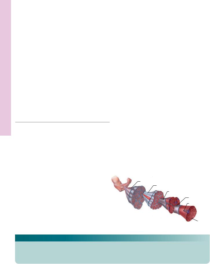
FIGURE 1. Myoneural junction. Tongue. Cat. Scanning electron microscopy. ×2,610.
The striations (arrows) of an isolated skeletal muscle fiber are
the nerve “twig” (N), which loops up and makes contact with the muscle at the myoneural junction (MJ). (Courtesy of Dr. L. Litke.)
clearly evident in this scanning electron micrograph. Note
M U S C L E 141
Microscopy Electron and Light pindle, S Muscle• 5-6 PLATE
FIGURE 1 and 2
FIGURE 1. Muscle spindle. Mouse. Plastic section. ×436.
Observe that the outer (oC) and inner (iC) capsules of the muscle spindle define the outer periaxial space (PS) and the inner axial space (asterisk). The inner capsule forms an envelope around the intrafusal fibers (IF). (From Ovalle W, Dow P. Comparative ultrastructure of the inner capsule of the muscle spindle and the tendon organ. Am J Anat 1983;166:343–357.)
FIGURE 2. Muscle spindle. Mouse. Electron microscopy. ×6,300.
Parts of the outer capsule (oC) may be observed at the corners of this electron micrograph. The periaxial space (PS) surrounds the slender inner capsule (iC), whose component cells form attenuated branches, subdividing the axial space (AS) into several compartments for the nuclear chain (NC) and nuclear bag (NB) intrafusal fibers and their corresponding sensory terminals (ST). Note that the attenuated processes of the inner capsule cells establish contact with each other (arrows). (From Ovalle W, Dow P. Comparative ultrastructure of the inner capsule of the muscle spindle and the tendon organ. Am J Anat 1983;166:343–357.)

Muscle mooth • S6-6 PLATE
142 M U S C L E
FIGURE 1. Smooth muscle. l.s. Monkey. Plastic section. ×270.
The longitudinal section of smooth muscle in this photomicrograph displays long fusiform smooth muscle cells (sM) with centrally located, elongated nuclei (N). Since the muscle fibers are arranged in staggered arrays, they can be packed very closely, with only a limited amount of intervening connective tissue (CT). Using hematoxylin and eosin, the nuclei appear bluish, whereas the cytoplasm stains a light pink. Each smooth muscle cell is surrounded by a basal lamina and reticular fibers, neither of which is evident in this figure. Capillaries are housed in the connective tissue separating bundles of smooth muscle fibers. The boxed area is presented at a higher magnification in Figure 2.
FIGURE 3. Smooth muscle. Uterine myometrium. x.s. Monkey. Plastic section. ×270.
The myometrium of the uterus consists of interlacing bundles of smooth muscle fibers, surrounded by connective tissue (CT) elements. Note that some of these bundles are cut in longitudinal section (1), others are sectioned transversely (2), and still others are cut obliquely (3). At low magnifications, such as in this photomicrograph, the transverse sections present a haphazard arrangement of dark nuclei (N) in a lightly staining region. With practice, it will become apparent that these nuclei are intracellular and that the pale circular regions represent smooth muscle fibers sectioned transversely. Note the numerous blood vessels (BV) traveling in the connective tissue between the smooth muscle bundles.
(Relaxed)
(Contracted)
Smooth muscle
FIGURE 2. Smooth muscle. l.s. Monkey. Plastic section. ×540.
This photomicrograph is a higher magnification of the boxed area of Figure 1. Observe that the nuclei (N) of the smooth muscle fibers are long, tapered structures located in the center of the cell. The widest girth of the nucleus is almost as wide as the muscle fiber. However, the length of the fiber is much greater than that of the nucleus. Note also that any line drawn perpendicular to the direction of the fibers will intersect only a few of the nuclei. Observe the difference between the connective tissue (CT) and smooth muscle (sM). The smooth muscle cytoplasm stains darker and appears smooth relative to the paleness and rough-appearing texture of the connective tissue. Observe capillaries (C) located in the connective tissue elements between bundles of muscle fibers. Inset.
Smooth muscle. Contracted. l.s. Monkey. Plastic section. ×540. This longitudinal section of smooth muscle during contraction displays the characteristic corkscrew-shaped nuclei (N) of these cells.
FIGURE 4a. Smooth muscle. x.s. Monkey. Plastic section. ×540.
To understand the three-dimensional morphology of smooth muscle as it appears in two dimensions, refer to Figure 2 directly above this photomicrograph. Once again note that the muscle fibers are much longer than their nuclei and that both structures are spindleshaped, being tapered at both ends. Recall also that at its greatest girth the nucleus is almost as wide as the cell. In transverse section, this would appear as a round nucleus surrounded by a rim of cytoplasm (asterisk). If the nucleus is sectioned at its tapered end, merely a small dot of it would be present in the center of a large muscle fiber (double asterisks). Sectioned anywhere between these two points, the nucleus would have varied diameters in the center of a large muscle cell. Additionally, the cell may be sectioned in a region away from its nucleus, where only the sarcoplasm of the large muscle cell would be evident (triple asterisks). Moreover, if the cell is sectioned at its tapered end, only a small circular profile of sarcoplasm is distinguishable (arrowhead). Therefore, in transverse sections of smooth muscle, one would expect to find only few cells containing nuclei of various diameters. Most of the field will be closely packed profiles of sarcoplasm containing no nuclei.
FIGURE 4b. Smooth muscle. Duodenum. Monkey. Plastic section. ×132.
This photomicrograph of the duodenum demonstrates the glandular portion (G) with its underlying connective tissue (CT). Deep to the connective tissue, note the two smooth muscle layers, one of which is sectioned longitudinally (1) and the other transversely (2).
KEY
BV |
blood vessel |
CT |
connective tissue |
N |
nucleus |
C |
capillary |
G |
glandular portion |
sM |
smooth muscle cell |

sM
sM |
CT |
N |
C |
sM
N
CT
N
|
|
sM |
FIGURE 1 |
|
FIGURE 2 |
BV |
a) |
b) |
2 |
|
G
CT 1
3
2 |
CT |
N |
1 |
BV
2
1
FIGURE 3 |
FIGURE 4 |
Muscle mooth • S6-6 PLATE

Microscopy Electron Muscle, mooth • S7-6 PLATE
144 M U S C L E
FIGURE 1. Smooth muscle. l.s. Mouse. Electron microscopy. ×15,120.
Smooth muscle does not display cross-bandings, transverse tubular systems, or the regularly arranged array of myofilaments characteristic of striated muscle. However, smooth muscle does possess myofilaments that, along with a system of intermediate filaments, are responsible for its contractile capabilities. Moreover, the plasma membrane appears to possess the functional, if not the structural, aspects of the T tubule. Observe that each smooth muscle is surrounded by an external lamina (EL), which is similar in appearance to basal lamina of epithelial cells. The sarcolemma (SL) displays the presence of numerous pino- cytotic-like invaginations, the caveolae (Ca), which are believed to act as T tubules of striated muscles in conducting impulses
into the interior of the fiber. Some suggest that they may also act in concert with the SR in modulating the availability of calcium ions. The cytoplasmic aspect of the sarcolemma also displays the presence of dense bodies (DB), which are indicative of the attachment of intermediate microfilaments (IM) at that point. Dense bodies, composed of a-actinin (Z disc protein found in striated muscle), are also present in the sarcoplasm (arrows). The nucleus (N) is centrally located and, at its pole, mitochondria (m) are evident. Actin and myosin are also present in smooth muscle but cannot be identified with certainty in longitudinal sections. Parts of a second smooth muscle fiber may be observed to the left of the cell described. A small capillary (C) is evident in the lower right-hand corner. Note the adherens junctions (AJ) between the two epithelial cells, one of which presents a part of its nucleus (N).
(Relaxed)
(Contracted)
Smooth muscle
KEY
AJ |
adherens junction |
DB |
dense body |
m |
mitochondrion |
C |
capillary |
EL |
external lamina |
N |
nucleus |
Ca |
caveola |
IM |
intermediate filament |
SL |
sarcolemma |

M U S C L E 145
Microscopy Electron Muscle, mooth • S7-6 PLATE
FIGURE 1

Muscle diac •Car8-6 PLATE
146 M U S C L E
FIGURE 1. Cardiac muscle. l.s. Human. Plastic section. ×270.
This low magnification of longitudinally sectioned cardiac muscle displays many of the characteristics of this muscle type. The branching (arrow) of the fibers is readily apparent, as are the dark and light bands (arrowheads) running transversely along the length of the fibers. Each muscle cell possesses a large, centrally located, oval nucleus (N), although occasional muscle cells may possess two nuclei. The intercalated discs (ID), indicating intercellular junctions between two cardiac muscle cells, clearly delineated in this photomicrograph, are not easily demonstrable in sections stained with hematoxylin and eosin. The intercellular spaces of cardiac muscle are richly endowed by blood vessels, especially capillaries. Recall that, in contrast to cardiac muscle, the long skeletal muscle fibers do not branch, their myofilaments parallel one another, their many nuclei are peripherally located, and they possess no intercalated discs. The boxed area appears at a higher magnification in Figure 2.
FIGURE 3. Cardiac muscle. x.s. Human. Plastic section. ×270.
Cross sections of cardiac muscle demonstrate polygon-shaped areas of cardiac muscle fibers (CM) with relatively large intercellular spaces whose rich vascular supply (BV) is readily evident. Note that the nucleus (N) of each muscle cell is located in the center, but not all cells display a nucleus. The clear areas in the center of some cells (arrows) represent the perinuclear regions at the poles of the nucleus. These regions are rich in SR, glycogen, lipid droplets, and an occasional Golgi apparatus. The numerous smaller nuclei in the intercellular areas belong to endothelial and connective tissue cells. In contrast to cardiac muscle, cross sections of skeletal muscle fibers display a homogeneous appearance with peripherally positioned nuclei. The connective tissue spaces between skeletal muscle fibers display numerous (frequently collapsed) capillaries.
FIGURE 2. Cardiac muscle. l.s. Human. Plastic section. ×540.
This is a higher magnification of the boxed area of Figure 1. The branching of the fibers (arrows) is evident, and the cross-striations, I and A bands (arrowheads), are clearly distinguishable. The presence of myofibrils (M) within each cell is well displayed in this photomicrograph, as is the “step-like” appearance of the intercalated discs (ID). The oval, centrally located nucleus (N) is surrounded by a clear area usually occupied by mitochondria. The intercellular areas are richly supplied by capillaries (C) supported by slender connective tissue elements.
FIGURE 4. Cardiac muscle. x.s. Human. Plastic section. ×540.
At high magnifications of cardiac muscle in cross section, several aspects of this tissue become apparent. Numerous capillaries (C) and larger blood vessels (BV) abound in the connective tissue spaces. Note the endothelial nuclei (EN) of these vessels as well as the white blood cells (WBC) within the venule in the upper righthand corner. Nuclei (N) of the muscle cells are centrally located, and the perinuclear clear areas (arrow) housing mitochondria are evident. The central clear zones at the nuclear poles are denoted by asterisks. Cross sections of myofibrils (arrowheads) are recognizable as numerous small dots of varying diameters within the sarcoplasm.
Cardiac muscle
|
Intercalated disk |
|
Endomysium |
|
Myofibril |
|
Sarcoplasm |
Nucleus in central |
|
sarcoplasm |
|
Endomysium |
Nucleus |
KEY
BV |
blood vessel |
EN |
endothelial nucleus |
N |
nucleus |
C |
capillary |
ID |
intercalated disc |
WBC |
white blood cell |
CM |
cardiac muscle fiber |
M |
myofibril |
|
|

ID
N
ID
N
FIGURE 1 |
FIGURE 2 |
EN
BV
WBC
N
EN
BV C
Muscle diac •Car8-6 PLATE
FIGURE 3 |
FIGURE 4 |

148 M U S C L E
Microscopy Electron Muscle, diac •Car9-6 PLATE
FIGURE 1
FIGURE 1. Cardiac muscle, l.s. Mouse. Electron microscopy. ×11,700.
The nucleus (N) of cardiac muscle cells is located in the center of the cell, as is evident from the location of the sarcolemma (Sl) in the upper part of the photomicrograph. The sarcoplasm is well endowed with mitochondria (m) and glycogen (Gl) deposits. Since this muscle cell is contracted, the I bands are not visible.
However, the Z discs (Z) are clearly evident, as are the individual myofibrils (M). Inset. Cardiac muscle. l.s. Mouse. Electron microscopy. ×20,700. An intercalated disc is presented in this electron micrograph. Note that this intercellular junction has two zones, the transverse portion (asterisk), composed mostly of desmosome-like junctions, and a longitudinal portion that displays extensive gap junctions (arrows).