

- •Preface
- •Acknowledgments
- •Reviewers
- •Contents
- •CHAPTER OUTLINE
- •CYTOPLASM
- •Plasmalemma
- •Mitochondria
- •Ribosomes
- •Endoplasmic Reticulum
- •Golgi Apparatus, cis-Golgi Network, and the trans-Golgi Network
- •Endosomes
- •Lysosomes
- •Peroxisomes
- •Proteasomes
- •Cytoskeleton
- •Inclusions
- •NUCLEUS
- •CELL CYCLE
- •CHAPTER OUTLINE
- •EPITHELIUM
- •Epithelial Membranes
- •GLANDS
- •Chapter Summary
- •CHAPTER OUTLINE
- •EXTRACELLULAR MATRIX
- •Fibers
- •Amorphous Ground Substance
- •Extracellular Fluid
- •CELLS
- •CONNECTIVE TISSUE TYPES
- •Chapter Summary
- •CHAPTER OUTLINE
- •CARTILAGE
- •BONE
- •Cells of Bone
- •Osteogenesis
- •Bone Remodeling
- •Chapter Summary
- •CHAPTER OUTLINE
- •FORMED ELEMENTS OF BLOOD
- •Lymphocytes
- •Neutrophils
- •PLASMA
- •COAGULATION
- •HEMOPOIESIS
- •Erythrocytic Series
- •Granulocytic Series
- •Chapter Summary
- •CHAPTER OUTLINE
- •SKELETAL MUSCLE
- •Sliding Filament Model of Muscle Contraction
- •CARDIAC MUSCLE
- •SMOOTH MUSCLE
- •Chapter Summary
- •CHAPTER OUTLINE
- •BLOOD-BRAIN BARRIER
- •NEURONS
- •Membrane Resting Potential
- •Action Potential
- •Myoneural Junctions
- •Neurotransmitter Substances
- •SUPPORTING CELLS
- •PERIPHERAL NERVES
- •Chapter Summary
- •CHAPTER OUTLINE
- •BLOOD VASCULAR SYSTEM
- •HEART
- •ARTERIES
- •Capillary Permeability
- •Endothelial Cell Functions
- •VEINS
- •LYMPH VASCULAR SYSTEM
- •Chapter Summary
- •CHAPTER OUTLINE
- •CELLS OF THE IMMUNE SYSTEM
- •Antigen-Presenting Cells
- •DIFFUSE LYMPHOID TISSUE
- •LYMPH NODES
- •TONSILS
- •SPLEEN
- •THYMUS
- •Chapter Summary
- •CHAPTER OUTLINE
- •PITUITARY GLAND
- •Pars Intermedia
- •Pars Nervosa and Infundibular Stalk
- •Pars Tuberalis
- •THYROID GLAND
- •Parathyroid Glands
- •Suprarenal Glands
- •Cortex
- •Medulla
- •Pineal Body
- •Chapter Summary
- •CHAPTER OUTLINE
- •SKIN
- •Epidermis of Thick Skin
- •Dermis
- •DERIVATIVES OF SKIN
- •Chapter Summary
- •CHAPTER OUTLINE
- •CONDUCTING PORTION OF THE RESPIRATORY SYSTEM
- •Extrapulmonary Region
- •Intrapulmonary Region
- •RESPIRATORY PORTION OF THE RESPIRATORY SYSTEM
- •MECHANISM OF RESPIRATION
- •Chapter Summary
- •CHAPTER OUTLINE
- •ORAL CAVITY AND ORAL MUCOSA
- •Oral Mucosa
- •Tongue
- •Teeth
- •Odontogenesis (See Graphic 13-2)
- •Chapter Summary
- •CHAPTER OUTLINE
- •REGIONS OF THE DIGESTIVE TRACT
- •Esophagus
- •Stomach
- •Small Intestine
- •Large Intestine
- •GUT-ASSOCIATED LYMPHOID TISSUE
- •DIGESTION AND ABSORPTION
- •Carbohydrates
- •Proteins
- •Lipids
- •Water and Ions
- •Chapter Summary
- •CHAPTER OUTLINE
- •MAJOR SALIVARY GLANDS
- •PANCREAS
- •LIVER
- •Exocrine Function of the Liver
- •Endocrine and Other Functions of the Liver
- •GALLBLADDER
- •Chapter Summary
- •CHAPTER OUTLINE
- •KIDNEY
- •Uriniferous Tubule
- •Nephron
- •Collecting Tubules
- •FORMATION OF URINE FROM ULTRAFILTRATE
- •EXTRARENAL EXCRETORY PASSAGES
- •Chapter Summary
- •CHAPTER OUTLINE
- •OVARY
- •Ovarian Follicles
- •Regulation of Follicle Maturation and Ovulation
- •Corpus Luteum and Corpus Albicans
- •GENITAL DUCTS
- •Oviduct
- •Uterus
- •FERTILIZATION, IMPLANTATION, AND THE PLACENTA
- •Fertilization and Implantation
- •Placenta
- •VAGINA
- •EXTERNAL GENITALIA
- •MAMMARY GLANDS
- •Chapter Summary
- •CHAPTER OUTLINE
- •TESTES
- •Spermatogenesis
- •GENITAL DUCTS
- •ACCESSORY GLANDS
- •PENIS
- •Erection and Ejaculation
- •Chapter Summary
- •CHAPTER OUTLINE
- •SENSORY ENDINGS
- •Chapter Summary
- •Terminology of Staining
- •Common Stains Used in Histology
- •Hematoxylin and Eosin
- •Wright Stain
- •Weigert Method for Elastic Fibers and Elastic van Gieson Stain
- •Silver Stain
- •Iron Hematoxylin
- •Bielschowsky Silver Stain
- •Masson Trichrome
- •Periodic Acid-Schiff Reaction (PAS)
- •Alcian Blue
- •von Kossa Stain
- •Sudan Red
- •Mucicarmine Stain
- •Safranin-O
- •Toluidine Blue

3 days after fertilization, the morula enters the lumen of the uterus.
•Once in the uterus, the cells of the morula rearrange themselves to form a hollow structure, the blastocyst, whose fluid-filled cavity also houses a small cluster of cells, the inner cell mass (embryoblasts) responsible for the formation of the embryo.
•Approximately 5 to 6 days after fertilization, the cells at the periphery of the blastocyst, the trophoblasts, proliferate and initiate the process of implantation into the endometrium. By the ninth day, implantation is complete.
•As the trophoblasts proliferate, they form an inner cellular layer, the cytotrophoblast, and an outer syncytial layer, the syncytiotrophoblast.
The syncytiotrophoblasts will initiate the formation of the embryonic portion of the placenta.
In response to the invasion of the syncytiotrophoblasts, the endometrium will initiate the formation of the maternal portion of the placenta.
Placenta
During pregnancy, the uterus participates in the formation of the placenta, a highly vascular structure that permits the exchange of various materials between the maternal and fetal circulatory systems (see Graphic 17-2). It must be stressed that the exchange occurs without the commingling of the maternal and fetal bloods and that the placenta is derived from both maternal and fetal tissues. The roles of the trophoblasts and the endometrium are as follows:
•The syncytiotrophoblasts and cytotrophoblasts form the chorion, the precursor of the chorionic plate from which the chorionic villi will arise.
•The endometrium in contact with the chorion becomes modified to form the decidua with its three regions:
Decidua basalis, the richly vascularized maternal portion of the placenta that induces the trophoblasts to form the chorionic villi.
Decidua capsularis, the tissue separating the lumen of the uterus from the embryo and will be known as the chorion laeve, and
Decidua parietalis, the endometrial tissue between the uterine lumen and the myometrium.
Initially, the chorionic villi are slender structures and are known as primary villi. Once they are invaded by mesenchymal cells and fetal capillary networks, they become more substantial and their population of cytotrophoblasts decreases because they become incorporated into the syncytiotrophoblasts; in this manner, the primary villi become known as secondary villi.
F E M A L E R E P R O D U C T I V E S Y S T E M 409
•As the placenta is forming, the decidua basalis develops large, blood-filled vascular channels, known as lacunae, and the secondary villi protrude into these “lakes” of maternal blood, supplied by maternal arterioles and drained by maternal venules.
•Secondary villi grow into these lacunae and some of the villi contact and fuse with the decidua basalis (anchoring villi), whereas other secondary villi (free villi) resemble fingers that are immersed in water.
The fetal capillary beds of the anchoring and free villi are located adjacent to the syncytiotrophoblasts and lie in close proximity of the maternal blood in the lacunae.
Oxygen and nutrients in the maternal blood diffuse through the villi to reach the fetal capillaries.
Carbon dioxide and waste products in the fetal blood also diffuse through the villi to reach the maternal blood in the lacunae.
The exchange of gases and material occurs by passing through the placental barrier whose components are listed in Table 17-3.
In addition to its role in the delivery of nutrients and oxygen to the fetus and exchanging it for the fetal waste products, the placenta also manufactures hormones and factors necessary for the maintenance of pregnancy and the delivery of the fetus (see Table 17-4).
VAGINA
The vagina, an 8- to 9-cm long muscular sheath, extending from the cervix of the uterus to the vestibule, is adapted for the reception of the penis during copulation and for the passage of the fetus from the uterus during birth. The wall of the vagina is composed of three layers: the mucosa, muscularis, and the adventitia.
•The mucosa consists of a stratified squamous epithelium and a loose, fibroelastic connective tissue layer, the lamina propria.
TABLE 17-3 • Components of the Placental Barrier
Endothelial cells of the fetal capillary
Basal lamina of the fetal endothelium
Connective tissue of the secondary villus
Basal lamina of the cytotrophoblasts
Cytotrophoblasts
Syncytiotrophoblasts

410 F E M A L E R E P R O D U C T I V E S Y S T E M
TABLE 17-4 • Principal Hormones and Factors Produced by the Various Components of the Placenta
Syncytiotrophoblasts |
Cytotrophoblasts |
Decidua Cells |
|
|
|
Estrogens |
Gonadotropin-releasing hormone |
Insulin-like growth factor binding proteins |
|
|
|
Progesterone |
Corticotropin-releasing hormone |
Relaxin |
|
|
|
Chorionic gonadotrophin |
Thyrotropin-releasing hormone |
Prolactin |
|
|
|
Chorionic somatotropin |
Growth hormone–releasing hormone |
Prostaglandins |
|
|
|
Placental growth hormone |
Inhibin |
|
|
|
|
Leptin |
Activin |
|
|
|
|
|
Leptin |
|
|
|
|
|
Insulin-like growth factors I and II |
|
|
|
|
Frequently, in a virgin, the external orifice of the vagina is partially occluded by the hymen, a thin, somewhat vascular connective tissue membrane, covered on both sides by stratified squamous epithelium.
•The muscularis is composed of a mostly longitudinally disposed smooth muscle layer interspersed with some circularly arranged fibers. At its external orifice, the muscularis of the vagina possesses a sphincter, composed of circularly arrayed smooth muscle fibers.
•The adventitia is a dense fibroelastic connective tissue that affixes the vagina to the surrounding pelvic connective tissue.
EXTERNAL GENITALIA
The external genitalia, composed of labia majora, labia minora, clitoris, and vestibular glands, are also referred to as the vulva. These structures are richly innervated and function during sexual arousal and copulation.
MAMMARY GLANDS
The mammary glands, highly modified sweat glands, are identical in males and females until the onset of puberty, when, due to hormonal influences, the female breasts develop.
•The mammary gland is composed of numerous individual compound glands, each of which is considered a lobe.
Each lobe is drained by a lactiferous duct that delivers milk, the secretion of the mammary glands, onto the surface of the nipple.
•The pigmented region of the skin surrounding the nipple, known as the areola, is richly endowed by sweat, sebaceous, and areolar glands.
•During pregnancy, several hormones interact to promote the development of the secretory units of the mammary gland. Cells of the terminal interalveolar ducts proliferate to form secretory alveoli.
The hormones involved in promoting this process are progesterone, estrogen, and human chorionic mammotropin from the placenta and lactogenic hormone (prolactin) from the acidophils of the adenohypophysis.
•Alveoli and terminal interalveolar ducts are surrounded by myoepithelial cells that contract as a result of the release of oxytocin from the neurohypophysis (in response to suckling), forcing milk out of the breast (milk ejection reflex).
Milk is composed of water, proteins, lipids, and lactose.
However, milk secreted during the first few days (colostrum) is different, in that it is rich in vitamins, minerals, lymphoid cells, and proteins, especially immunoglobulin A, providing antibodies for the neonate for the first few months of life.
F E M A L E R E P R O D U C T I V E S Y S T E M 411
CLINICAL CONSIDERATIONS
Papanicolaou Smear |
Endometriosis |
The Papanicolaou (Pap) smear is performed as part of routine gynecological examination to examine stained exfoliative cells of the lining of the cervix and vagina. Evaluation of the smeared cells permits the recognition of precancerous conditions as well as cancer of the cervix. An annual smear test is recommended since cervical cancer is relatively slow growing and the Pap smear is an extremely cost-effective procedure that has been responsible for the early detection of cervical cancer and for saving lives of affected individuals.
Gonorrhea
Gonorrhea is a sexually transmitted bacterial infection caused by the gram-negative diplococcus Neisseria gonorrhoeae. In the United States, over a million cases of gonorrhea occur annually. Frequently, this sexually transmitted disease is responsible for pelvic inflammatory disease (PID) and for acute salpingitis.
Pelvic Inflammatory Disease
PID an infection of the cervix, uterus, fallopian tubes, and/or ovary, is usually a sequel to microbial infection. Individuals suffering from PID exhibit tenderness and pain in the lower abdominal region, fever, unpleasantsmelling vaginal discharge, and episodes of abnormal bleeding.
Adenomyosis
Adenomyosis is a common condition in which the endometrial glands invade the myometrium and cause the uterus to enlarge, occasionally becoming two or three times its normal dimensions. In most women, adenomyosis has no symptoms, and it is only on gynecological examination that the condition is discovered. When it becomes symptomatic, the woman is usually between 35 and 50 years of age, she may experience pain during intercourse, and she notices an increase in menstrual flow as well as bleeding between periods. Although the condition is benign, if the symptoms are severe and uncontrollable, hysterectomy may be indicated.
Endometriosis is distinguished by the presence of ectopic endometrial tissue dispersed to various sites along the peritoneal cavity. Occasionally, the tissues may migrate to extraperitoneal areas, including the eyes and brain. The etiology of this disease is not known. In some cases the lesions of endometriosis involve small cysts attached separately or in small clumps on the visceral or parietal peritoneum.
This photomicrograph is from the fallopian tube of a female patient with endometriosis. Observe that uterine glands and stroma occupy the lumen of the oviduct. (Reprinted with permission from Mills SE, Carter D, et al., eds. Sternberg’s Diagnostic Surgical Pathology, 5th ed. Philadelphia: Lippincott Williams & Wilkins, 2010, p. 2377.)

412 F E M A L E R E P R O D U C T I V E S Y S T E M
Endometrial Carcinoma
Endometrial carcinoma is a malignancy of the uterine endometrium usually occurring in postmenopausal women. The most common type of endometrial cancer is adenocarcinoma. Since during the early stages the cancer cells do not invade the cervix, Pap smears are not very effective in diagnosing this disease until it has entered its later stages. The major symptom of endometrial cancer is abnormal uterine bleeding.
Hydatidiform Mole
Occasionally, an ovum does not develop normally and instead of becoming a fetus forms a mass of tissue that
initially mimics pregnancy, or in some patients, after delivery, remnants of placental tissue may proliferate. Known as a hydatidiform mole, these growths increase in size much faster than would a fetus. When the physician does not hear a heartbeat, the patient’s abdomen swells more than expected, and the patient complains of vomiting and severe nausea, a hydatidiform mole should be suspected. This is especially true in individuals who complain of a vaginal discharge of grape-like clusters of tissue. In most of the cases, the hydatidiform mole resorbs on its own. Only in about 20% of the cases does it become invasive, and in very rare cases does it becomes malignant (then it is known as a choriocarcinoma).
This photomicrograph is from the uterus of a female patient with Grade 1 carcinoma of the endometrium. Top: Observe that the uterine glands are very crowded with a scant amount of connective tissue between the glands. Bottom: The cells of the gland are interspersed with malignant cells displaying cytologic atypia. (Reprinted with permission from Mills SE, Carter D, et al., eds. Sternberg’s Diagnostic Surgical Pathology, 5th ed. Philadelphia: Lippincott Williams & Wilkins, 2010, p. 2208.)

F E M A L E R E P R O D U C T I V E S Y S T E M 413
Paget’s Disease of the Nipple
Paget’s disease of the nipple usually occurs in elderly women and is associated with breast cancer of ductal origin. Initially, the disease manifests as scaly or crusty
nipple frequently accompanied by a fluid discharge from the nipple. Usually, the patient has no other symptoms and frequently neglects the condition.
This photomicrograph is from the nipple of a female patient with Paget’s disease of the nipple. Note the large Paget’s cells throughout the basal aspect of the stratified squamous keratinized epithelium, with their light pink cytoplasm, vesicular-appearing nuclei, and large nucleoli. (Reprinted with permission from Mills SE, Carter D, et al., eds. Sternberg’s Diagnostic Surgical Pathology, 5th ed. Philadelphia: Lippincott Williams & Wilkins, 2010, p. 293.)

System Reproductive Female • 1-17 GRAPHIC
414 F E M A L E R E P R O D U C T I V E S Y S T E M
Uterine (Fallopian) tube
Ovary
Ovarian ligament
Broad ligament
Uterus
Cervical canal
Cervix
Lateral cervical ligament
Vagina
PRIMORDAL
FOLLICLE:
Follicular cells
Oocyte
Intramural portion of uterine tube
 Isthmus of uterine tube
Fimbriae
Endometrium
Myometrium Wall of uterus
Adventitia
|
|
|
|
|
|
|
2º FOLLICLE: |
UNILAMINAR |
MULTILAMINAR |
|
|
|
Theca folliculi |
||
PRIMARY |
|
|
|
|
Granulosa cells |
||
PRIMARY |
FOLLICLE: |
|
|
|
|
|
Zona pellucida |
FOLLICLE: |
Theca folliculi |
|
|
|
|
|
Oocyte |
|
|
|
|
|
|
||
|
|
|
|
|
|
Basement |
|
Granulosa |
Zona pellucida |
|
|
|
|
|
|
|
|
|
|
|
membrane |
||
cells |
|
|
|
|
|
|
|
|
|
|
|
|
|
|
|
|
|
|
|
|
|
|
|
|
|
|
|
|
|
|
|
|
|
|
|
|
|
|
|
|
|
|
|
|
|
|
|
Ampulla
Infundibulum
GRAAFIAN
FOLLICLE:
Membrana granulosa
Cumulus oophorus
Zona pellucida
Oocyte
Corona radiata (around oocyte)
Corpus albicans
Corpus Theca lutein  luteum Granulosa lutein
Each follicle houses a primary oocyte
arrested in the prophase of the first meiotic division. The most developed Graafian follicle releases its oocyte during
ovulation. As that primary oocyte is being released, it finishes its
first meiotic division, becomes a secondary oocyte, and is arrested in
the metaphase stage of the second meiotic division. Subsequent to ovulation the Graafian follicle differentiates into the corpus luteum, which will eventually degenerate into the corpus albicans.
Antrum
Theca interna
Theca externa
 Corona radiata
  Oocyte
Oocyte nucleus

F E M A L E R E P R O D U C T I V E S Y S T E M 415
Placental
Structure
After delivery, the decidua detaches at this point
Ovarian cycle
 Chorionic plate
Anchoring (primary) villi
Chorionic (secondary) villi
Branch (tertiary) villi
Placental septum
Decidua basalis
Stratum compactum
Stratum spongiosum
Myometrium
Hypothalamus
FSHRF LHRF
Anterior pituitary
FSH LH
The human placenta is composed of a maternally derived and a fetally derived region. It is constructed in such a fashion that the mother’s blood does not come in contact with the blood of the fetus, yet it permits the exchange of nutrients, gases, and waste products between them. The maternal portion of the placenta is composed of the decidua basalis, whereas the fetal portion consists of the chorionic plate and its extensions. There are three types of villi arising from the chorionic plate: those that contact the decidua basalis (anchoring or primary villi), those that arise directly from the chorionic plate but do not contact the decidua basalis (chorionic or secondary villi), and branches arising from the secondary villi (branch or tertiary villi).
Primary |
Secondary |
Graafian |
|
Corpus |
Corpus |
follicle |
follicle |
follicle |
Ovulation |
luteum |
albicans |
Endometrial |
|
|
Progesterone |
|
|
changes |
Estrogens |
|
|
|
|
|
|
and estrogens |
|
||
|
|
|
|
||
Stratum functionalis
Stratum |
Menses |
Preovulatory phase Ovulation Postovulatory phase |
Menses |
basalis |
|||
Myometrium |
|
Days |
|
|
|
|
The effects of hypothalamic and adenohypohyseal hormones on the ovarian cortex and uterine endometrium.
Cycle Hormonal and Placenta• 2-17 GRAPHIC

ary • Ov1-17 PLATE
416 F E M A L E R E P R O D U C T I V E S Y S T E M
FIGURE 1. Ovary. Monkey. Plastic section. ×14.
The ovary is subdivided into a medulla (Me) and a cortex (Co). The medulla houses large blood vessels (BV) from which the cortical vascular supply is derived. The cortex of the ovary contains numerous ovarian follicles, most of which are very small (arrows); a few maturing follicles have reached the Graafian follicle (GF) stage. The thick, fibrous connective tissue capsule, tunica albuginea (TA), is shown to advantage; the germinal epithelium (GE) is evident occasionally. Observe that the mesovarium (Mo) not only suspends the ovary but also conveys the vascular supply to the medulla. A region similar to the boxed area is presented at a higher magnification in Figure 2.
FIGURE 3. Primary follicles. Monkey. Plastic section. ×270.
Primary follicles differ from primordial follicles not only in size but also in morphology and number of follicular cells. The unilaminar primary follicle of the inset (×270) displays a single layer of cuboidal follicular cells (FC) that surround the relatively small primary oocyte (PO), whose nucleus (N) is clearly evident. The multilaminar primary follicle displays a primary oocyte (PO) that has increased in size. The follicular cells (FC) now form a stratified layer around the oocyte, being separated from it by the intervening zona pellucida (ZP). The stroma (St) is being reorganized around the follicle to form the theca interna (TI). Note the presence of a basal membrane (BM) between the follicular cells and the theca interna.
FIGURE 2. Ovary. Monkey. Plastic section. ×132.
This photomicrograph is a higher magnification of a region similar to the boxed area of Figure 1. Observe that the germinal epithelium (GE) covers the collagenous capsule, the tunica albuginea (TA). This region of the cortex (Co) houses numerous primordial follicles (PF). Observe that the connective tissue of the ovary is highly cellular and is referred to as the stroma (St). Inset. Ovary. Cortex. Monkey. Plastic section. ×540. The primordial follicle is composed of a primary oocyte (PO), whose nucleus (N) and nucleolus (arrow) are evident. Observe the single layer of flat follicular cells (FC) surrounding the oocyte. The tunica albuginea (TA) and the germinal epithelium (GE) are also shown to advantage in this photomicrograph.
FIGURE 4. Secondary follicle. Rabbit. Paraffin section. ×132.
Secondary follicles are very similar to primary multilaminar follicles, the major difference being their larger size. Moreover, the stratification of the follicular cells (FC) has increased, displaying more layers, and more important, a follicular fluid (FF) begins to appear in the intercellular spaces, which coalesces into several Call-Exner bodies. Note also that the stroma immediately surrounding the follicular cells is rearranged to form a cellular theca interna (TI) and a more fibrous theca externa (TE).
|
Primary |
|
|
Oocyte |
follicles |
Secondary follicle |
|
|
|
|
|
Follicular |
|
|
Graafian follicle |
|
|
||
|
|
|
|
cells |
|
|
Theca interna |
|
|
|
|
Blood vessels |
|
Theca externa |
|
|
|
||
Medulla |
|
|
|
|
Cortex |
|
|
Ovary
KEY
BM |
basal membrane |
GF |
Graafian follicle |
St |
stroma |
BV |
blood vessel |
Me |
medulla |
TA |
tunica albuginea |
Co |
cortex |
Mo |
mesovarium |
TE |
theca externa |
FC |
follicular cell |
N |
nucleus |
TI |
theca interna |
FF |
follicular fluid |
PF |
primordial follicle |
ZP |
zona pellucida |
GE |
germinal epithelium |
PO |
primary oocyte |
|
|

ary • Ov1-17 PLATE
FIGURE 1 |
FIGURE 2 |
FIGURE 3 |
FIGURE 4 |

Luteum Corpus and vary • O2-17 PLATE
418 F E M A L E R E P R O D U C T I V E S Y S T E M
FIGURE 1. Graafian follicle. Paraffin section. ×132.
The Graafian follicle is the most mature of all ovarian follicles and is ready to release its primary oocyte in the process of ovulation. The follicular fluid (FL) fills a single chamber, the antrum, which is surrounded by a wall of granulosa (follicular) cells known as the membrana granulosa (MG). Some of the granulosa cells, which surround the primary oocyte (PO), jut into the antrum as the cumulus oophorus (CO). Observe the basal membrane (BM), which separates the granulosa cells from the theca interna (TI). The fibrous theca externa (TE) merges almost imperceptibly with the surrounding stroma. The boxed area is presented at a higher magnification in Figure 2.
FIGURE 3. Corpus luteum. Human. Paraffin section. ×14.
Subsequent to ovulation, the Graafian follicle becomes modified to form a temporary structure, the corpus hemorrhagicum, which will become the corpus luteum. The cells comprising the membrana granulosa enlarge, become vesicular in appearance, and are referred to as granulosa lutein cells (GL), which become folded; the spaces between the folds are occupied by connective tissue elements, blood vessels, and cells of the theca interna (arrows). These theca interna cells also enlarge, become glandular, and are referred to as the theca lutein cells. The remnants of the antrum are filled with fibrin and serous exudate that will be replaced by connective tissue elements. A region similar to the boxed area is presented at a higher magnification in Figure 4.
FIGURE 2. Graafian follicle. Cumulus oophorus. Paraffin section. ×270.
This photomicrograph is a higher magnification of the boxed area of Figure 1. Observe that the cumulus oophorus houses the primary oocyte (PO), whose nucleus (N) is just visible in this section. The zona pellucida (ZP) surrounds the oocyte, and processes (arrows) of the surrounding follicular cells extend into this acellular region. The single layer of follicular cells appears to radiate as a crown at the periphery of the primary oocyte and is referred to as the corona radiata (CR). Note the basal membrane (BM) as well as the theca interna (TI) and the theca externa (TE).
FIGURE 4. Corpus luteum. Human. Paraffin section. ×132.
This photomicrograph is a higher magnification of a region similar to the boxed area of Figure 3. The granulosa lutein cells (GL) of the corpus luteum are easily distinguished from the connective tissue (CT) elements, since the former display round nuclei (N), mostly in the center of large round cells (arrowheads). The center of the field is occupied by a fold, housing theca lutein cells (TL) amid numerous connective tissue (CT) and vascular (BV) elements. A region similar to the boxed area is presented at a higher magnification in Figure 1 of the next plate.
|
|
Membrana granulosa |
|
|
|
Cumulus oophorus |
|
|
|
Zona pellucida |
|
|
|
Oocyte |
Graafian |
|
|
follicle |
|
|
|
|
|
|
|
Corona radiata |
|
|
|
(around oocyte) |
|
Corpus |
Theca lutein |
Antrum |
|
luteum |
Granulosa lutein |
|
|
Ovary
KEY
BM |
basal membrane |
FL |
follicular fluid |
TE |
theca externa |
BV |
vascular elements |
GL |
granulosa lutein cells |
TI |
theca interna |
CO |
cumulus oophorus |
MG |
membrana granulosa |
TL |
theca lutein cells |
CR |
corona radiata |
N |
nucleus |
ZP |
zona pellucida |
CT |
connective tissue |
PO |
primary oocyte |
|
|

Luteum Corpus and vary • O2-17 PLATE
FIGURE 1 |
FIGURE 2 |
FIGURE 3 |
FIGURE 4 |

Oviduct and vary • O3-17 PLATE
420 F E M A L E R E P R O D U C T I V E S Y S T E M
FIGURE 1. Corpus luteum. Human. Paraffin section. ×540.
This photomicrograph is similar to the boxed area of Figure 4 of the previous plate. Observe the large granulosa lutein cells (GL), whose cytoplasm appears vesicular, representing the spaces occupied by lipids in the living tissue. Note that the nuclei (N) of these cells are farther away from each other than the nuclei of the smaller theca lutein cells (TL), which also appear to be darker staining (arrowheads). The flattened nuclei (arrows) belong to various connective tissue cells.
FIGURE 2. Corpus albicans. Human. Paraffin section. ×132.
As the corpus luteum involutes, its cellular elements degenerate and undergo autolysis. The corpus luteum becomes invaded by macrophages that phagocytose the dead cells, leaving behind relatively acellular fibrous tissue (FT). The previously rich vascular supply (BV) also regressed, and the entire corpus albicans appears pale in comparison to the relatively dark staining of the surrounding ovarian stroma (St). The corpus albicans will regress until it becomes a small scar on the surface of the ovary.
FIGURE 3. Oviduct. x.s. Human. Paraffin section. ×14.
The oviduct, also referred to as the fallopian or uterine tube, extends from the ovary to the uterine cavity. It is suspended from the body wall by the broad ligament (BL), which conveys a rich vascular supply (BV) to the serosa (S) of the oviduct. The thick muscularis (M) is composed of ill-defined inner circular and outer longitudinal muscle layers. The mucosa (Mu) is thrown into longitudinal folds, which are so highly exaggerated in the infundibulum and ampulla that they subdivide the lumen (L) into labyrinthine spaces. A region similar to the boxed area is presented at a higher magnification in Figure 4.
FIGURE 4. Oviduct. x.s. Monkey. Plastic section. ×132.
This photomicrograph is a higher magnification of a region similar to the boxed area of Figure 3. The entire thickness of the wall of the oviduct displays its vascular (BV) serosa (S) that envelops the thick muscularis, whose outer longitudinal (OL) and inner circular (IC) layers are not very well delineated. The mucosa (Mu) is highly folded and is lined by a simple columnar epithelium (Ep). The loose connective tissue of the lamina propria (LP) is richly vascularized (arrows). The boxed area is presented in a higher magnification in Figure 1 in the following plate.
Corpus albicans
Corpus |
Theca lutein |
luteum |
Granulosa lutein |
Ovary
KEY
BL |
broad ligament |
IC |
inner circular muscle |
N |
nucleus |
BV |
vascular supply |
L |
lumen |
OL |
outer longitudinal muscle |
Ep |
epithelium |
LP |
lamina propria |
S |
serosa |
FT |
fibrous tissue |
M |
muscularis |
ST |
stroma |
GL |
granulosa lutein cell |
Mu |
mucosa |
TL |
theca lutein cell |
Oviduct and vary • O3-17 PLATE
FIGURE 1 |
FIGURE 2 |
FIGURE 3 |
FIGURE 4 |

Microscopy Electron and Light viduct, • O4-17 PLATE
422 F E M A L E R E P R O D U C T I V E S Y S T E M
FIGURE 1. Oviduct. x.s. Monkey. Plastic section. ×270.
This photomicrograph is a higher magnification of the boxed area of Figure 4 of the previous plate. Observe the inner circular muscle (IC) layer of the muscularis. The lamina propria (LP) is very narrow in this region (arrows) but presents longitudinal epithelially lined folds. The core of these folds is composed of a vascular (BV), loose but highly cellular connective tissue (CT). The simple columnar epithelium (Ep) lines the labyrinthine lumen (L) of the oviduct. A region similar to the boxed area is presented at a higher magnification in Figure 2.
FIGURE 3. Oviduct epithelium. Human. Electron microscopy. ×4,553.
The human oviduct at midcycle (day 14) presents two types of epithelial cells, the peg cell (PC) and the ciliated cell (CC). The former are secretory cells, as indicated by their extensive Golgi apparatus (GA) situated in the region of the cell apical to the nucleus
(N). Observe the electron-dense secretory products (arrows) in the expanded, apical free ends of these cells. Note also that some ciliated cells display large accumulations of glycogen (Gl) at either pole of the nucleus. (From Verhage H, Bareither M, Jaffe R, Akbar M. Cyclic changes in ciliation, secretion and cell height of the oviductal epethelium in women. Am J Anat 1979;156:505–522.)
FIGURE 2. Oviduct. x.s. Monkey. Plastic section. ×540.
This photomicrograph is a higher magnification of a region similar to the boxed area of Figure 1. The lamina propria (LP) is a highly cellular, loose connective tissue that is richly vascularized. The basal membrane (BM) separating the connective tissue from the epithelial lining is clearly evident. Note that the epithelium is composed of two different cell types, a thinner peg cell (PC), which bears no cilia but whose apical extent bulges above the ciliated cells. These bulges (arrowheads) contain nutritive materials that nourish gametes. The second cell type of the oviduct epithelium is a ciliated cell (CL), whose cilia move in unison with those of neighboring cells, propelling the nutrient material toward the uterine lumen.
KEY
BV |
vascular elements |
Ep |
epithelium |
L |
lumen |
BM |
basal membrane |
GA |
Golgi apparatus |
LP |
lamina propria |
CC |
ciliated cell |
GL |
glycogen |
N |
nucleus |
CT |
connective tissue |
IC |
inner circular muscle |
PC |
peg cell |

Microscopy Electron and Light viduct, • O4-17 PLATE
FIGURE 1 |
FIGURE 2 |
FIGURE 3

erus • Ut5-17 PLATE
424 F E M A L E R E P R O D U C T I V E S Y S T E M
FIGURE 1. Uterus. Follicular phase. Human. Paraffin section. ×14.
The uterus is a thick-walled organ whose wall consists of three layers. The external serosa (or in certain regions, adventitia) is unremarkable and is not presented in this photomicrograph. The thick myometrium (My) is composed of smooth muscle, subdivided into three poorly delineated layers: outer longitudinal (OL), middle circular (MC), and inner longitudinal (IL). The endometrium
(En) is subdivided into a basal layer (B) and a functional layer
(F). The functional layer varies in thickness and constitution and passes through a sequence of phases during the menstrual cycle. Note that the functional layer is in the process of being built up and that the forming glands (GL) are straight. The deeper aspects of some of these glands display branching (arrow). The boxed area is presented at a higher magnification in Figure 2.
FIGURE 3. Uterus. Luteal phase. Human. Paraffin section. ×14.
The myometrium (My) of the uterus remains constant during the various endometrial phases. Observe its three layers, noting especially that the middle circular layer of smooth muscle is richly vascularized and is therefore frequently referred to as the stratum vasculare (SV). The endometrium (En) is richly endowed with glands (GL) that become highly tortuous in anticipation of the blastocyst that will be nourished by secretions of these glands subsequent to implantation. A region similar to the boxed area is presented at a higher magnification in Figure 4.
FIGURE 2. Uterus. Follicular phase. Human. Paraffin section. ×132.
This photomicrograph is a higher magnification of the boxed area of Figure 1. Note that the functional layer (F) of the endometrium is lined by a simple columnar epithelium (Ep) that is displaying mitotic activity (arrows). The forming glands (GL) also consist of a simple columnar epithelium (Ep) whose cells are actively dividing. The stroma (St) is highly cellular, as evidenced by the numerous connective tissue cell nuclei visible in this field. Note also the rich vascular supply (BV) of the endometrial stroma.
FIGURE 4. Uterus. Early luteal phase. Human. Paraffin section. ×132.
This photomicrograph is a higher magnification of a region similar to the boxed area of Figure 3. The functional layer of the endometrium is covered by a simple columnar epithelium (Ep), separating the endometrial stroma (St) from the uterine lumen (L). Note that the glands (GL), also composed of simple columnar epithelium, are more abundant than those in the follicular phase (Figure 2, above). Observe also that these glands appear more tortuous and are dilated and their lumina contain a slight amount of secretory product (arrow).
Uterine (Fallopian) tube |
|
Intramural portion of uterine tube |
|||||||||||||
|
|
Isthmus of uterine tube |
|||||||||||||
Ovarian ligament |
|
|
|
|
|
||||||||||
|
|
|
|
|
|
|
|
Ampulla |
|||||||
Ovary |
|
|
|
|
|
|
|
|
|
|
|
|
|
||
|
|
|
|
|
|
|
|
|
|
|
|
|
|
Infundibulum |
|
|
|
|
|
|
|
|
|
|
|
|
|
|
|||
|
|
|
|
|
|
|
|
|
|||||||
Broad ligament |
|
|
|
|
|
|
|
|
|
|
|
|
|
||
|
|
|
|
|
|
|
|
|
|
|
|
|
|||
|
|
|
|
|
|
|
|
|
|
|
|
||||
|
Uterus |
|
|
|
|
|
|
|
|
|
|
||||
|
|
|
|
|
|
|
|
|
|
|
|||||
Cervical canal |
|
|
|
|
Fimbrae |
||||||||||
|
Cervix |
|
|
Endometrium |
|||||||||||
|
|
|
Myometrium Wall of uterus |
||||||||||||
Lateral cervical ligament |
|
|
|
|
|||||||||||
|
|
|
|
Adventitia |
|||||||||||
|
|
|
|
||||||||||||
|
|
|
|
Vagina |
|
|
|
|
|
|
|
|
|||
|
|
|
|
|
|
|
|
|
|
|
|
||||
Female reproductive system
KEY
B |
basal layer |
GL |
gland |
OL |
outer longitudinal muscle |
BV |
vascular supply |
IL |
inner longitudinal muscle |
St |
stroma |
En |
endometrium |
L |
lumen |
SV |
stratum vasculare |
Ep |
epithelium |
MC |
middle circular muscle |
|
|
F |
functional layer |
My |
myometrium |
|
|

erus • Ut5-17 PLATE
FIGURE 1 |
FIGURE 2 |
FIGURE 3 |
FIGURE 4 |

erus • Ut6-17 PLATE
426 F E M A L E R E P R O D U C T I V E S Y S T E M
FIGURE 1. Uterus. Midluteal phase. Human. Paraffin section. ×270.
During the midluteal phase, the endometrial glands (GL) become quite tortuous and corkscrew-shaped, and the simple columnar cells (CC) accumulate glycogen (arrow). Observe that during this phase of the endometrium, the glycogen is basally located, displacing the nucleus (N) toward the center of the cell. Note also that the stroma (St) is undergoing a decidual reaction in that some of the connective tissue cells enlarge as they become engorged with lipid and glycogen. A helical artery (HA) is evident as several cross sections.
FIGURE 2. Uterus. Late luteal phase. Human. Paraffin section. ×132.
During the late luteal phase of the endometrium, the glands assume a characteristic ladder (or sawtooth) shape (arrows). The simple columnar epithelial cells (CC) appear pale, and interestingly, the position of the glycogen is now apical (arrowheads) rather than basal. The apical location of the glycogen imparts a ragged, torn appearance to the free surface of these cells. Note that the lumina (L) of the glands are filled with a glycogen-rich, viscous fluid. Observe also that the stroma (St) is infiltrated by numerous leukocytes (Le).
FIGURE 3. Uterus. Menstrual phase. Human. Paraffin |
|
FIGURE 4. Uterus. Menstrual phase. Human. Paraffin |
section. ×132. |
|
section. ×270. |
The menstrual phase of the endometrium is characterized by periodic constriction and sequential opening of helical arteries (HA), resulting in ischemia with subsequent necrosis of the superficial aspect of the functional layer. Due to these spasmodic contractions, sudden spurts of arterial blood detach necrotic fragments (NF) of the superficial layers of the endometrium that are then discharged as menstrual flow. The endometrial stroma becomes engorged with blood, increasing the degree of ischemia, and eventually, the entire functional layer is desquamated. Observe that the lumen (L) no longer possesses a complete epithelial lining (arrowheads). The boxed area is presented at a higher magnification in Figure 4.
This photomicrograph is a higher magnification of the boxed area of Figure 3. Observe that some of the endometrial glands (GL) are torn and a necrotic fragment (NF) has been detached from the functional layer (F) of the endometrium. The stroma (St) is infiltrated by leukocytes, whose dense nuclei (N) mask most of the endometrial cells. Note that some of the endometrial cells are still enlarged, indicative of the decidual reaction.
Uterine (Fallopian) tube |
|
Intramural portion of uterine tube |
|||||||||||||
|
|
Isthmus of uterine tube |
|||||||||||||
Ovarian ligament |
|
|
|
|
|
||||||||||
|
|
|
|
|
|
|
|
Ampulla |
|||||||
Ovary |
|
|
|
|
|
|
|
|
|
|
|
|
|
||
|
|
|
|
|
|
|
|
|
|
|
|
|
|
Infundibulum |
|
|
|
|
|
|
|
|
|
|
|
|
|
|
|||
|
|
|
|
|
|
|
|
|
|||||||
Broad ligament |
|
|
|
|
|
|
|
|
|
|
|
|
|
||
|
|
|
|
|
|
|
|
|
|
|
|
|
|||
|
|
|
|
|
|
|
|
|
|
|
|
||||
|
|
|
|
|
|
|
|
|
|||||||
|
Uterus |
|
|
|
|
|
|
|
|
|
|
||||
|
|
|
|
|
|
|
|
|
|
|
|||||
Cervical canal |
|
|
|
|
Fimbrae |
||||||||||
|
|
|
|
|
|
|
|
|
|
|
|
|
|||
|
Cervix |
|
|
Endometrium |
|||||||||||
|
|
|
Myometrium Wall of uterus |
||||||||||||
Lateral cervical ligament |
|
|
|
|
|||||||||||
|
|
|
|
Adventitia |
|||||||||||
|
|
|
|
||||||||||||
|
|
|
|
Vagina |
|
|
|
|
|
|
|
|
|||
|
|
|
|
|
|
|
|
|
|
|
|
||||
Female reproductive system
KEY
CC |
columnar cell |
HA |
helical artery |
N |
nucleus |
F |
functional layer |
L |
lumen |
NF |
necrotic fragment |
GL |
gland |
Le |
leukocyte |
St |
stroma |

erus • Ut6-17 PLATE
FIGURE 1 |
FIGURE 2 |
FIGURE 3 |
FIGURE 4 |

Vagina and Placenta• 7-17 PLATE
428 F E M A L E R E P R O D U C T I V E S Y S T E M
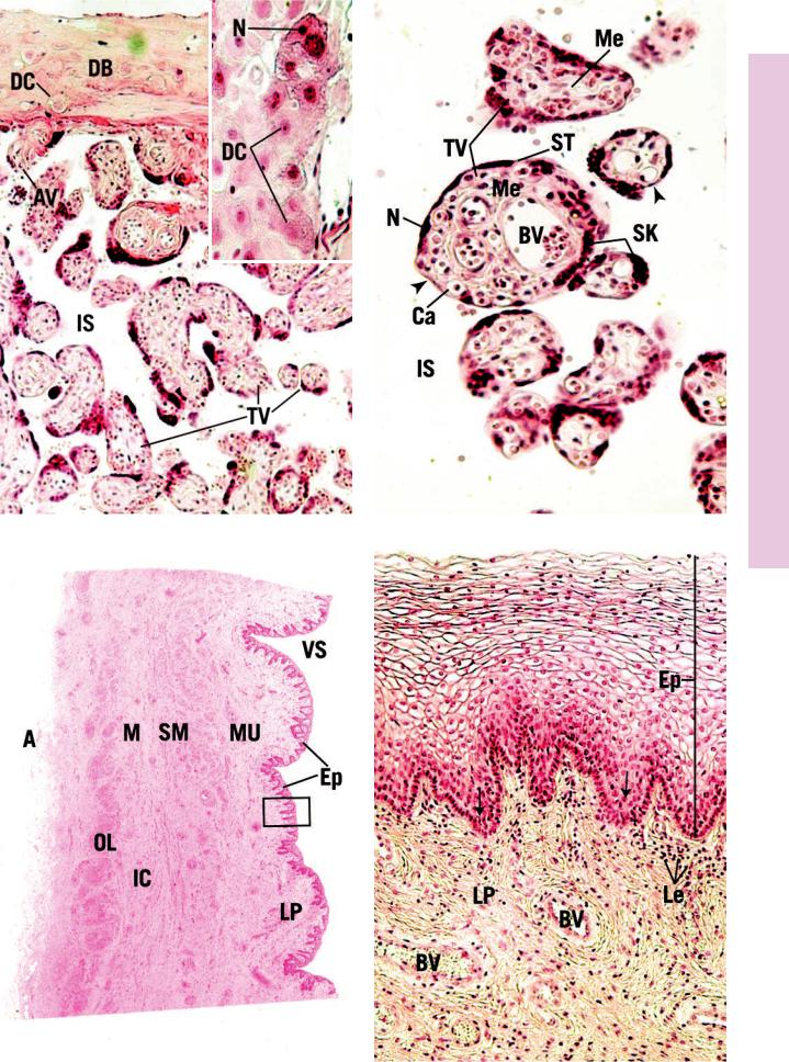
FIGURE 1. Placenta. Human. Paraffin section. ×132.
The human placenta is intimately associated with the uterine endometrium. At this junction, the decidua basalis (DB) is rich in clumps of large, round to polygonal decidual cells (DC), whose distended cytoplasm is filled with lipid and glycogen. Anchoring chorionic villi (AV) are attached to the decidua basalis; other villi are blindly ending in the intervillous space (IS). These are the most numerous and are referred to as terminal villi (TV), most of which are cut in cross or oblique sections. These villi are freely branching and, in the mature placenta, are smaller in diameter than in the immature placenta. Inset. Placenta. Human. Paraffin section.
×270. Note that the decidual cells (DC) are round to polygonal in shape. Their nuclei (N) are more or less centrally located, and their cytoplasm appears vacuolated due to the extraction of glycogen and lipids during histologic preparation.
FIGURE 2. Placenta. Human. Paraffin section. ×270.
Cross sections of terminal villi (TV) are very simple in the mature placenta. They are surrounded by the intervillous space (IS) that, in the functional placenta, is filled with maternal blood. Hence, the cells of the villus act as a placental barrier. This barrier is greatly reduced in the mature placenta, as presented in this photomicrograph. The external layer of the terminal villus is composed of syncytial trophoblasts (ST), whose numerous nuclei (N) are frequently clustered together as syncytial knots (SK). The core of the villus houses numerous fetal capillaries (Ca) that are located usually in regions of the villus void of syncytial nuclei (arrowheads). Larger fetal blood vessels (BV) are also found in the core, surrounded by mesoderm (Me). The cytotrophoblasts and phagocytic Hofbauer cells of the immature placenta mostly disappear by the end of the pregnancy.
FIGURE 3. Vagina. l.s. Monkey. Plastic section. ×14.
The vagina is a fibromuscular tube, whose vaginal space (VS) is mostly obliterated since its walls are normally in contact with each other. This wall is composed of four layers: mucosa (Mu), submucosa (SM), muscularis (M), and adventitia (A). The mucosa consists of an epithelium (Ep) and underlying lamina propria (LP). Deep to the mucosa is the submucosa, whose numerous large blood vessels impart to it an erectile tissue appearance. The smooth muscle of the muscularis is arranged in two layers, an inner circular (IC) and a thicker outer longitudinal (OL). A region similar to the boxed area is presented at a higher magnification in Figure 4.
FIGURE 4. Vagina. l.s. Human. Paraffin section. ×132.
This photomicrograph is a higher magnification of a region similar to the boxed area in Figure 3. The stratified squamous nonkeratinized epithelium (Ep) of the vagina is characterized by the empty appearance of the cells, comprising most of its thickness. This is due to the extraction lipids and glycogen during histologic preparation. Observe that the cells in the deeper aspect of the epithelium possess fewer inclusions; therefore, their cytoplasm appears normal. Note also that the lamina propria (LP) is richly vascularized (BV) and always possesses numerous leukocytes (Le) (arrows). Finally, note the absence of glands and muscularis mucosae.
Anchoring (primary) villi |
|
|
|
|
|
|
|
|
Chorionic plate |
||
|
|
|
|
|
|
|
|||||
|
|
|
|
|
|
|
|
|
|||
|
|
|
|
|
|
|
|
|
|||
Chorionic (secondary) villi |
|
|
|
|
|
|
|
|
|
|
Placental septum |
|
|
|
|
|
|||||||
Branch (tertiary) villi |
|
|
|
|
|
|
|
|
|
Decidua basalis |
|
|
|
|
|
||||||||
|
|
|
|
|
|
|
|
|
|
|
Stratum compactum |
|
|
|
|
|
|
|
|
|
|
|
Stratum spongiosum |
|
|
|
|
|
|
|
|
|
|
|
|
|
|
|
|
|
|
|
|
|
|
|
Myometrium |
|
|
|
|
|
|
|
|
|
|
|
|
Placenta
KEY
A |
adventitia |
IC |
inner circular muscle |
N |
nucleus |
AV |
anchoring chorionic villus |
IS |
intervillous space |
OL |
outer longitudinal muscle |
BV |
blood vessel |
Mu |
mucosa |
SK |
syncytial knot |
Ca |
capillary |
Le |
leukocyte |
SM |
submucosa |
DB |
decidua basalis |
LP |
lamina propria |
ST |
syncytial trophoblast |
DC |
decidual cell |
M |
muscularis |
TV |
terminal villus |
Ep |
epithelium |
Me |
mesoderm |
VS |
vaginal space |
Vagina and Placenta• 7-17 PLATE
FIGURE 1 |
FIGURE 2 |
FIGURE 3 |
FIGURE 4 |

Gland y Mammar• 8-17 PLATE
430 F E M A L E R E P R O D U C T I V E S Y S T E M
FIGURE 1. Mammary gland. Resting. Human. Paraffin section. ×132.
The mammary gland is a modified sweat gland that, in the resting stage, presents ducts (D) with occasional buds of alveoli (BA) branching from the blind ends of the duct. The remainder of the breast is composed of dense collagenous connective tissue
(dCT) interspersed with lobules of fat. However, in the immediate vicinity of the ducts and buds of alveoli, the connective tissue (CT) is more loosely arranged. It is believed that this looser CT is derived from the papillary layer of the dermis. Compare this photomicrograph with Figure 2.
FIGURE 2. Mammary gland. Lactating. Human. Paraffin section. ×132.
During pregnancy, the ducts (D) of the mammary gland undergo major development, in that the buds of alveoli proliferate to form lobules (Lo) composed of numerous alveoli (Al). The interlobular connective tissue (CT) becomes reduced to thin sheets in regions; elsewhere it maintains its previous character to support the increased weight of the breast. Observe that the CT in the immediate vicinity of the ducts and lobules (arrows) retains its loose consistency. Compare this photomicrograph with Figure 1.
FIGURE 3. Mammary gland. Lactating. Human. Paraffin section. ×132.
The active mammary gland presents numerous lobules (Lo) of alveoli (Al) that are tightly packed so that the connective tissue (CT) elements are greatly compressed. This photomicrograph clearly illustrates the crowded nature of this tissue. Although this tissue bears a superficial resemblance to the histology of the thyroid gland, the presence of ducts and branching alveoli (arrows), as well as the lack of colloid material, should assist in distinguishing this tissue as the active mammary gland. Inset. Mammary gland. Active. Human. Paraffin section. ×270. Observe the branching (arrows) of this alveolus, some of whose simple cuboidal epithelial cells (Ep) appear vacuolated (arrowheads). Note also that the lumen (L) contains fatty secretory product (milk).
FIGURE 4. Mammary gland. Nipple. Human. Paraffin section. ×14.
The large, conical nipple of the breast is covered by a thin epidermis (Ed), composed of stratified squamous keratinized epithelium. Although the nipple possesses neither hair nor sweat glands, it is richly endowed with sebaceous glands (SG). The dense irregular collagenous connective tissue (CT) core displays numerous longitudinally positioned lactiferous ducts that pierce the tip of the nipple to convey milk to the outside. The lactiferous ducts are surrounded by an extensive network of smooth muscle fibers (SM) that are responsible for the erection of the nipple, elevating it to facilitate the suckling process. The region immediately surrounding the nipple is known as the areola (Ar).
KEY
AL |
alveolus |
D |
duct |
L |
lumen |
Ar |
areola |
DcT |
dense connective tissue |
Lo |
lobule |
BA |
buds of alveoli |
Ed |
epidermis |
SM |
smooth muscle |
CT |
connective tissue |
Ep |
epithelium |
SG |
sebaceous gland |
Gland y Mammar• 8-17 PLATE
FIGURE 1 |
FIGURE 2 |
FIGURE 3 |
FIGURE 4 |