- •Preface
- •Acknowledgments
- •Reviewers
- •Contents
- •CHAPTER OUTLINE
- •CYTOPLASM
- •Plasmalemma
- •Mitochondria
- •Ribosomes
- •Endoplasmic Reticulum
- •Golgi Apparatus, cis-Golgi Network, and the trans-Golgi Network
- •Endosomes
- •Lysosomes
- •Peroxisomes
- •Proteasomes
- •Cytoskeleton
- •Inclusions
- •NUCLEUS
- •CELL CYCLE
- •CHAPTER OUTLINE
- •EPITHELIUM
- •Epithelial Membranes
- •GLANDS
- •Chapter Summary
- •CHAPTER OUTLINE
- •EXTRACELLULAR MATRIX
- •Fibers
- •Amorphous Ground Substance
- •Extracellular Fluid
- •CELLS
- •CONNECTIVE TISSUE TYPES
- •Chapter Summary
- •CHAPTER OUTLINE
- •CARTILAGE
- •BONE
- •Cells of Bone
- •Osteogenesis
- •Bone Remodeling
- •Chapter Summary
- •CHAPTER OUTLINE
- •FORMED ELEMENTS OF BLOOD
- •Lymphocytes
- •Neutrophils
- •PLASMA
- •COAGULATION
- •HEMOPOIESIS
- •Erythrocytic Series
- •Granulocytic Series
- •Chapter Summary
- •CHAPTER OUTLINE
- •SKELETAL MUSCLE
- •Sliding Filament Model of Muscle Contraction
- •CARDIAC MUSCLE
- •SMOOTH MUSCLE
- •Chapter Summary
- •CHAPTER OUTLINE
- •BLOOD-BRAIN BARRIER
- •NEURONS
- •Membrane Resting Potential
- •Action Potential
- •Myoneural Junctions
- •Neurotransmitter Substances
- •SUPPORTING CELLS
- •PERIPHERAL NERVES
- •Chapter Summary
- •CHAPTER OUTLINE
- •BLOOD VASCULAR SYSTEM
- •HEART
- •ARTERIES
- •Capillary Permeability
- •Endothelial Cell Functions
- •VEINS
- •LYMPH VASCULAR SYSTEM
- •Chapter Summary
- •CHAPTER OUTLINE
- •CELLS OF THE IMMUNE SYSTEM
- •Antigen-Presenting Cells
- •DIFFUSE LYMPHOID TISSUE
- •LYMPH NODES
- •TONSILS
- •SPLEEN
- •THYMUS
- •Chapter Summary
- •CHAPTER OUTLINE
- •PITUITARY GLAND
- •Pars Intermedia
- •Pars Nervosa and Infundibular Stalk
- •Pars Tuberalis
- •THYROID GLAND
- •Parathyroid Glands
- •Suprarenal Glands
- •Cortex
- •Medulla
- •Pineal Body
- •Chapter Summary
- •CHAPTER OUTLINE
- •SKIN
- •Epidermis of Thick Skin
- •Dermis
- •DERIVATIVES OF SKIN
- •Chapter Summary
- •CHAPTER OUTLINE
- •CONDUCTING PORTION OF THE RESPIRATORY SYSTEM
- •Extrapulmonary Region
- •Intrapulmonary Region
- •RESPIRATORY PORTION OF THE RESPIRATORY SYSTEM
- •MECHANISM OF RESPIRATION
- •Chapter Summary
- •CHAPTER OUTLINE
- •ORAL CAVITY AND ORAL MUCOSA
- •Oral Mucosa
- •Tongue
- •Teeth
- •Odontogenesis (See Graphic 13-2)
- •Chapter Summary
- •CHAPTER OUTLINE
- •REGIONS OF THE DIGESTIVE TRACT
- •Esophagus
- •Stomach
- •Small Intestine
- •Large Intestine
- •GUT-ASSOCIATED LYMPHOID TISSUE
- •DIGESTION AND ABSORPTION
- •Carbohydrates
- •Proteins
- •Lipids
- •Water and Ions
- •Chapter Summary
- •CHAPTER OUTLINE
- •MAJOR SALIVARY GLANDS
- •PANCREAS
- •LIVER
- •Exocrine Function of the Liver
- •Endocrine and Other Functions of the Liver
- •GALLBLADDER
- •Chapter Summary
- •CHAPTER OUTLINE
- •KIDNEY
- •Uriniferous Tubule
- •Nephron
- •Collecting Tubules
- •FORMATION OF URINE FROM ULTRAFILTRATE
- •EXTRARENAL EXCRETORY PASSAGES
- •Chapter Summary
- •CHAPTER OUTLINE
- •OVARY
- •Ovarian Follicles
- •Regulation of Follicle Maturation and Ovulation
- •Corpus Luteum and Corpus Albicans
- •GENITAL DUCTS
- •Oviduct
- •Uterus
- •FERTILIZATION, IMPLANTATION, AND THE PLACENTA
- •Fertilization and Implantation
- •Placenta
- •VAGINA
- •EXTERNAL GENITALIA
- •MAMMARY GLANDS
- •Chapter Summary
- •CHAPTER OUTLINE
- •TESTES
- •Spermatogenesis
- •GENITAL DUCTS
- •ACCESSORY GLANDS
- •PENIS
- •Erection and Ejaculation
- •Chapter Summary
- •CHAPTER OUTLINE
- •SENSORY ENDINGS
- •Chapter Summary
- •Terminology of Staining
- •Common Stains Used in Histology
- •Hematoxylin and Eosin
- •Wright Stain
- •Weigert Method for Elastic Fibers and Elastic van Gieson Stain
- •Silver Stain
- •Iron Hematoxylin
- •Bielschowsky Silver Stain
- •Masson Trichrome
- •Periodic Acid-Schiff Reaction (PAS)
- •Alcian Blue
- •von Kossa Stain
- •Sudan Red
- •Mucicarmine Stain
- •Safranin-O
- •Toluidine Blue
whose members seed various lymphoid organs, and are responsible for the humoral immune response.
•As the B cell is becoming immunocompetent, it manufactures IgM or IgD and places them on their cell membrane in such a fashion that the epitope binding sites are located in the extracellular space and the Fc moiety of the surface immunoglobulins (SIGs) is embedded in the plasmalemma in association with two pairs of integral proteins, Igb and Iga.
•The SIGs of a particular B cell target the same epitope. Unlike T cells, B cells have the capability of acting as APCs and present their MHC II-epitope complex to TH1 cells.
•When the newly formed B cell binds to its epitope, the Igb and the Iga transduce the information with the resultant activation of the B cell. Once activated, B cells manufacture and release IL-12, a cytokine that
promotes the formation of TH1 cells. B cells proliferate during a humoral immune response to form plasma cells and B memory cells.
Plasma cells are differentiated cells that do not possess SIGs but are “antibody factories” that synthesize and release an enormous number of identical copies of the same antibody that is specific against a particular epitope (although it may cross-react with similar epitopes).
•Antibodies, once released, bind to a specific antigen. In some instances,
binding inactivates the antigen, whereas in others
the attachment of antibodies to antigens may enhance phagocytosis (opsonization) or activate the complement cascade, resulting in chemotaxis of neutrophils and, frequently, lysis of the invader.
B memory cells are similar to T memory cells in that they are long-lived, circulating cells that are added to and increase the number of cells of the original clone. They possess SIGs so that they can be activated by an appropriate antigen during a secondary immune response. Thus, it is this increase in the size of the clone that is responsible for the anamnestic response against a subsequent encounter with the same antigen.
DIFFUSE LYMPHOID TISSUE
Diffuse lymphoid tissue occurs throughout the body, especially under moist epithelial membranes, where the loose connective tissue is infiltrated by lymphoid cells, such as lymphocytes, plasma cells, macrophages, and reticular cells. Therefore, these are referred to as mucosaassociated lymphoid tissue (MALT).
•MALT is particularly evident in the lamina propria of the digestive tract and in the subepithelial connective tissue of the respiratory tract, where they are known as
LYMPHOID TISSUE 203
gut-associated lymphoid tissue (GALT) and
bronchus-associated lymphoid tissue (BALT), respectively.
It may be noted that the lymphoid cells are not arranged in any particular pattern but are scattered in a haphazard manner. Frequently, lymphoid nodules, transitory structures that are a denser aggregation of lymphoid tissue composed mainly of lymphocytes, may be observed. Lymphoid nodules may be primary or secondary, where the secondary lymphoid nodules present the characteristic appearance of a lighter germinal center and a darker, peripherally located corona, indicating activation by antigen. The germinal centers are sites of plasma cell production, whereas the corona is produced by mitosis from existing B lymphocytes.
LYMPH NODES
Lymph nodes are ovoidto kidney-shaped organs through which lymph is filtered by exposure to large numbers of lymphoid cells (see Graphic 9-2).
•They possess a convex surface, which receives afferent lymph vessels, and
•a hilum, where blood vessels leave and enter and efferent lymph vessels leave and drain lymph from the organ.
•Lymphocytes enter lymph nodes via the afferent lymph vessels as well as via arterioles that penetrate the lymph node at the hilum, travel to the paracortex within connective tissue trabeculae, and form high endothelial vessels (postcapillary venules).
Each lymph node has a dense, irregular, collagenous connective tissue capsule and septa, derived from the capsule, subdividing the cortex into incomplete compartments. Attached to the septa and the internal aspect of the capsule is a network of reticular tissue and associated reticular cells that act as a framework for housing the numerous free and migratory cells, mostly lymphocytes, antigen-presenting cells, and macrophages, occupying the organ.
•The cortex of the lymph node houses the capsular and cortical sinuses, as well as lymphoid nodules, composed mainly of B lymphocytes, APCs, macrophages, and reticular cells.
•Between the cortex and the medulla is the paracortex, populated by T lymphocytes, APCs, and macrophages.
•The medulla consists of medullary cords and medullary sinusoids.
The medullary cords are composed mainly of
T cells, B cells, and plasma cells that arise in the cortex and paracortex and migrate into the medulla.
204LYMPHOID TISSUE
The medullary sinusoids are continuous with the capsular and cortical sinuses.
T cells and B cells enter the sinusoids and leave the lymph node via efferent lymph vessels.
Additional cell components of lymph nodes are macrophages, antigen-presenting cells, and some granulocytes. Aside from functioning in the maintenance and production of immunocompetent cells, lymph nodes also filter lymph.
•The filtering procedure is facilitated by the elongated processes of reticular cells that span the sinuses of the node and thus disturb and retard lymph flow, providing more time for the resident macrophages to phagocytose antigens and other debris.
TONSILS
Tonsils are aggregates of incompletely encapsulated lymphoid tissue situated at the entrances to the oral pharynx and to the nasal pharynx. Participating in the formation of the tonsillar ring are the
•palatine,
•pharyngeal, and
•lingual tonsils.
The tonsils produce antibodies against the numerous antigens and microorganisms that abound in their vicinity. There are additional, smaller tonsils, such as the tubal and lingual tonsils, that function in the same manner.
SPLEEN
The spleen is the largest lymphoid organ of the body (see Graphic 9-2). Its principal functions are to filter blood, phagocytose senescent red blood cells and invading microorganisms, supply immunocompetent T and B lymphocytes, and manufacture antibodies. Unlike lymph nodes, the spleen is not divided into cortical and medullary regions, nor is it supplied by afferent lymphatic vessels. Blood vessels enter and leave the spleen at its hilum and travel within the parenchyma via trabeculae derived from its connective tissue capsule.
•The spleen is subdivided into white and red pulps.
White pulp is composed of lymphoid tissue that is arranged in a specific fashion, either as periarterial lymphatic sheaths (PALS) composed of T lymphocytes or as lymphoid nodules consisting of B lymphocytes.
The red pulp consists of pulp cords (of Billroth) interposed between a spongy network of sinusoids lined by unusual elongated endothelial cells displaying large intercellular spaces, supported by a
thick, discontinuous, hoop-like basement membrane. Reticular cells and reticular fibers associated with these sinusoids extend into the pulp cords to contribute to the cell population that consists of macrophages, plasma cells, and extravasated blood cells.
A region of smaller sinusoids forms the interface between the white and red pulps, and this interface is known as the marginal zone. Capillaries arising from the central arteries deliver their blood to sinusoids of the marginal zone, which is rich in arterial vessels and avidly phagocytic macrophages. APCs of the marginal zone monitor this blood for the presence of antigens and foreign substances.
Understanding splenic organization depends on knowing the vascular supply of the spleen.
•The splenic artery entering at the hilum is distributed to the interior of the organ via trabeculae as trabecular arteries.
•On leaving a trabecula, the vessel enters the parenchyma to be surrounded by the periarterial lymphatic sheaths (PALS) and occasional lymphoid nodules and is termed the central artery.
•Central arteries enter the red pulp by losing their PALS and subdivide into numerous small, straight vessels known as penicillar arteries.
•Penicillar arteries possess three regions: pulp arterioles, sheathed arterioles, and terminal arterial capillaries. Whether these terminal arterial capillaries drain directly into the sinusoids (closed circulation) or terminate as open-ended vessels in the pulp cords (open circulation) has not been determined conclusively; however, in humans, the open circulation is believed to predominate.
•It is during this passage of red blood cells from the splenic cords into the sinusoids that damaged and aging red blood cells are eliminated.
•Sinusoids are drained by pulp veins, which lead to trabecular veins and eventually join the splenic vein.
THYMUS
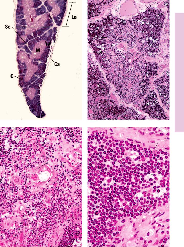
The thymus is an endodermally derived, bilobed, encapsulated lymphoid organ located in the mediastinum, overlying the great vessels of the heart (see Graphic 9-2). The thymus attains its greatest development shortly after birth, but subsequent to puberty, it begins to involute and becomes infiltrated by adipose tissue; however, even in the adult, the thymus retains its ability to form a reduced number of T lymphocytes. The thin connective tissue capsule of the thymus sends septa deep into the organ, incompletely subdividing it into lobules.
The thymus possesses no lymphoid nodules; instead, it is divided into an
•outer darker staining cortex, composed of epithelial reticular cells, macrophages, and small T lymphocytes (thymocytes), and an
•inner lighter staining medulla consisting of large T lymphocytes, epithelial reticular cells, and thymic (Hassall’s) corpuscles (see Table 9-4).
The major functions of the thymus are the formation, potentiation, and destruction of T lymphocytes.
•Immunoincompetent (immature) T-lymphocyte precursors enter the corticomedullary junction of the thymus, where they become known as thymocytes, and migrate to the outer cortex where they are activated by cytokines released by epithelial reticular cells to express certain T-cell markers.
•The markers that thymocytes express do not include CD4, CD8, or the CD3-TCR complex and become known as double negative thymocytes. These cells migrate into the inner cortex and express pre-TCRs (pre–T-cell receptors) that trigger their propagation.
•The progeny of the pre–TCR-bearing thymocytes express both CD4 and CD8 molecules as well as a limited number of CD3-TCR molecules and are known as double-positive thymocytes.
•Cortical epithelial reticular cells assess if doublepositive thymocytes are able to recognize self-MHC- self-epitope complexes. About 90% of double-positive thymocytes are unable to recognize these complexes, and they undergo apoptosis. The remaining 10% of these double-positive thymocytes that do recognize the self-MHC-self-epitope complexes mature, express many more TCRs, and lose either CD8 or CD4 molecules from their cell surface.
LYMPHOID TISSUE 205
•Thymocytes that express many TCRs and either CD4 or CD8 molecules are known as single-positive thymocytes, which pass through the corticomedullary border to enter the medulla.
•Dendritic cells and epithelial reticular cells of the medulla assess the abilities of single-positive thymocytes to initiate an immune response against the self.
Single-positive thymocytes that can initiate an immune response against the self undergo apoptosis (clonal deletion) due to the effect of thymic stromal lymphopoietin, released by epithelial reticular cells of Hassall’s corpuscles.
Single-positive thymocytes that are unable to attack the self are released from the thymus as naïve T lymphocytes. These naïve T cells migrate to the secondary lymphoid organs to set up clones of T cells.
Blood vessels gain entrance to the medulla by traveling in the connective tissue septa, which they exit at the corticomedullary junction, where they provide capillary loops to the cortex.
•The capillaries that enter the cortex are the continuous type and are surrounded by epithelial reticular cells that isolate them from the cortical lymphocytes, thus establishing a blood-thymus barrier, providing an antigen-free environment for the potentiation of the immunocompetent T lymphocytes.
•The blood vessels of the medulla are not unusual and present no blood-thymus barrier.
•The thymus is drained by venules in the medulla, which also receives blood from the cortical capillaries.
•Epithelial reticular cells form a specialized barrier between the cortex and medulla to prevent medullary material from gaining access to the cortex.
TABLE 9-4 • |
Thymic Epithelial Reticular Cells |
|
Cell Type |
Location |
Function |
|
|
|
Type I |
Cortex |
Surround blood vessels and isolate cortex from capsule and septa |
|
|
|
Type II |
Midcortex |
Form a boundary around and present MHC I, MHC II, and self-antigen |
|
|
molecules to thymocytes |
|
|
|
Type III |
Corticomedullary junction |
Present MHC I, MHC II, and self-antigen molecules to thymocytes |
|
|
|
Type IV |
Corticomedullary junction |
Isolate type III epithelial reticular cells from the medulla |
|
|
|
Type V |
Medulla |
Form the cellular scaffolding of the medulla |
|
|
|
Type VI |
Medulla |
Form Hassall’s corpuscles; release the cytokine thymic stromal lymphopoietin |
|
|
responsible for clonal deletion |
|
|
|
206 LYMPHOID TISSUE
CLINICAL CONSIDERATIONS
Hodgkin’s Disease
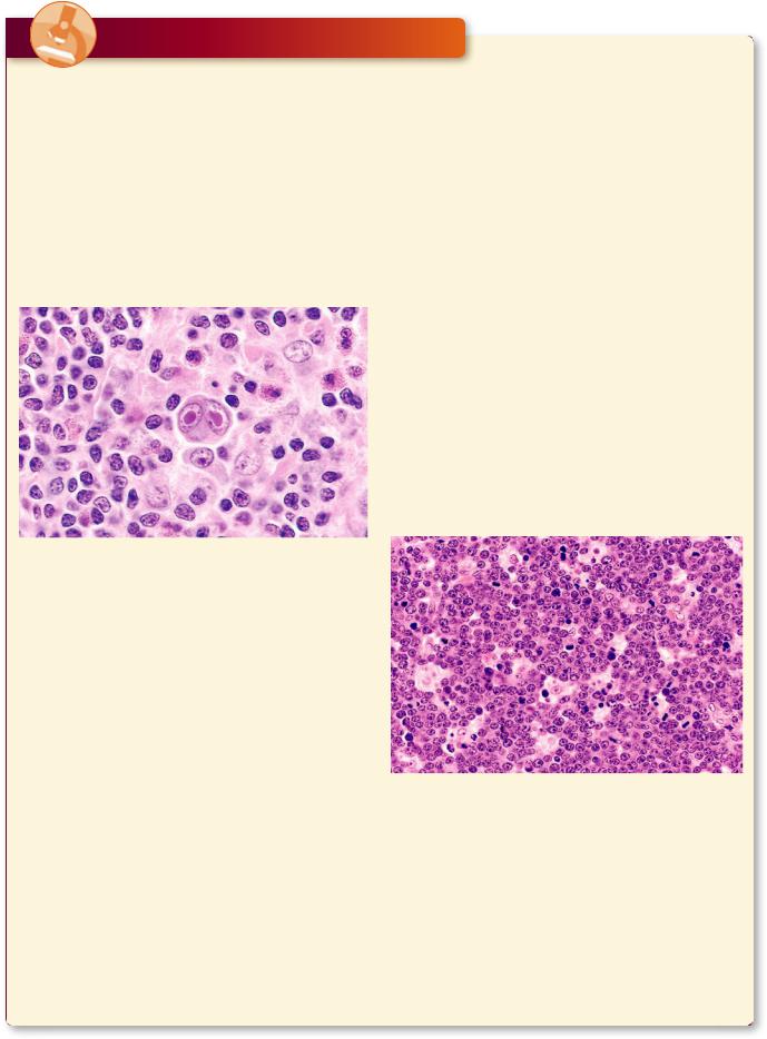
Hodgkin’s disease is a neoplastic transformation of lymphocytes that is prevalent mostly in young males. Its clinical signs are asymptomatic initially because the swelling of the liver, spleen, and lymph nodes are not accompanied by pain. Other manifestations include the loss of weight, elevated temperature, diminished appetite, and generalized weakness. Histopathologic characteristics include the presence of Reed-Sternberg cells, easily recognizable by their large size, and the presence of two large, pale, oval nuclei in each cell.
or curtailed. Most individuals with this syndrome die in early childhood as a result of uncontrollable infections.
Lymph Nodes During Infection
In a healthy patient with a normal amount of adipose tissue, the lymph nodes are small, soft structures that cannot be palpated easily. However, during an infection, the regional lymph nodes become enlarged and hard to the touch due to the large number of lymphocytes that are being formed within the node.
Burkitt’s Lymphoma
Burkitt’s lymphoma is a very rapidly growing non– Hodgkin’s lymphoma that has its origins in B cells. It is relatively rare in the United States but is more common in Central Africa, where it affects young males infected with the Epstein-Barr virus. It is also prevalent in people afflicted with the HIV. The lymphoma cells proliferate quickly and spread to the lymph nodes and the small intestine. In more severe cases, the lymphoma cells can invade the central nervous system, bone marrow, and blood. If untreated, the disease is fatal, but treatment, especially in the early stages of the disease, has a very good prognosis.
This photomicrograph is from the lymph node of a patient with Hodgkin’s lymphoma displaying the characteristic binucleate Reed-Sternberg cell in the center of the field. Note the distinguishing eosinophilic nuclei that resemble nuclear inclusions. (Reprinted with permission from Mills SE, Carter D, et al., eds. Sternberg’s Diagnostic Surgical Pathology, 5th ed. Philadelphia: Lippincott Williams & Wilkins, 2010, p. 701.)
Wiskott-Aldrich Syndrome
Wiskott-Aldrich syndrome is an immunodeficiency disorder occurring only in boys and is characterized by eczema (dermatitis), lowered platelet count, and lymphocytopenia (abnormally low levels of lymphocytes, both B- and T-cell populations). The immunosuppressed state of these children leads to recurring bacterial infections, hemorrhage, and death at an early age. Most children who survive the first decade of life are stricken with leukemia or lymphoma.
DiGeorge’s Syndrome
DiGeorge’s syndrome is the name of the congenital disorder when the thymus fails to develop and the patient is unable to produce T lymphocytes. These patients cannot mount a cellularly mediated immune response, and some of their humorally mediated responses are also disabled
This photomicrograph is from a lymph node of a patient with Burkitt’s lymphoma. Note the presence of several mitotic figures in the field. The image resembles a “starry sky” due to the presence of an abundance of tingible-body macrophages. (Reprinted with permission from Mills SE, Carter D, et al., eds. Sternberg’s Diagnostic Surgical Pathology, 5th ed. Philadelphia: Lippincott Williams & Wilkins, 2010, p. 722.)
Peripheral T-cell Lymphoma in the Spleen
A relatively rare disease, peripheral T-cell lymphomas in the spleen are derived from T cells and T-cell precursors that proliferate and invade various organs, including the skin and the spleen. When the spleen is affected, the
LYMPHOID TISSUE 207
cells are large and aggressive with clear cytoplasms. They congregate in the vicinity of the periarterial lymphatic sheaths (PALSs). The prognosis of patients with peripheral T-cell lymphomas depends on whether or not the
invading cells express the protein anaplastic lymphoma kinase (ALK). Patients whose cells express ALK respond to treatment much better than patients whose cells do not express this protein.
This photomicrograph is of the spleen of a patient with peripheral T-cell lymphoma. The large, clear cells surround the PALS and the B-cell–rich germinal center appears unaffected. (Reprinted with permission from Mills SE, Carter D, et al., eds. Sternberg’s Diagnostic Surgical Pathology, 5th ed. Philadelphia: Lippincott Williams & Wilkins, 2010, p. 755 Fig. 18-17.)
Tissues Lymphoid • 1-9 GRAPHIC
208 LYMPHOID TISSUE
Cervical nodes
Tracheobronchial nodes
Axillary nodes
Thoracic duct
Aortic nodes
Peyer’s patches (ileum)
Iliac nodes
Inguinal nodes
Tonsils
Thymus
Spleen
Lymphoid tissue consists of several encapsulated organs, lymph nodes, tonsils, thymus, and spleen, as well as diffuse lymphoid tissue, composed of loose conglomerates of the lymphoid cells: B lymphocytes, T lymphocytes, plasma cells, macrophages, and antigen-presenting cells. Frequently, these lymphoid cells are collected as lymphatic nodules that appear as they are needed, although they are always present in the gut (GALT, gut-associate lymphoid tissue, and Peyer’s patches), in the bronchial tubes (BALT, bronchus-associated lymphoid tissue), and certain mucosae (MALT, mucosa-associated lymphoid tissue).
T lymphocytes originate in the bone marrow and then migrate to the thymus to become immunologically competent T cells.
T cell |
Thymus |
B cell |
|
|
B lymphocytes |
|
are believed to |
Bone |
remain in the bone |
marrow |
marrow to become |
|
immunologically |
|
competent B cells. |
|
Lymph node |
These immunocompetent T and B cells then seed lymphoid tissues, especially the spleen, lymph nodes, and lymphatic nodules, and are capable of becoming activated (mature) and responding to an antigenic challenge.
Mature and immunocompetent cells circulate among the various lymphoid tissues, using blood and lymph vessels.
|
|
|
|
LYMPHOID TISSUE |
209 |
|
Germinal center |
} |
|
|
|
Efferent |
|
|
|
|
|
lymphatic vessel |
Lymphatic nodule |
Cortex |
|
|
|
|
Cortical sinus |
|
|
|
|
|
|
Paracortex |
|
|
|
|
|
Medullary cord |
} Medulla |
|
|
|
|
Medullary sinus |
|
||
|
|
Trabecula |
|
|
|
|
|
Afferent lymphatic vessel |
|
||
Lymph nodes |
|
Capsule |
|
|
|
|
|
|
|
|
|
function in T and |
|
Adipose tissue |
|
|
|
B cell formation, |
|
|
|
|
|
as well as in the |
|
|
|
|
|
clearing of lymph. |
|
|
|
|
|
|
|
|
Thymic capsule |
|
|
|
|
|
Capsular vein |
|
Cortex
Medulla
Hassall’s (thymic) corpuscles
The thymus is responsible for the |
|
|
maturation of T cells. T helper cells |
|
|
play a pivotal role in the |
|
|
development and maintenance of |
|
|
the immune response. They |
|
|
interact with antigen-presenting |
|
|
cells and release cytokines, |
|
|
resulting in the generation of |
|
|
plasma cells for the humoral and T |
|
Capsular arteries |
killer (cytotoxic) cells for the |
|
|
cell-mediated response. |
|
Red pulp |
|
|
|
Splenic |
|
White pulp |
Vein |
Splenic sinusoid |
|
vein |
|
|
|
|
|
|
Artery |
Trabeculae |
|
|
Splenic artery
The spleen cleanses the blood, eliminates defunct red blood cells, forms T cells and B cells, and in
some animals but not Capsule humans, stores red blood
cells.
Spleen and Thymus, Node, Lymph • 2-9 GRAPHIC
Formation Cell Plasma and Memory B • 3-9 GRAPHIC
210 LYMPHOID TISSUE
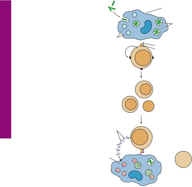
Antigen-dependent cross linking of the surface antibodies activates the B cell which places the epitope-MHC II complex on the external aspect of its plasmalemma.
The TCR and CD4 molecules of the TH2 cell recognize the B cell’s MHC II-epitope complex. Additionally, binding of the B cell’s CD40 molecule to the TH2 cell’s CD40 receptor induces the B cell to proliferate and the TH2 cell to release of IL4, IL5, and IL6.
IL4, IL5, and IL6 induce the activation of B cells and their differentiation into B memory and plasma cells.
Antigen
B cell
Antibodies
Class II MHC- CD40 epitope complex
|
B cell |
CD4 molecule |
CD 40 |
|
|
T cell receptor |
receptor |
TH2 cell
Cytokines IL4,
IL5, and IL6
B memory cell |
Plasma cell |
|
Antibodies
The T cell receptor (TCR) and CD4 molecule of the TH1 cell binds to the epitope and the MHC II of the antigen-presenting cell (APC), respectively. The binding induces the
APC to express B7 molecules on its plasmalemma, which then binds to the CD28 molecule of the TH1 cell, inducing that cell to release IL2.
The same APC expresses the MHC I-epitope complex, which is recognized by the CD8 molecule and the TCR of the cytotoxic T lymphocyte (CTL).
Additionally, the CD28 molecule of the
CTL binds with the B7 molecule on the APC plasmalemma. These interactions induce the expression of IL2 receptors on the CTL plasma membrane. Binding of IL2 (released by the TH1 cell) to the
IL2 receptors of the CTL induces that cell to proliferate.
The plasmalemma of virally transformed cells expresses MHC I-epitope complex, which is recognized by the CD8 molecule and TCR of the newly formed cytotoxic T lymphocytes. The binding of the CTL induces these cells to secrete perforins and fragmentins. The former assemble to form pores in the plasma membrane of the transformed cell, and framentin drives the transformed cell into apoptosis.
LYMPHOID TISSUE 211
IL2
TH1 cell
CD28 |
T cell receptor |
|
molecule |
||
CD4 molecule |
||
B7 |
||
Class II MHC-epitope complex |
||
molecule |
||
|
||
|
Antigen- |
|
|
presenting |
|
|
cell |
Class I MHC- |
|
epitope complex |
Cytotoxic T |
CD8 molecule |
lymphocyte |
|
|
|
CTL |
Fragmenting
Virus-
transformed
cell
Perforins
Cell Transformed Virally of Killing and Activation Cell-T Cytotoxic• 4-9 GRAPHIC
Cells 1 T by Activation Macrophage • 5-9 GRAPHIC
H
212 LYMPHOID TISSUE
Bacteria-infected macrophages bear MHC II-epitope complexes on their plasmalemma that, if recognized by the CD4 molecule and TCR of TH1 cells, activates these T cells, causing them to release IL2 and to express IL2 receptors on their plasma membrane.
Binding of IL2 to the IL2 receptors induces proliferation of the TH1 cells.
The TCR and CD4 molecules of the newly formed TH1 cells recognize and bind to the MHC II-epitope complexes of bacteria-infected macrophages. The binding causes activation of these TH1 cells so that they release -interferon, a cytokine that encourages the macrophages to destroy their endocytosed bacteria.
Bacteria |
Macrophage |
Lysosomes
IL2
-IFN
Activated lysosome
Macrophage
Bacteria proliferating
in phagosomes
Class II MHC-epitope complex
CD4 molecule
T cell receptor
TH1 cell
TH1 cell
Nodule Lymphatic Infiltration, Lymphatic • 1-9 PLATE
214 LYMPHOID TISSUE
FIGURE 1. Lymphatic infiltration. Monkey duodenum. Plastic section. ×540.
The connective tissue (CT) deep to moist epithelia is usually infiltrated by loosely aggregated lymphocytes (Ly) and plasma cells (PC), evident from their clockface nuclei. Observe that the simple columnar epithelium (E) contains not only the nuclei (N) of epithelial cells but also dark, dense nuclei of lymphocytes (arrows), some of which are in the process of migrating from the lamina propria (connective tissue) into the lumen of the duodenum. Note also the presence of a lacteal (La), a blindly ending, lymph-filled lymphatic channel unique to the small intestine. These vessels can be recognized by the absence of red blood cells, although nucleated white blood cells may frequently occupy their lumen.
FIGURE 3. Lymphatic nodule. Monkey. Plastic section. ×270.
This is a higher magnification of a lymphatic nodule from Peyer’s patches in the monkey ileum. Note that the lighter staining germinal center (Gc) is surrounded by the corona (Co) of darker staining cells possessing only a limited amount of cytoplasm around a dense nucleus. These cells are small lymphocytes (Ly). Germinal centers form in response to an antigenic challenge and are composed of lymphoblasts and plasmablasts, whose nuclei stain much lighter than those of small lymphocytes. The boxed area is presented at a higher magnification in the following figure.
FIGURE 2. Lymphatic nodule. Monkey. Plastic section. ×132.
The gut-associated lymphatic nodule in this photomicrograph is part of a cluster of nodules known as Peyer’s patches (PP) and is taken from the monkey ileum. The lumen (L) of the small intestine is lined by a simple columnar epithelium (E) with numerous goblet cells (GC). However, note that the epithelium is modified over the lymphoid tissue into a follicle-associated epithelium
(FAE), whose cells are shorter, infiltrated by lymphocytes, and display no goblet cells. Observe that this particular lymphatic nodule presents no germinal center but is composed of several cell types, as recognized by nuclei of various sizes and densities. These are described in Figures 3 and 4. Although this lymphatic nodule is unencapsulated, the connective tissue (CT) between the smooth muscle (SM) and the lymphatic nodule is free of infiltrate.
FIGURE 4. Lymphatic nodule. Monkey. Plastic section. ×540.
This is a higher magnification of the boxed area of the previous figure. Observe the small lymphocytes (Ly) at the periphery of the germinal center (Gc). The activity of this center is evidenced by the presence of mitotic figures (arrows) as well as the lymphoblasts (LB) and plasmablasts (PB). The germinal center is the site of production of small lymphocytes that then migrate to the periphery of the lymphatic nodule to form the corona.
KEY
Co |
corona |
GC |
goblet cell |
N |
nucleus |
CT |
connective tissue |
L |
lumen |
PB |
plasmablast |
E |
epithelium |
La |
lacteal |
PC |
plasma cell |
FAE |
follicle-associated epithelium |
LB |
lymphoblast |
PP |
Peyer’s patch |
Gc |
germinal center |
Ly |
small lymphocyte |
SM |
smooth muscle |
CT
E
PC
PC
N Ly
La
E
FIGURE 1
Gc
Co
Ly
FIGURE 3
L
E
FAE
GC
PP
CT
SM
FIGURE 2
Ly
LB
Gc LB
Ly |
PB |
PB
FIGURE 4
Nodule Lymphatic Infiltration, ymphatic • 1L -9 PLATE
Node Lymph • 2-9 PLATE
216 LYMPHOID TISSUE
FIGURE 1. Lymph node. Paraffin section. ×14.
Lymph nodes are kidney-shaped structures possessing a convex and a concave (hilar) surface. They are invested by a connective tissue capsule (Ca) that sends trabeculae (T) into the substance of the node, thereby subdividing it into incomplete compartments. The compartmentalization is particularly prominent in the cortex (C), the peripheral aspect of the lymph node. The lighter staining central region is the medulla (M). The zone between the medulla and cortex is the paracortex (PC). Observe that the cortex displays numerous lymphatic nodules (LN), many with germinal centers (Gc). This is the region of B lymphocytes, whereas the paracortex is particularly rich in T lymphocytes. Note that the medulla is composed of sinusoids (S), trabeculae (T) of connective tissue conducting blood vessels, and medullary cords (MC). The medullary cords are composed of lymphocytes, macrophages, reticular cells, and plasma cells. Lymph enters the lymph node, and as it percolates through sinuses and sinusoids, foreign substances and nonself antigenic elements are removed from it by phagocytic activity of macrophages.
FIGURE 3. Lymph node. Monkey. Plastic section. ×132.
The cortex of the lymph node is composed of numerous lymphatic nodules, one of which is presented in this photomicrograph. Observe that the lymph node is usually surrounded by adipose tissue (AT). The thin connective tissue capsule (Ca) sends trabeculae
(T) into the substance of the lymph node. Observe that the lymphatic nodule possesses a dark staining corona (Co), composed mainly of small lymphocytes (Ly) whose heterochromatic nuclei are responsible for their staining characteristics. The germinal center (Gc) displays numerous cells with lightly staining nuclei, belonging to dendritic reticular cells, plasmablasts, and lymphoblasts.
FIGURE 2. Lymph node. Monkey. Plastic section. ×270.
Afferent lymphatic vessels (AV) enter the lymph node at its convex surface. These vessels bear valves (V) that regulate the direction of flow. Lymph enters the subcapsular sinus (SS), which contains numerous macrophages (Ma), lymphocytes (Ly), and antigen-transporting cells. These sinuses are lined by endothelial cells (EC), which also cover the fine collagen fibers that frequently span the sinus to create a turbulence in lymph flow. Lymph from the subcapsular sinus enters the cortical sinus and then moves into the medullary sinusoids. It is here that lymphocytes also migrate into the sinusoids, leaving the lymph node via the efferent lymph vessels eventually to enter the general circulation.
FIGURE 4. Lymph node. Human. Silver stain. Paraffin section. ×132.
The hilum of the human lymph node displays the collagenous connective tissue capsule (Ca), from which numerous trabeculae
(T) enter into the substance of the lymph node. Observe that the region of the hilum is devoid of lymphatic nodules but is particularly rich in medullary cords (MC). Note that the basic framework of these medullary cords, as well as of the lymph node, is composed of thin reticular fibers (arrows), which are connected to the collagen fiber bundles of the trabeculae and capsule.
|
Germinal center |
} |
|
Efferent |
Lymphatic nodule |
Cortex |
|
lymphatic vessel |
|||
|
Cortical sinus |
|
|
|
Medullary cord |
} Medulla |
|
|
Medullar sinus |
||
|
Trabecula |
|
|
|
Afferent lymphatic vessel |
||
|
Capsule |
|
|
Lymph node |
Adipose tissue |
|
KEY
AT |
adipose tissue |
Gc |
germinal center |
PC |
paracortex |
AV |
afferent lymphatic vessel |
LN |
lymphatic nodule |
S |
sinusoid |
C |
cortex |
Ly |
small lymphocyte |
SS |
subcapsular sinus |
Ca |
capsule |
M |
medulla |
T |
trabeculae |
Co |
corona |
Ma |
macrophage |
V |
valve |
EC |
endothelial cell |
MC |
medullary cord |
|
|
Ly
Ma
SS
V
AV
EC
FIGURE 1 |
FIGURE 2 |
MC
Gc
Ca
Ly
Co |
T |
|
Ca
MC
AT
T
FIGURE 3 |
FIGURE 4 |
Node ymph • 2L -9 PLATE
Tonsils Node, ymph • 3L -9 PLATE
218 LYMPHOID TISSUE
FIGURE 1. Lymph node. Paraffin section. ×132.
The medulla of the lymph node is rich in endothelially lined sinusoids (S), which receive lymph from the cortical sinuses. Surrounding the sinusoids are many medullary cords (MC), packed with macrophages, small lymphocytes, and plasma cells, whose nuclei (arrows) stain intensely. Both T and B lymphocytes populate the medullary cords, since they are in the process of migrating from the paracortex and cortex, respectively. Some of these lymphocytes will leave the lymph node using the sinusoids and efferent lymphatic vessels at the hilum. The medulla also displays connective tissue trabeculae (T), connective tissue elements that are conduits for blood vessels (BV), which enter and leave the lymph node at the hilum.
FIGURE 3. Palatine tonsil. Human. Paraffin section. ×14.
The palatine tonsil is an aggregate of lymphatic nodules (LN), many of which possess germinal centers (Gc). The palatine tonsil is covered by a stratified squamous nonkeratinized epithelium (E) that lines the deep primary crypts (PCr) that invaginate deeply into the substance of the tonsil. Frequently, secondary crypts (SCr) are evident, also lined by the same type of epithelium. The crypts frequently contain debris (arrow) that consists of decomposing food particles as well as lymphocytes that migrate from the lymphatic nodules through the epithelium to enter the crypts. The deep surface of the palatine tonsil is covered by a thickened connective tissue capsule (Ca).
FIGURE 2. Lymph node. Monkey. Plastic section. ×540.
High magnification of a sinusoid (S) and surrounding medullary cords (MC) of a lymph node medulla. Note that the medullary cords are populated by macrophages, plasma cells (PC), and small lymphocytes (Ly). The sinusoids are lined by a discontinuous endothelium (EC). The lumen contains lymph, small lymphocytes (Ly), and macrophages (Ma). The vacuolated appearance of these macrophages is indicative of their active phagocytosis of particulate matter.
FIGURE 4. Pharyngeal tonsil. Human. Paraffin section. ×132.
The pharyngeal tonsil, located in the nasopharynx, is a loose aggregate of lymphatic nodules, often displaying germinal centers (Gc). The epithelial lining (E) is pseudostratified ciliated columnar with occasional patches of stratified squamous nonkeratinized epithelium (asterisk). The lymphatic nodules are located in a loose, collagenous connective tissue (CT) that is infiltrated by small lymphocytes (Ly). Note that lymphocytes migrate through the epithelium (arrows) to gain access to the nasopharynx.
|
Germinal center |
} |
|
Efferent |
|
Cortex |
|
Lymphatic nodule |
|
||
lymphatic vessel |
|
||
|
|
|
|
|
Medullary cord |
||
|
Trabecula |
|
|
|
Afferent lymphatic vessel |
||
|
Capsule |
|
|
Lymph node
KEY
BV |
blood vessel |
Gc |
germinal center |
PC |
plasma cell |
Ca |
capsule |
LN |
lymphatic nodule |
PCr |
primary crypt |
CT |
connective tissue |
Ly |
lymphocyte |
S |
sinusoid |
E |
epithelium |
Ma |
macrophage |
T |
trabeculae |
EC |
endothelial cell |
MC |
medullary cord |
SCr |
secondary crypt |
BV
T
MC
S
S
MC
FIGURE 1
|
Ma |
|
|
PC |
|
Ly |
EC |
S |
|
||
|
|
Ly |
MC
PC
FIGURE 2
E
* CT
Ly
Gc
Tonsils Node, ymph • 3L -9 PLATE
FIGURE 3 |
FIGURE 4 |
Microscopy Electron Node, ymph • 4L -9 PLATE
220 LYMPHOID TISSUE
FIGURE 1. Popliteal lymph node. Mouse. Electron microscopy. ×8,608.
Electron micrograph of a mouse lymph node. Immediately deep to the capsule (Ca) lies the subcapsular sinus occupied by three lymphocytes, one of which is labeled (L), as well as the process
(P) of an antigen-transporting (antigen-presenting) cell, whose cell body (arrowheads) and nucleus are in the cortex, deep to the sinus. The process enters the lumen of the subcapsular sinus via a
pore (arrows) in the epithelial lining of its floor (FL). It is believed that antigen-transporting cells are nonphagocytic and that they trap antigens at the site of antigenic invasion and transport them to lymphatic nodules of lymph nodes, where they mature to become dendritic reticular cells. (From Szakal A, Homes K, Tew J. Transport of immune complexes from the subcapsular sinus to lymph node follicles on the surface of nonphagocytic cells, including cells with dendritic morphology. J Immunol 1983;131:1714–1717.)
Subscapular sinus
Capsule
Lymph node
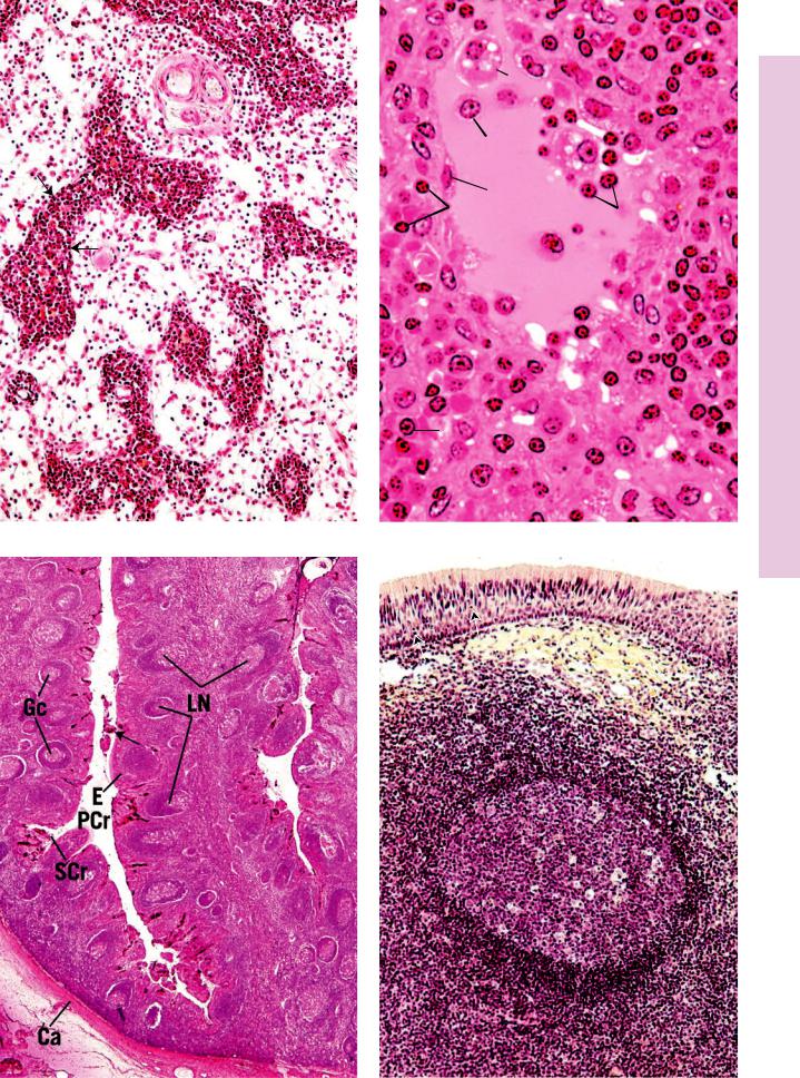
PLATE 9-4L • ymph Node, Electron Microscopy
FIGURE 1
hymus • T5-9 PLATE
222 LYMPHOID TISSUE
FIGURE 1. Thymus. Human infant. Paraffin section. ×14.
The thymus of a prepubescent individual is a well-developed organ that displays its many characteristics to advantage. This photomicrograph presents a part of one lobe. It is invested by a thin connective tissue capsule (Ca) that incompletely subdivides the thymus into lobules (Lo) by connective tissue septa (Se). Each lobule possesses a darker staining peripheral cortex (C) and a lighter staining medulla (M). The medulla of one lobule, however, is continuous with that of other lobules. The connective tissue capsule and septa convey blood vessels into the medulla of the thymus. The thymus begins to involute in the postpubescent individual, and the connective tissue septa become infiltrated with adipocytes.
FIGURE 3. Thymus. Monkey. Plastic section. ×270.
The center of this photomicrograph is occupied by the medulla
(M) of the thymus, presenting a large thymic (Hassall’s) corpuscle (TC), composed of concentrically arranged epithelial reticular cells (ERC). The function, if any, of this structure is not known. The thymic medulla houses numerous blood vessels (BV), macrophages, lymphocytes (Ly), and occasional plasma cells.
FIGURE 2. Thymus. Monkey. Plastic section. ×132.
The lobule of the thymus presented in this photomicrograph appears to be completely surrounded by connective tissue septa (Se); three-dimensional reconstruction would reveal this lobule to be continuous with surrounding lobules (Lo). Observe the numerous blood vessels (BV) in the septa as well as the darker staining cortex (C) and the lighter staining medulla (M). The characteristic light patches of the cortex correspond to the high density of epithelial reticular cells and macrophages (arrows). The darker staining structures are nuclei of the T-lymphocyte series. The medulla contains the characteristic thymic corpuscles (TC) as well as blood vessels, macrophages, and epithelial reticular cells.
FIGURE 4. Thymus. Monkey. Plastic section. ×540.
The cortex of the thymus is bounded externally by collagenous connective tissue septa (Se). The substance of the cortex is separated from the septa by a zone of epithelial reticular cells (ERC), recognizable by their pale nuclei. Additional ERC form a cellular reticulum; in whose interstices, lymphocytes (Ly) develop into mature T lymphocytes. Numerous macrophages (Ma) are also evident in the cortex. These cells phagocytose lymphocytes destroyed in the thymus.
Thymic capsule
Capsular vein
Cortex
Medulla
Thymic corpuscles (Hassal’s corpuscles)
Thymus
KEY
BV |
blood vessel |
Lo |
lobule |
Ma |
macrophage |
C |
cortex |
Ly |
lymphocyte |
Se |
septum |
Ca |
capsule |
M |
medulla |
TC |
thymic corpuscle |
ERC |
epithelial reticular cell |
|
|
|
|
FIGURE 1
M
ERC
TC
BV
Ly
FIGURE 3
BV
C |
9 PLATE |
|
T5- |
|
• |
|
hymus |
TC
Se
M
Lo
FIGURE 2
Se
ERC
Ma
Ly
ERC
FIGURE 4
pleen • S6-9 PLATE
224 LYMPHOID TISSUE
FIGURE 1. Spleen. Human. Paraffin section. ×132.
The spleen, the largest lymphoid organ, possesses a thick collagenous connective tissue capsule (Ca). Since it lies within the abdominal cavity, it is surrounded by a simple squamous epithelium (E). Connective tissue septa (SE), derived from the capsule, penetrate the substance of the spleen, conveying blood vessels (BV) into the interior of the organ. Histologically, the spleen is composed of white pulp (WP) and red pulp (RP). White pulp is arranged as a cylindrical, multilayered sheath of lymphocytes (Ly) surrounding a blood vessel known as the central artery (CA). The red pulp consists of sinusoids (S) meandering through a cellular tissue known as pulp cords (PC). The white pulp of the spleen is found in two different arrangements. The one represented in this photomicrograph is known as a periarterial lymphatic sheath (PALS), composed mostly of T lymphocytes. The zone of lymphocytes at the junction of the PALS and the red pulp is known as the marginal zone (MZ).
FIGURE 2. Spleen. Monkey. Plastic section. ×132.
Lying within the periarterial lymphatic sheaths (PALS) of the spleen, a second arrangement of white pulp may be noted, namely, lymphatic nodules (LN) bearing a germinal center (Gc). Lymphatic nodules frequently occur at the branching of the central artery (CA). Nodules are populated mostly by B lymphocytes (arrows), which account for the dark staining of the corona (CO). The germinal center is the site of active production of B lymphocytes during an antigenic challenge. The marginal zone (MZ), also present around lymphatic nodules, is the region where lymphocytes leave the small capillaries and first enter the connective tissue spaces of the spleen. It is from here that T lymphocytes migrate to the PALSs, whereas B lymphocytes seek out lymphatic nodules. Both the marginal zone and the white pulp are populated with numerous macrophages and antigen-presenting cells (arrowheads), in addition to lymphocytes.
FIGURE 3. Spleen. Monkey. Plastic section. ×540.
The red pulp of the spleen, presented in this photomicrograph, is composed of splenic sinusoids (S) and pulp cords (PC). The splenic sinusoids are lined by a discontinuous type of epithelium, surrounded by an unusual arrangement of basement membrane (BM) that encircles the sinusoids in a discontinuous fashion. Sinusoids contain numerous blood cells (BC). Nuclei (N) of the sinusoidal lining cells bulge into the lumen. The regions between sinusoids are occupied by pulp cords, rich in macrophages, reticular cells, and plasma cells. The vascular supply of the red pulp is derived from penicillar arteries, which give rise to arterioles (AR), whose endothelial cells (EC) and smooth muscle (SM) cells are evident in the center of this field.
Splenic vein
Vein
Artery
Splenic artery
FIGURE 4. Spleen. Human. Silver stain. Paraffin section. ×132.
The connective tissue framework of the spleen is demonstrated by the use of silver stain, which precipitates around reticular fibers. The capsule (Ca) of the spleen is pierced by blood vessels (BV) that enter the substance of the organ via trabeculae. The white pulp (WP) and red pulp (RP) are clearly evident. In fact, the lymphatic nodule presents a well-defined germinal center (Gc) as well as a corona (CO). The central artery (CA) is also evident in this preparation. Reticular fibers (RF), which form an extensive network throughout the substance of the spleen, are attached to the capsule and to the trabeculae.
Red pulp
White pulp
Splenic sinusoid
Trabecula
Capsule
Spleen
KEY
AR |
arteriole |
EC |
endothelial cell |
PC |
pulp cord |
BC |
blood cell |
Gc |
germinal center |
RF |
reticular fiber |
BM |
basement membrane |
LN |
lymphatic nodule |
RP |
red pulp |
BV |
blood vessel |
Ly |
lymphocyte |
S |
sinusoid |
Ca |
capsule |
MZ |
marginal zone |
SE |
septum |
CA |
central artery |
N |
nucleus |
SM |
smooth muscle |
CO |
corona |
PALS |
periarterial lymphatic |
T |
trabeculae |
E |
epithelium |
|
sheath |
WP |
white pulp |
E
Ca
PC
SE
RP
S
BV
WP
Ly
CA
PALS
MZ
FIGURE 1
N
S BM
PC
AR
EC SM
BC
PALS
LN |
CA |
GC
CO
MZ
|
FIGURE 2 |
Ca |
BV |
|
RP
CO
WP
RF
GC
CA
pleen • S6-9 PLATE
FIGURE 3 |
FIGURE 4 |