


Decreased Opacity without |
13 |
Cystic Airspace |
Mosaic Attenuation
Definition
Mosaic attenuation pattern appears as patchwork of regions of differing attenuation that may represent (a) patchy interstitial disease, (b) obliterative small airway disease, or (c) occlusive vascular disease [1, 2]. Mosaic attenuation pattern caused by the latter two disease categories is called mosaic perfusion (Figs. 13.1 and 13.2). The combination of mixed lung attenuations (combination of ground-glass opacity, normal lung, and reduced lung attenuation as a result of mosaic perfusion) often gives the lung a geographic appearance and has been termed the head-cheese sign.
Diseases Causing the Mosaic Attenuation Pattern
Causes of mosaic attenuation pattern include inÞltrative lung disease, airway disease, and vascular disease. Mosaic attenuation can be seen in a variety of airway diseases including bronchiectasis, cystic fibrosis (Fig. 13.2), allergic bronchopulmonary aspergillosis (ABPA), asthma, and constrictive bronchiolitis (Fig. 13.1). Vascular causes of mosaic perfusion include chronic pulmonary thromboembolism (Fig. 13.3) and pulmonary arterial hypertension (Fig. 13.4) (Table 13.1). Various interstitial lung diseases characterized by patchy areas of ground-glass opacity (GGO) are also the causes of mosaic attenuation pattern. Please note Chaps. 20 and 21 areas of GGO with or without reticulation. Mixed inÞltrative and obstructive diseases (hypersensitive pneumonitis, sarcoidosis, atypical infection with associated bronchiolitis) also cause mosaic attenuation pattern.
Distribution
In cystic Þbrosis, proximal or perihilar bronchi are always involved when bronchiectasis is present. All lobes are
typically involved, although early in the disease, abnormalities show often predominantly upper lobe predominance in their distribution [3]. Although there are some overlaps, areas of mosaic perfusion in cystic Þbrosis correspond to pulmonary lobules or subsegments [4], whereas those in chronic thromboembolism are typically segmental or subsegmental in distribution [5]. In subacute hypersensitivity pneumonitis, areas of GGO are usually diffuse, bilateral, and symmetric. However, areas of mosaic perfusion are multifocal and usually have a conÞguration consistent with involvement of single or multiple adjacent pulmonary lobules [6].
Clinical Considerations
Cystic Þbrosis results from an autosomal-recessive genetic defect in the structure of the cystic Þbrosis transmembrane regulator protein, which leads to abnormal chloride transport across epithelial membranes [7]. ABPA results from both type I and type III hypersensitivity reactions to the endobronchial growth of Aspergillus species and is characteristically associated with eosinophilia, symptoms of asthma, and typical imaging Þndings [8]. Conditions associated with constrictive bronchiolitis include heartÐlung or lung transplantation, chronic allograft rejection, allogeneic bone marrow transplantation with chronic graft-versus- host disease, and collagen vascular disease, especially rheumatoid arthritis [9]. Pulmonary arterial hypertension may be idiopathic or arise in association with chronic pulmonary thromboembolism; pulmonary embolism caused by tumor cells, parasitic material, or foreign material; parenchymal lung disease; liver disease; vasculitis; human immunodeÞciency virus infection; or a left-to-right cardiac shunt [5]. Most cases of hypersensitivity pneumonitis develop only after many years of inhaling allergens, which include microbes, animal or plant proteins, and certain chemicals [10].
K.S. Lee et al., Radiology Illustrated: Chest Radiology, Radiology Illustrated, |
117 |
DOI 10.1007/978-3-642-37096-0_13, © Springer-Verlag Berlin Heidelberg 2014 |
|
118 |
13 Decreased Opacity without Cystic Airspace |
|
|
a |
b |
d
c
e |
f |
|
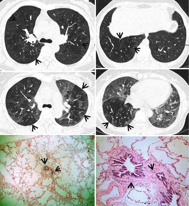
Fig. 13.1 Constrictive bronchiolitis in a 45-year-old woman. (a, b) Lung window images of thin-section (1.5-mm section thickness) CT scans obtained at levels of main bronchi (a) and liver dome (b), respectively, show patchy areas of mosaic perfusion (arrows) in both lungs. (c, d) Expiratory CT scans obtained at similar levels to (a, c) and (b, d), respectively, demonstrate air trapping (arrows) more clearly in both
lungs. (e) Low-magniÞcation (×40) photomicrograph of pathologic specimen obtained with surgical lung biopsy displays bronchiolar col- lagen-type Þbrosis inducing luminal narrowing (arrows) of a membranous bronchiole. (f) High-magniÞcation photomicrograph (×200) discloses Þbrous thickening of lamina propria between epithelium and muscularis mucosa (arrows)

Mosaic Attenuation |
119 |
|
|
Key Points for Differential Diagnosis
1.Regardless of its cause, when mosaic perfusion is present, pulmonary vessels in the areas of decreased opacity often appear smaller than vessels in relatively dense areas of the lung [11, 12]. This discrepancy can be quite helpful in distinguishing mosaic perfusion from patchy appearance of GGO. In patients with GGO, vessels usually appear equal in size throughout the lungs.
2.In patients with mosaic perfusion resulting from airway diseases, abnormally dilated or thick-walled airways may be visible in the relatively lucent lung
a
b
regions [2], and lobular areas of low attenuation are common [13]. Air trapping on expiratory scans is often helpful in conÞrming the diagnosis. Attenuation differences are accentuated scans obtained on expiration [14].
3.In patients with mosaic perfusion resulting from vascular diseases, areas of low attenuation are usually larger than lobules. In patients with mosaic perfusion occurring in association with chronic pulmonary embolism or pulmonary arterial hypertension, enlargement of the main pulmonary arteries may be visible.
c
Fig. 13.2 Cystic Þbrosis in a 24-year-old man who underwent lung transplantation. (a, b) Lung window images of thin-section (2.5-mm section thickness) CT scans obtained at levels of aortic arch (a) and main bronchi (b), respectively, show extensive areas of bronchiectasis and cellular bronchiolitis (arrowheads) in both lungs. Also note bilateral patchy areas of mosaic attenuation (arrows). (c) Coronal reformatted CT image (2.0-mm section thickness) demonstrates bronchiectasis
and cellular bronchiolitis in both lungs. Also note patchy areas of mosaic attenuation (arrows), in which oligemia (decreased caliber of vessels) Þndings are clearly visualized. (d) Gross pathology and photomicrographs at corresponding area indicated by bars of explanted lungs disclose pus in bronchiectatic or bronchiolectatic airways. In a portion of right lung, airway wall Þbrosis and resultant postobstructive airway dilatation are seen (open arrows)
120 |
13 Decreased Opacity without Cystic Airspace |
|
|
d
Fig. 13.2 (continued)
4.Characteristic CT vascular signs including webs or bands, intimal irregularities, abrupt narrowing, or complete obstruction of the pulmonary arteries with enlarged main pulmonary arteries enable the diagnosis of chronic pulmonary thromboembolism in patients with mosaic perfusion [15].
5.Data from electrocardiographically (ECG)-gated multidetector CT studies show that functional parameters such as right pulmonary artery distensibility, systolicÐdiastolic right ventricular outßow tract dimensions, and diastolic wall thickness can be measured with good interobserver agreement and used as reliable criteria for a diagnosis of pulmonary hypertension [16]. Mosaic lung perfusion is found signiÞcantly more often among those with
pulmonary arterial hypertension due to vascular disease than among those with pulmonary arterial hypertension due to cardiac or lung disease (74 % [17 of 23] vs. 8 % [3 of 38] of the patients in one series) [17].
6.The head-cheese sign is usually indicative of mixed inÞltrative and obstructive disease, usually associated with bronchiolitis. In patients with this appearance, the presence of GGO is caused by lung inÞltration, whereas the presence of mosaic perfusion with decreased vessels sign is usually caused by small airway disease. The combination of GGO on inspiratory scans and air trapping on expiratory scans is considered indicative of a mixed inÞltrative and obstructive disease such as hypersensitivity pneumonitis [14].