- •Adrenaline (again)
- •Adrenergic receptor agonists and antagonists
- •Acetylcholine receptors
- •Acetylcholine
- •Cholinergic receptor subtypes
- •Nicotinic receptors
- •Muscarinic receptors
- •Nicotinic receptors are ion channels
- •Architecture of the nicotinic receptor
- •Other ligand-gated ion channels
- •The 7TM superfamily of G-protein-linked receptors
- •Categories of 7TM receptor
- •Receptor diversity: variation and specialization
- •Binding of low-molecular-mass ligands
- •Calcium sensors and metabotropic receptors
- •Proteinase-activated receptors (PARs)
- •The adhesion receptor subfamily
- •Frizzled
- •Receptor–ligand interaction and receptor activation
- •A two-state equilibrium description of receptor activation
- •Receptor dimerization
- •Transmitting signals into cells
- •The receptor and the effector: one and the same or separate entities?
- •Mixing and matching receptors and effectors
- •Intracellular 7TM receptor domains and signal transmission
- •Adrenaline (yet again)
- •References

Signal Transduction
stalk,57 and a cysteine box.58 These appear to mediate cellular adhesion and for two members of this family, EMR2 and CD97, the cellular ligand has been identified as chondroitin sulfate, a sulfated glycosaminoglycan.59 Interestingly, it is uncertain whether signalling due to the subgroup of EGF-containing LNB-7TM receptors is conveyed through GTP-binding proteins, or even if it is mediated through the 7TM domain. For other LNB-7TM molecules that are likely also to mediate cellular interactions through their large extracellular domains, a role for GTP-binding proteins in signal transmission seems
likely.60 Included in this family are also the adhesion molecules that qualify as cadherins (see Figure 13.10, page 389).
Frizzled
Frizzled, the family of receptors for the Wnt family of ligands, plays a role in the regulation of cellular differentiation signal through a number of different routes only one of which, the canonical Wnt pathway, involves heterotrimeric G proteins (Go) (see page 426 and Figure 14.8, page 429).
Receptor–ligand interaction and receptor activation
A two-state equilibrium description of receptor activation
It is well recognized that ion channels are proteins that can exist in discrete states, typically ‘open’ or ‘closed’. For ligand-activated channels the open-state probability is increased enormously when the ligand (e.g. acetylcholine) is bound.
A similar two-state equilibrium applies to the activation of the 7TM receptors. However, since Langley’s first description of a ‘receptive substance’ towards the end of the 19th century,61 it has been generally accepted that activation requires the actual binding of an agonist. Fundamental to this thinking is that the extent of the biological response is determined by the law of mass action linking the stimulus and the reactive tissue (i.e. the ligand and the empty binding site). The binding of a ligand is understood to induce a change in receptor conformation, such that only in this state does it communicate with its affiliated GTP-binding protein.
Receptors may be activated in the absence of ligand
The activity of adenylyl cyclase does not decline to zero in the absence of stimulating hormones. Of course, it is difficult to be quite sure that stimulating hormones are fully excluded even when a system has been extensively washed and then doped with inhibitors. Even so, there always remains a residual
66

Receptors
low level of basal (or constitutive) activity. Now we know that this is for real. However, even if activating ligands are not absolutely required, the coupling of a receptor to the cyclase is obligatory. Quite simply, the unoccupied receptors themselves provide the necessary stimulus. For example, the synthesis of cAMP in Sf9 insect cells, which are normally unresponsive to catecholamines, can be enhanced by transfecting them with 2-adrenergic receptors. These cells now generate cAMP at a rate which is directly proportional to the level of receptor expression and they do this in the absence of any stimulating ligand62 (see Figure 4.24, page 118), though of course the activity of the system is greatly increased if catecholamines are provided. (We return to the matter of reconstituting the adrenergic receptor/cyclase system in Sf9 cells in Chapter 4.)
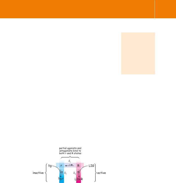
The activation of enzyme activity in the absence of stimulating ligands can be understood if the receptors exist in two conformational states, one of which can initiate downstream events (R), and the other cannot (r). The equilibrium between these two states exists regardless of the presence or absence of a stimulating ligand (Figure 3.18). Conventional agonists are those which bind to the active conformation (R) and as a result, increase the proportion of active receptors (R LIG.R). Conversely, there are‘inverse’agonists (lig) that bind selectively to
the receptor in its inactive conformation. These increase the proportion in the form (r lig.r) in which they are unable to transmit signals. Thus the number of active receptors is reduced and the activity of the system is suppressed below its normal basal state. In between the extremes of full (conventional) and inverse agonists are those agonists that bind to receptors in both the active and inactive conformations. These are the partial agonists. Such compounds are unable to achieve maximal stimulation of effector enzymes (e.g. of adenylyl cyclase) even when all the receptor binding sites are occupied, because they bind to both the activating and the inhibitory states of the receptor.
Fig 3.18 Receptor states and inverse agonists.
This diagram illustrates the equilibrium between the inactive (blue, r) and active (magenta, R) states of a receptor. Agonistic ligands (LIG) bind exclusively to the active form of the receptor and thus increase the proportion and the total amount (R LIG.R). Conversely, inverse agonists (lig) bind exclusively to the inactive form of the receptor and thus increase the total amount of the receptor (r lig.r) in the form that is incapable of transmitting signals.
Sf9 insect cells and the baculovirus transfection system are used as an alternative to bacteria. Being eukaryotic and moreover animal cells, newly synthesized proteins are posttranslationally modified as in other multicellular organisms.
67

Signal Transduction
The classification of drugs as agonists or antagonists, conventional, neutral, or inverse, is
a pharmacological minefield. In practice, many of the compounds in use in the clinic and in the laboratory, and depending on the circumstances, have activities that could enable them to be
classified both as agonists and antagonists. A further complicating factor is that the spectrum of activities varies from tissue to tissue. For the right atrium of the (rat) heart, in which2-adrenergic receptors play a greater role than
in other cardiac regions, it appears that almost all the beta blockers behave as inverse agonists.
Other diseases that might be amenable to treatment with appropriate inverse
agonists include inherited conditions such as retinitis pigmentosa, congenital night blindness (both due to mutations of rhodopsin), familial male precocious puberty, and familial hyperthyroidism. An inverse agonist active
at the receptor for parathyroid hormone, recognized as being constitutively activate in
Inverse agonism offers the potential of developing new drugs that attenuate the effects of mutant receptors that are constitutively activated. The neutral antagonists ( -blockers such as pindolol) bind to both active and inactive conformations and are better regarded as passive antagonists. These impede the binding of both agonists and inverse agonists.
Neutral antagonists prevent the stimulation of adenylyl cyclase by catecholamines, but they also oppose the inhibitory effect of inverse agonists. Examples of inverse agonists for -adrenergic receptors are propranolol and timolol, originally classified as -blockers (see Figure 3.2) and used as such in the treatment of glaucoma and hypertension. However, unlike the -blockade due to neutral antagonists, which mainly affects the heart during exercise and stress, these compounds also suppress the resting heart rate.63,64
Cell activation by over-expression of receptors
From this, one might imagine that if a receptor could be sufficiently overexpressed in a responsive tissue, the system would be fully activated even in the absence of a stimulating agonist. The equilibrium between the inactive and active species (r and R) would be unaltered, but the increased total would ensure a sufficient level of the active form to induce cell activation. Indeed, 200-fold over-expression of the 2-adrenergic receptor in the hearts of transgenic mice is sufficient to maximize cardiac function in the absence of
any stimulus.65 Under these conditions, no hormone is required to increase the level of active receptors to the point at which they can stimulate the tissue.
There are indications that receptor over-expression may play a role in the aetiology of some disease states, and this offers the prospect that inverse agonists could provide more specific therapeutic approaches than the currently available neutral antagonists. Indeed, some forms of schizophrenia may be associated with an elevation of the D4 (dopamine) receptor in the frontal cortex.66–68 Although the measured extent of the elevation is not large, about three-fold compared with controls, it is possible that this could be a reflection of much greater changes in limited focal regions, which could be of importance in determining the disease state. A number of compounds (raclopride, haloperidol) that find widespread application as antipsychotic drugs, appear to possess inverse agonist activity at dopamine receptors.69–71 Similarly, the action of progesterone in reducing oxytocin-induced uterine excitability, essential for the maintenance of pregnancy (see Chapter 2), is also best understood as the action of an inverse agonist rather than as a conventional inhibitor.72
Of course, there is one receptor that is expressed at a level out of all proportion to all others. This is opsin, the major element of the visual pigment rhodopsin. It is legitimate to ask by what mechanism the photoreceptor system ensures that dark really means dark, that we see nothing when the lights are turned off. Even if the equilibrium favoured the inactive state to a
68

Receptors
degree much greater than any hormone receptor, the presence of such an enormous amount of rhodopsin would surely elicit some activating response, however minimal. Yet, dark really does mean dark. This important question is discussed in connection with the mechanism of visual transduction in Chapter 6.
Receptor dimerization
It is increasingly apparent that many (if not most) 7TM receptors function not as monomers, but as dimers or oligomeric clusters. The first hints concerning oligomerization of -adrenergic receptors emerged more than a quarter of a century ago.74
The metabotropic GABAB receptor is incapable of transmitting signals unless two subtypes of the receptor (splice variants GABAB1 and GABAB2) are both expressed.75–77 They are understood to operate as a dimeric unit, regulating the activity of G-protein-activated K channel (GIRK). In the relevant neural tissues, both are present so there is no problem. However, successful reconstitution of K channel activity in cells that lack these receptors requires the expression of both subtypes. The GABAB receptor operates as a obligatory heterodimer (or larger oligomer). Only the B1 component binds the agonist,78 while onward signalling (to the GTP-binding proteins Gi and Go, see Chapter 4) is conveyed through the four intracellular segments of the B2 component.79 (These are the three intracellular loops and the C-terminal stretch.) Indeed, coexpression of GABAB1 and GABAB2 is not only a requirement for effective ligand binding and signal propagation, but is also a prerequisite for maturation and transport of the receptor to the cell surface.
Homodimers
It is likely that the GABAB1–GABAB2 heterodimer exists as a stable entity and is unaffected by the state of receptor activation. Other receptors, such as those for glutamate (mGluR580), vasopressin (V281), adenosine, opiates ( -opioid 82) and even the well investigated 2-adrenergic81 and muscarinic receptors may be linked as homodimers. For some of these at least, the monomer–dimer equilibrium appears to reflect the state of receptor activation.
Stimulation of 2-adrenergic receptors with catecholamines favours the formation of dimers81 while, conversely, the application of an inverse agonist, timolol, favours monomer formation. When isolated, the dimers remain stable even under the denaturing conditions applied for analysis by SDS gel electrophoresis. Of the 7TM helices, the F helix (Figure 3.16) may offer the site of intermolecular attachment determining dimer formation. A synthetic peptide based on the sequence of this segment, that is therefore expected to
impede the access of the second protein molecule, was found to reduce dimer formation.81 It also inhibits the activation of adenylyl cyclase by isoprenaline.
patients suffering from Jansen-type metaphyseal chondrodysplasia,
has recently been described.73
69
