


Receptors
Channels regulated by glutamate are the major excitatory receptors of the CNS. Their amino acid sequences and membrane topology are quite different from the Cys-loop proteins. They are tetrameric and their subunits possess only three transmembrane segments with the consequence that the N- and C-termini are expressed on either side of the membrane. In these receptors, the equivalent M2 segment that is presumed to line the channel is rudimentary.
It enters into the membrane and re-emerges on the same side (cytoplasm).26 ATP-gated channels differ yet again from the other families, possessing three homologous subunits, each of which has two transmembrane segments.
Just as acetylcholine interacts with two distinct forms of receptor, ionotropic (nicotinic) or metabotropic (muscarinic), so does GABA and glutamate. On the one hand, there are the metabotropic GABAB and glutamate (mGlu) receptors, which communicate through G proteins to activate PLC and mobilize intracellular Ca2 . On the other hand there are the ionotropic receptors GABAA, GABAC, and glutamate (iGlu), which are ligand-gated ion channels. Again, the ionotropic receptors and the metabotropic receptors for GABA and glutamate are antagonized by quite distinct inhibitors.
The three classes of ionotropic glutamate receptors are classified according to their susceptibility to inhibition by the synthetic glutamate analogues NMDA, AMPA, and kainate. For the NMDA receptor, opening of the non-selective cation channel requires glutamate (or aspartate) together with glycine (coagonist). If the glycine binding site is blocked (or unoccupied), glutamate alone is insufficient
to effect channel opening. The AMPA receptors, present on all neurons in the central nervous system, are responsible for the fastest transmission of excitatory synaptic responses.27 Opening of the cation channel requires the occupation of both agonist binding sites by glutamate, with relatively low affinity.
Glutamate appears to be involved in the pathogenesis of a number of severe diseases and condition such as stroke and epilepsy, due either to excessive release, reduced uptake, or altered receptor function. Antagonists at these receptors have been used as general anaesthetics (particularly in veterinary practice) and, as widely, as drugs of recreation (e.g. ketamine and phencyclidine).
The 7TM superfamily of G-protein-linked receptors
The muscarinic acetylcholine receptors and the GABAB, mGlu, and catecholamine receptors are members of the same protein superfamily. Their structures are closely related to the visual pigment rhodopsin and very distantly related to the bacteriorhodopsins, which constitute ion pumps in the membranes of certain archaeans, most prominently Halobacterium. The feature that relates all these structures is the topological organization of the
is that GABA acts either as an excitatory or as an inhibitory
neurotransmitter. During the day, when the concentration of Cl
in the SCN is in excess of about 15 mmol l 1, GABA enhances the firing rate (8–10 Hz) and causes excitation. At night, opening of GABAA channels allows an influx of Cl- which causes membrane hyperpolarization and a reduction in the firing rate (2–4 Hz).
NMDA is N-methyl- d-aspartic acid, AMPA is -amino-3- hydroxy-5-methyl-4-
isoxazolepropionic acid, and kainate, derived from algae, is 2-carboxy- 3-carboxymethyl-4- isopropenyl-pyrrolidine.
More than 50% of the pharmaceutical products currently on the market (or in development) are targeted at the 7TM class of receptors.28
55
Signal Transduction
Fig 3.11 Hydropathy plots of rhodopsin and the -adrenergic receptor.
The hydropathy plot of rhodopsin is shown on the left, the 2-adrenergic receptor on the right. For the derivation of the hydrophobicity see legend to Figure 3.8. The sequence data were taken from the Swissprot database, accession numbers P08100 and P07550.
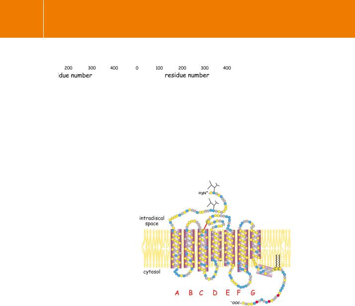
Fig 3.12 Membrane topology of bovine rhodopsin based on hydropathy data and X-ray diffraction analysis. The membrane organization of rhodopsin based on the hydropathy profile of its amino acid sequence (see Figure 3.11). The discs indicate hydrophobic residues (yellow), non-polar residues (grey), and polar residues (blue). The amino
acids comprising the seven transmembrane spans (A–G) are predominantly hydrophobic, though non-polar and even charged residues are not entirely absent. The residues making contact with the ligand (11-cis retinal) are indicated (single letter code). The incidence of polar and charged residues is much greater in the exposed loops and terminal domains. The residues shown as red discs are targets for phosphorylation by rhodopsin kinase.
From Ohguro et al.29
single peptide chain which in all cases traverses the membrane seven times. This is indicated by hydropathy plots, such as those shown in Figure 3.11 for rhodopsin and the -adrenergic receptor. Topological diagrams are depicted in Figure 3.12 and Figure 3.13. The membrane-spanning segments are linked by
56
Receptors
Fig 3.13 Membrane topology of the 2-adrenergic receptor.
The membrane organization of the 2-adrenergic receptor based on the hydropathy profile shown in Figure 3.11. The discs indicate hydrophobic residues (yellow), non-polar residues (grey) and polar residues (blue). The residues making contact with adrenaline are indicated (single-letter code). Residues shown as red discs are targets for phosphorylation by the 2-adrenergic receptor kinase (GRK2). The black discs indicate the serine residues (in the consensus sequence -RRSS-) that are targets for phosphorylation by PKA.
From Fredericks et al.30
three exposed loops on either side of the membrane, with the N-terminus projected to the outside and the C-terminus in the cytosol.
Although they share obvious features of structural organization,31 it is far from certain whether the archaean bacteriorhodopsins and the 7TM
receptor proteins of eukaryotes are derived from a common ancestor. The similarity might arise from convergent rather than divergent evolution. For the receptors and the visual pigment rhodopsin, the organization of the 7TM chains was deduced mainly by analysis of the hydropathy plots of the amino acid sequences. The other essential feature that relates the 7TM receptors and the visual pigments is that nearly all of them interact inside cells, with GTPbinding proteins. Possible exceptions are members of the subgroup of EGF module-containing adhesion receptors (see page 65).
We now have the crystal structure of bovine rhodopsin, and it is clear that the organization of the transmembrane chains and the linking loops is more or less as had been inferred.37 We may also be confident that other related 7TM receptors share the same basic structural organization. However, the idea that rhodopsin offers a predictive template for other 7TM receptors, for which structures are not yet available, may be over-optimistic.38 Indeed,
there are only a very few highly conserved residues in the whole superfamily, such as the two cysteines forming the disulfide bridge (see Figure 3.12).
Stop Press, 2008. Technical advances have recently enabled the crystallization of the 2-adrenergic receptor, and a highresolution structure has now been obtained.32–34 The receptor was
locked into its inactive configuration by the use of an inverse agonist. The overall conclusion is that rhodopsin and the2-adrenergic receptor share the main structural features with binding pockets for their ligands located deep within the transmembrane helices. However, the three extracellular loops of the receptor are clearly
structured and this might give a clue of how its diffusible ligands can gain access to their binding site (for brief reviews,
see Ranganathan35 and Sprang.36
57