 CHAPTER 19
328 IMMUNOTOXICITY
|
Stimulations |
|
Suppression |
|
Allergy |
|
Infection |
||
|
Immune |
|
|
|
|
System |
|
|
|
Autoimmunity |
|
Neoplasia |
||
Xenobiotic
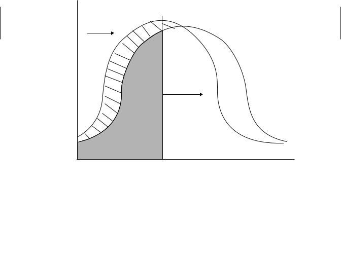
Figure 19.1 Potential consequences of immunotoxicity.
Table 19.1 Leukocytes
Granulocytes (polymorphonuclear leukocytes)
Neutrophils
Eosinophils
Basophils/mast cellsa
Monocytes
Lymphocytes
Monocytes/macrophagesa
Natural killer cells
a Found in blood/more activated form found in tissues.
cells, undergo differentiation and maturation in the thymus. Leukocytes circulate throughout the body in blood and lymph and populate other lymphoid tissues including the spleen, lymph nodes (scattered throughout the body), tonsils, and adenoids, as well as aggregates of lymphoid tissue in the lung, gut, and skin, which are referred to as bronchus-, gutand skin-associated lymphoid tissue (BALT, GALT, and SALT). Also immune cells can be recruited to almost any tissue in the body where there is injury or infection. Accumulation of leukocytes in tissues in response to injury is known as inflammation. Cytokines (e.g., interleukins, interferons, and chemokines), soluble mediators produced by immune cells as well as cells outside the immune system, control the maturation, differentiation, and mobilization of immune cells. Immune responses are divided into innate responses directed nonspecifically against foreign substances, and acquired responses directed against specific antigens. There is considerable interaction between these two types of immunity.
Innate immunity provides a rapid, although usually incomplete, antimicrobial defense. Granulocytes, natural killer cells, and macrophages are important mediators of innate immunity. Granulocytes have the capacity to phagocytize (engulf) infectious agents or other types of particles and to destroy or remove them from the tissue. They release a variety of soluble mediators that can kill invading organisms, increase vascular permeability, and recruit more leukocytes to the tissue. Natural killer cells are large granular lymphocytes that nonspecifically kill tumor and virus-infected cells. Macrophages are also phagocytic, can release chemotactic and cytotoxic cytokines, and, when activated, can kill tumor or virus-infected cells. Mediators released from


330 IMMUNOTOXICITY
in the response to subsequent exposures. IgE acts as a mediator of allergy and parasitic immunity. IgA is found in secretions such as mucous, tears, saliva, and milk, as well as serum, and acts locally to block entrance of pathogens through mucous membranes. IgD is mainly membrane bound on B cells. Little is known about the function of this isotype. It does not appear to have a unique role that affects host immunity.
A given B cell will form antibody against just one single antigen; however, during the lifetime of this cell, it can switch to make a different class of antibody. Isotype switching is mediated by T helper cells. B cells recognize two types of antigen: T- independent antigens, which activate the cell without T cell help (predominantly an IgM response), and T-dependent antigens, which required T cell help in order to activate B cells. Most antigens belong to this latter category. Antibodies that specifically recognize microbial antigens can, in combination with plasma proteins known as complement, lyse bacterial cells or neutralize virus. Also microbes complexed with antibody are more readily phagocytized.
T cells recognize antigen that is presented via an antigen-presenting cell (APC) such as macrophages or dendritic cells. APCs process and present short peptide fragments complexed with major histocompatibility (MHC) molecules on the surface of the APC. This processing and presentation is required for T cell activation. There are two major divisions of T cells that are distinguished by expression of different cell surface markers (CD4 and CD8). CD-4 cells are also know as T-helper cells because they provide help for B cell activation. CD-8 cells are also known as cytotoxic T cells because they lyse cells expressing specific viral or tumor antigens.
As indicated above the thymus plays a key role in T cell differentiation. Pre-T cells migrate from the bone marrow to the thymus. As relatively immature cells, T cells express both CD4 and CD8 molecules. As maturation progresses these cells undergo both positive and negative selection. During positive selection only cells that bind to MHC with a certain affinity survive. As a result of this process T cells become MHC restricted; that is, they will only respond to antigen presented in association with MHC. Cells that survive positive selection are potentially able to respond to self proteins. However, before T cells leave the thymus negative selection occurs during which self-reactive cells are removed or functionally inactivated. During the course of positive and negative selection CD4+ CD8+ cells down-regulate the expression of one of these molecules such that mature T cells express only CD4 or CD8. Mature T cells leave the thymus and populate secondary lymphoid organs.
19.3IMMUNE SUPPRESSION
Experimental studies in laboratory rodents have demonstrated that a diverse array of chemical exposures suppress immune function (Table 19.2). In addition a limited number of clinical and epidemiologic studies have reported suppression of immune function and/or increased frequency of infectious and/or neoplastic disease following exposure of humans to some of these agents. From the description above it is clear there are a number of cellular and molecular targets for chemicals that act as immunosuppressants. Clearly, a chemical that disrupts cell proliferation would affect clonal expansion. Disruption of T cell maturation in the thymus is another potential mechanism for immune suppression. Chemicals may also interfere with receptor ligand binding at the cell

IMMUNE SUPPRESSION |
331 |
Table 19.2 Selected Examples of Immunosuppressive Agents
Drugs
Cyclosporin A, cyclophosphamide, glucocorticoids (Dexamethazone), azothioprine
Metals
Lead, cadmium, methylmercury, organotinsa
Pesticides
Chlorodanea , DDTa , Dieldrina
Industrial compounds
2,3,7,8-Tetrachlorodibenzo-p-dioxin (TCDD), polychlorinated and polybrominated biphenyls (PCBs and PBBs), benzene, poly aromatic hydrocarbonsa
Addictive substances
Cocaine, ethanol, opiates, cannabinoids, nicotine
Air pollutants
Environmental tobacco smoke, ozone, nitrogen dioxide
Microbial toxins
Aflatoxin,b ochratoxin A,b trichothecenes T-2 toxinb
Radiation
Ionizing, UV
Other
Asbestos, diethylstilbestrol (DES), dimethylnitrosamine
a Effects in humans are unknown; for all other compound without superscripts changes have been demonstrated in both rodents and humans.
b Effects in humans unknown, but veterinary clinicians have noted immunosuppression in livestock ingesting mycotoxins at levels below those that cause overt toxicity.
surface and/or the cascade of signals that lead to transcription of genes responsible for generating and regulating the appropriate immune responses.
Because of the complexity of the immune system, tiered approaches to testing chemicals for immunosuppressive potential have been developed. Like other types of toxicity testing, the first level of the tier (Table 19.3) frequently relies solely on structural end points, including changes in the weight of thymus and other lymphoid organs, histopathology of these organs, or differential blood cell counts. This type of evaluation is convenient because it can be carried out along with an evaluation for other organ systems during routine toxicity testing using one set of animals. However, although these nonfunctional endpoints may be effective in identifying gross (high dose) immunotoxic effects, they are not very accurate in predicting changes in immune function or alterations in susceptibility to challenge with infectious agents or tumor cells at lower chemical doses. Hence the first testing tier (Table 19.3) often includes functional end points designed to assess (1) antibody-mediated responses, (2) T-cell-mediated responses, and (3) NK cell activity. The most commonly used immune function assay in laboratory animals assesses the ability of a mouse or rat to respond to challenge with an antigen, usually sheep red blood cells (SRBC) (Figure 19.3). The response is assessed by determining the number of antigen specific antibody (IgM)
332 IMMUNOTOXICITY
Table 19.3 Tier I Tests (Screen) for Immune Suppression Using Laboratory Rodents
Immunopathology |
Hematology: Complete blood count and differential |
|
Weights: body, spleen, thymus |
|
Histology: Spleen, thymus, lymph node |
Antibody-mediated immunity |
IgM plaque-forming cell (PFC) response to T cell-dependent |
|
antigen (e.g., SRBC) |
Cell-mediated immunity |
Lymphoproliferative response: T cell mitogens (Con A and |
|
PHA) Allogeneic mixed leukocyte response (MLR) |
Nonspecific immunity |
Natural killer (NK) cell activity |
Note: For details on specific assays see M. I. Luster et al., Fund Appl. Tox. 10: 2–19, 1988.
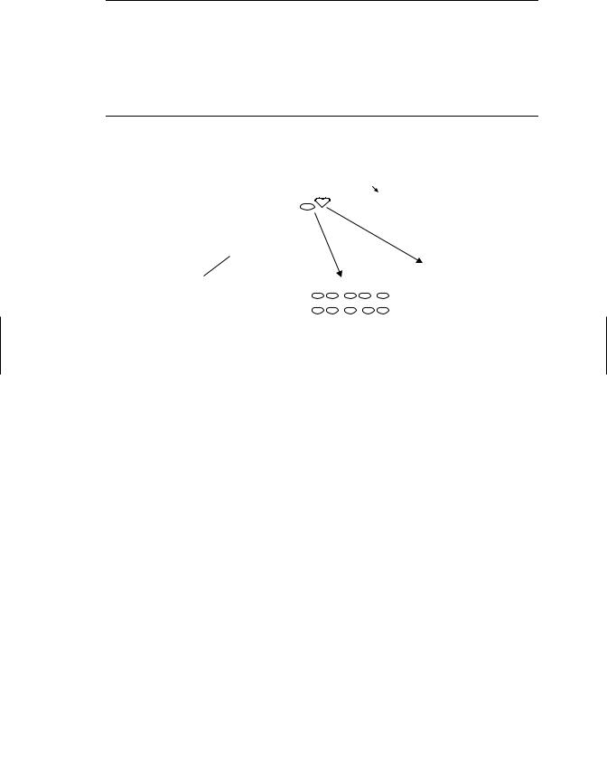
IgM Response to SRBCs
|
2. Remove |
|
|
|
spleen 4 |
2. Or draw |
|
|
days |
||
|
blood for |
||
1. Inject SRBC IV |
|
|
|
|
|
ELISA |
|
|
|
||
|
|
|
|
3. Assess antibody forming cells
Figure 19.3 Assessing chemicals for immunosuppressive effects. The most common approach to accomplish this goal is to inject chemical and vehicle treated mice or rats with antigen and assess the antibody response. Most often the antigen injected is sheep red blood cells (SRBC); four days later slides are made with a single cell suspension of spleen cells, sheep red blood cells, and complement immobilized in agar. Slides are incubated and spleen cells making antibody against SRBC lyse the surrounding RBCs generating plaques. Plaques are counted to determine the number of antibody forming cells. Alternatively, serum can be obtained and an ELISA assay performed to detect SRBC specific antibody.
forming cells (AFC) in the spleen (Jerne assay) or by assessing antigen specific antibodies in serum using an enzyme-linked immunosorbent assay (ELISA). Because the SRBC is a T-dependent antigen, T and B cells, as well as antigen presenting cells, must be functional to have a successful immunization. Suppression of this response is highly predictive of suppression of other immune function tests and also correlates well with tests that assess resistance to challenge with an infectious agent or tumor cells. The disadvantage to this test is that it usually requires a dedicated set of animals because of the antigen challenge. The most common approach has been to treat the animals for 14 to 28 days with the xenobiotic of interest, inject the antigen at the end of that exposure, and collect spleen or serum 4 to 5 days later. Unlike the tests for antibody-mediated immunity, tier 1 tests for cell-mediated immunity, and natural killer cell activity can be done ex vivo and do not require a dedicated set of animals. However, these tests focus on one cell type and are not as predictive of overall immunocompetence as the antibody assays.

|
IMMUNE SUPPRESSION |
333 |
Table 19.4 Tier II More Indepth Evaluation of Immunosuppressive Chemicals |
|
|
|
|
|
Immunopathology |
Quantitation of B and T cell numbers using flow cytometry |
|
Antibody-mediated immunity |
IgG PFC to SRBC |
|
|
IgM PFC to T cell-independent antigen (e.g., TNP-LPS) |
|
Cell-mediated immunity |
Cytotoxic T lymphocyte (CTL) cytolysis |
|
|
Delayed hypersensitivity response (DHR) |
|
Nonspecific immunity |
Macrophage: phagocytosis, bactericidal/tumoricidal activity) |
|
|
Neutrophil: function (phagocytosis and bactericidal activity) |
|
Host resistance models |
Response to challenge with infectious agent or tumor cells |
|
Note: For details on specific assays see M. I. Luster et al., Fund Appl. Tox. 10: 2–19, 1988.
When immunosuppressive effects are noted in tier 1, an in-depth evaluation using more sophisticated tests may be carried out (tier 2, Table 19.4). This might include enumeration of lymphocyte subsets (B cells, total T cells, and CD4+ and CD8+) using flow cytometry or assessment of the IgM response to a T-independent antigen in an effort to determine what portion of the immune response is the actual target. Unlike tier 1, tests of cell-mediated immunity in tier 2 require administration of an antigen and subsequent test for cytotoxic T cells (e.g., against an immunizing tumor cell) or a delayed type hypersensitivity response (similar to the response to a tuberculin test). In order to understand the mechanism’s underlying immune suppression, a host of other tests can be carried out, including expression of an assortment of cytokines.
Tier 2 also include host resistance models, tests in which an animal is exposed to a xenobiotic and then challenged with an infectious agent or tumor cells. This is considered the ultimate test for an adverse effect on the immune system. However, it should be noted that the amount of immune suppression that can be tolerated is greatly dependent on the dose and virulence of the challenging agent, as well as the genetics of the host. Manipulation of these variables can affect greatly results obtained in host resistance tests.
As in animal studies, human clinical data obtained from routine hematology (differential cell counts) and clinical chemistry (serum immunoglobulin levels) may provide general information on the status of the immune system in humans. However, as with the animal studies, these may not be as sensitive nor as informative as assays that target specific components of the immune system and/or assess function. The assessment of certain lymphocyte surface antigens has been successfully used in the clinic to detect and monitor the progression or regression of leukemias, lymphomas, and HIV infections, all diseases associated with severe immunosuppression. However, there is considerable variability in the “normal” human population, such that the clinical significance of slight to moderate quantitative changes in the numbers of immune cell populations is difficult to interpret. There is consensus within the immunotoxicology community that tests that measure the response to an actual antigen challenge are likely to be more reliable predictors of immunotoxicity than flow cytometric assays for cell surface markers because the latter generally only assesses the state of the immune system at rest. For ethical reasons it is not possible to immunize humans with SRBC. One approach under consideration is assessing responses to vaccines in chemically exposed populations. This approach has been used successfully to demonstrate a link between mild, stress-induced suppression of the antibody response to influenza vaccine and enhanced risk of infectious disease.