

16 INTRODUCTION TO BIOCHEMICAL AND MOLECULAR METHODS IN TOXICOLOGY
DYE INFLUX |
MEMBRANE LEAKAGE |
Trypan Blue |
Cytoplasmic LDH, Cr2− |
Propidium lodide |
|
|
Plasma Membrane |
ucleu |
|
N |
s |
Nucleus
↓DNA, RNA Synthesis
H+
|
|
|
|
|
|
yso |
|
|
|
|
|
|
|
|
|
L |
so |
|
|
|
|
|
|
|
|
|
|
m |
|
|
|
|
|
|
|
Neutral |
e |
||
|
|
|
|
|
|
|
|||
|
|
|
|
|
|
Lysosomes |
Red |
|
|
|
|
|
|
|
|
|
|
|
|
|
|
|
|
|
|
↓ Neutral Red |
|
|
|
|
|
|
|
|
|
Uptake |
|
|
|
|
|
|
|
|
|
Endoplasmic |
|
|
|
|
|
|
|
|
|
Reticulum |
|
|
|
|
|
|
|
|
↓ Protein |
|
|
|
|
|
|
|
|
|
|
Synthesis |
|
|
|
|
|
|
|
|
dria |
Mitochondria |
|
||
|
|
|
|
n |
|
|
|
|
|
|
|
|
o |
|
|
|
|
|
|
|
|
h |
|
|
|
|
|
|
|
|
c |
|
|
|
↓ MTT Reduction |
||||
o |
|
|
|
|
|||||
it |
|
|
|
|
|
||||
M |
|
|
|
|
|
↓ Rhodamine 123 |
|||
|
|
|
|
|
|
||||
|
|
|
|
|
|
Retention |
|||
|
|
|
|
|
ATP/ADP |
|
|
|
|
Microfilaments Cortical
BLEB
Figure 2.1 Idealized diagram of a cell to illustrate parameters often used to measure cytotoxicity and the corresponding affected subcellular organelle. (From An Introduction to Biochemical Toxicology, 3rd ed., E. Hodgson and R. C. Smart, eds., Wiley, 2001.)
evaluated by examination of end points that indicate effects on cellular organelles such as leakage of cell constituents into the medium, uptake of dyes into the cell and the formation of surface “blebs.” This is illustrated in Figure 2.1.
Longer term assessments of cell toxicity are highly dependent on the relevant toxic end point. They may include measurement of growth competence, apoptosis, and/or necrosis, incorporation of radioactive precursors into essential cellular constituents such as RNA, DNA, and protein and specialized cellular functions. Some examples of the use of cultured cell lines in the study of toxicity effects are shown in Table 2.1.
2.3MOLECULAR TECHNIQUES
Recombinant DNA techniques, including molecular cloning, have provided recent dramatic advances in many areas of both fundamental and applied biology, toxicology not excepted. Responses to toxicants may involve changes in gene expression and the new microarray techniques enable the simultaneous examination of the level of expression of many genes. The completion of the Human Genome Project will permit toxic effects in humans to be investigated and will facilitate extrapolation from experimental animals. The human genome will also provide the essential genetic background information for studies of polymorphisms in xenobiotic-metabolizing and other enzymes. Such polymorphisms have already been shown to be very important in individual sensitivity to clinical drugs and in the definition of populations and/or individuals at increased risk from particular toxicants. Chemically induced mutations, particularly in oncogenes and tumor-suppressor genes are important in chemical carcinogenesis. The

|
|
|
|
|
|
|
|
|
|
|
|
|
|
|
|
|
|
|
|
|
|
MOLECULAR TECHNIQUES |
17 |
||||
|
Promoter region containing |
|
|
|
|
|
|
|
|
|
|
|
|
|
|
|
|
|
|
|
|
|
|
||||
|
proximal and distal elements |
EXON |
INTRON EXON |
INTRON |
|
|
|
EXON |
|
||||||||||||||||||
|
and enhancers |
|
|
|
|
|
|
|
|||||||||||||||||||
|
|
|
|
|
|
|
|
|
|
|
|
|
|
|
|
|
|
|
|
|
|
|
|
|
|
||
|
|
|
|
|
|
|
|
|
GT |
AG |
|
|
|
|
|
GT |
AG |
|
|
|
|
|
|
|
|
||
5′ |
|
|
|
|
|
..ATG.. |
..TTT... |
|
|
..TAA.....AATAA |
|
3′ |
|||||||||||||||
|
|
|
|
|
|
|
|
|
|
|
|
|
|
|
|
|
|
|
|
|
|
|
|
|
|
||
|
DNA sense strand |
|
|
|
splice |
|
|
|
|
|
splice |
|
|
|
|
|
|
|
|||||||||
|
|
|
|
|
|
|
|
|
|
|
|
|
|
|
|
|
|
||||||||||
|
|
|
|
|
|
|
|
|
|
|
|
|
|
|
|
|
|
|
|
|
|||||||
|
|
|
cap site |
|
|
|
|
|
|
polyadenylation signal |
|||||||||||||||||
|
|
|
|
junctions |
|
|
|
|
|
junctions |
|||||||||||||||||
|
|
|
translation initiation codon |
|
|
|
|
|
|
|
|
translation termination codon |
|
||||||||||||||
|
|
|
|
|
|
|
|
|
|
|
|
|
|
|
TRANSCRIPTION |
|
|
|
|
|
|||||||
|
|
hnRNA |
|
|
|
|
|
GU |
|
|
|
|
|
GU |
AG |
|
|
|
|
|
|
AAAAAAA |
|
||||
|
|
|
|
|
|
|
|
|
|
|
|
|
|
|
|
|
|
|
|
||||||||
|
|
|
|
|
..AUG.. |
AG...UUU... |
|
..UAA....AAUAA |
|
||||||||||||||||||
|
|
|
|
|
|
|
|
|
|
|
|
|
|
|
|
|
|
|
|
|
|
|
|
|
|
||
|
|
|
|
|
|
|
|
|
|
|
|
|
|
|
PROCESSING |
|
|
|
|
|
|
|
|
||||
|
|
|
|
|
|
|
|
|
|
|
|
|
|
|
|
|
|
|
|
|
|
|
|||||
|
|
|
|
|
|
5' |
|
|
|
|
|
|
|
|
|
|
|
|
AAAAAAAA 3' |
|
|||||||
|
|
|
|
|
|
|
|
|
|
|
|
|
|
|
|
|
|
|
|
|
|
||||||
|
|
mRNA |
|
|
|
..AUG... |
..UUU....UAA...AAUAA |
|
|
|
|||||||||||||||||
|
|
|
|
|
|
|
|
|
|
|
|
|
|
|
|
|
|
|
|
|
|
|
|
|
|
||
|
|
|
|
|
|
|
|
|
|
|
|
|
|
|
TRANSLATION |
|
|
|
|
|
|||||||
|
|
|
|
|
|
|
|
|
|
|
|
|
|
|
|
|
|
|
|
||||||||
|
|
|
|
|
|
|
|
||||||||||||||||||||
|
|
Protein |
Amino terminus |
Met..........Phe.....Stop |
Carboxy terminus |
|
|||||||||||||||||||||
Figure 2.2 Transcription, mRNA processing, and translation. DNA sense strand is designated by bold lines, hnRNA and mRNA by thinner lines. Exons are shown as rectangles and introns as the intervening spaces between exons. (From An Introduction to Biochemical Toxicology, 3rd edition, E. Hodgson and R. C. Smart, eds., Wiley, 2001.)
ability to develop “knockout” animals lacking a particular gene and transgenic animals with an additional transgene is also proving important in toxicological studies.
Gene structure and any of the processes involved in DNA expression including transcription, mRNA processing and translation and protein synthesis (Figure 2.2) can all be examined by molecular techniques. In toxicology this may include toxic effects on these processes or the role of the processes in the mechanism of toxic action.
2.3.1Molecular Cloning
The basic principle of molecular cloning is the insertion of a DNA segment into a suitable vector. The vector is an autonomously replicating DNA molecule and the inserted DNA segment may be as large as a gene or a small as a few nucleotides. The vector containing the DNA is inserted into a cell such as yeast, where it can be replicated many times, and either the DNA or the expressed protein subsequently isolated (Figure 2.3).
2.3.2cDNA and Genomic Libraries
cDNA or genomic libraries are collections of DNA fragments incorporated into a recombinant vector and transformed into an appropriate host cell. In the case of cDNA libraries, the cDNAs complementary to all of the mRNAs in the tissue or cell sample are synthesized in a procedure using reverse transcriptase, before incorporation into the vector. With genomic DNA libraries the genomic DNA is digested, before cloning into the vector, with a restriction enzyme to produce an overlapping set of DNA fragments of some 12 to 20 kb.
18 INTRODUCTION TO BIOCHEMICAL AND MOLECULAR METHODS IN TOXICOLOGY
Eco-RI
|
|
|
|
|
|
|
|
A |
|
|
|
A |
|
|
|
|
|
|
|
|
|
|
|
|
|
|
A |
|
|
|
|
|
|
|
|
|
|
A |
|
|
|
|
T |
|
|
|
|
G |
|
|
|
T |
T |
|
|
|
|
|
|
|
|
|
|
|
|
|
|
|
|
|
C |
|
||
|
|
|
|
|
|
|
|
|
|
|
|
G |
|
|
|
|
|
|
|
|
C |
|
|
|
|
|
|
T |
|
Eco-RI |
|
|
|
|
|
|
|
|
|
|
|
AATTC |
G |
|
Amp R |
|
|
|
|
|
|
|
Amp R |
|
|
|
G |
CTTAA |
|
|
|
|
|
|
|
|
|
|
|
|
|
|||
O-RI |
|
|
|
|
|
|
|
|
O-RI |
|
|
|||
|
|
|
|
|
|
|
|
|
|
|
|
|
|
|
|
|
|
|
|
|
|
C |
G |
A |
|
|
|||
|
|
|
|
|
T |
|
|
|
|
|
||||
|
|
|
T |
|
|
G |
C |
|
|
A |
|
|
||
|
|
|
|
|
|
|
T |
|
||||||
|
|
A |
|
|
A |
T |
|
|
||||||
|
|
|
|
|
|
A |
|
|
||||||
|
|
|
|
|
|
|
T |
|
|
|
||||
|
A |
|
|
T |
A |
|
|
|
|
|
|
C |
|
|
G |
|
|
T |
|
|
|
|
|
|
|
|
|
T |
|
|
|
C |
|
|
|
|
|
|
|
|
|
A |
|
|
|
|
|
|
|
|
|
|
|
|
|
G |
|
||
|
|
|
|
|
|
|
|
|
|
|
|
|
||
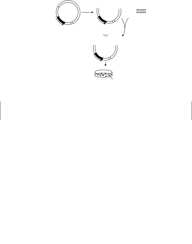
Amp R
O-RI
Transform into E.coli and select with ampicillin
Bacterial colonies containing plasmid DNA
Figure 2.3 Molecular cloning using a plasmid vector. (From An Introduction to Biochemical Toxicology, 3rd ed., E. Hodgson and R. C. Smart, eds., Wiley, 2001.)
These libraries are used in many screening procedures and many transgenic proteins now routinely available were obtained by their use. Although in some applications the use of cDNA and genomic libraries has been superceded by other methods, particularly those based on PCR, they are still used to advantage in many applications.
2.3.3Northern and Southern Blot Analyses
Northern analysis is usually used to identify and quantitate specific mRNAs in a sample. Southern analysis is used to determine whether or not a gene of interest is present as well as its copy number. Other uses for Southern analysis include identifying restriction fragment length polymorphisms and changes in heterozygosity.
In both Southern and Northern analyses restriction-digested DNA fragments, mRNA, and polyA mRNA are separated by size when electrophoresed on agarose gel. The separated molecules are transferred, by electroblotting or capillary blotting, on to a nylon or nitrocellulose membrane. The immobilized RNA or DNA is reacted with a radiolabeled, chemiluminescent, or fluorescent probe that is complementary to the DNA/RNA of interest, unbound probe is washed off, and the membrane exposed, in the case of radioactive probes, to radioautographic film to visualize the sample of interest.
2.3.4Polymerase Chain Reaction (PCR)
PCR is a powerful technique that can, starting with amounts of DNA as small as those found in single cells, amplify the DNA until large amounts are available for many