

Understanding the Human Machine - A Primer for Bioengineering - Max E. Valentinuzzi
.pdf138 |
Understanding the Human Machine |
fluid at 300 mOsm/L gets in shifting the whole column by a certain length. Again the active mechanism restores the osmolar difference (stage 4), but observe that the region of the bent (right side in the figure, right upper panel) begins to increase its concentration creating a difference with respect to the entrance. Successive fluid shifts followed always by the basic transversal osmolar gradient build-up plus the countercurrent flow give rise to a longitudinal much larger gradient between the entrance (at 300 mOsm/L) and the medullar region (reaching there 1,200– 1,400 mOsm/L). The mechanism is most interesting and ingenious, so much that it can be qualified as outstanding. A neat design of the Great Engineer, indeed! Rash (1984) simulated with a microprocessor this multiplying phenomenon, which may even have technological applications. With the present technology, such simulation should be easier and a real challenge for biomedical engineering students.
2.4.5.2. Countercurrent exchanger
The medullo-cortical concentration gradient, however, tends to equilibrate, as a ball placed on a ramp tends to roll down unless something is done either to prevent it or at least to partially brake it. Another mechanism is needed to conserve such gradient. The vasa recta of the peritubular capillaries that run parallel to Henle’s limbs take care of that function (Figure 2.53). Notice the specialization of the medullary nephrons. Blood gets into the vasa recta at 300 mOsm/L and, as it moves down in their descending branches, water traverses the capillary walls because of the osmotic pull from outside while active osmotic particles get into the blood, also due to a small transverse gradient. As blood goes up following the ascending limbs of the vasa recta the opposite shifts take place, that is, water gets into blood and solutes go out because at any level the ascending side has a slightly higher concentration than the descending one. Thus, the countercurrent exchanger reduces excessive loss of osmotically active solutes from the inner medulla. Blood in the vasa recta remove sodium and water. Loss of the medullocortical osmotic gradient would be disastrous for animal or human life.
This mechanism is similar to the countercurrent heater exchangers widely used in industrial plants, it has been amply studied both theoretically and experimentally and its better efficiency has been fully demonstrated as compared to heater exchangers with parallel streams in the
Chapter 2. Source: Physiological Systems and Levels |
139 |
same direction. Penguins have in their legs a circulatory arrangement of the same kind that helps them in keeping a temperature gradient from the trunk to the feet. Does it make the feet warmer? No, but the upper legs, the abdomen and the upper body better conserve body temperature. The extremities of the sloth also have the same circulatory arrangement, however, its function is unknown, especially if we recall that they live in tropical regions, like NE Brasil. Another stimulating subject for the inquisitive mind.
2.4.5.3. Osmotic exchanger
The distal convoluted tubules and the collecting ducts (more the latter than the former) regulate the final osmotic urine adjustment by the action or not of the antidiuretic hormone (ADH), which, as mentioned before, controls their wall permeability to water. Most of the water is recovered and only a minor amount is excreted, either as hypotonic or hypertonic urine (measured with respect to the plasmatic 300 mOsm/L), and depending on the hydration degree of the subject. Sodium is also recovered along this final pathway along with other ions (such as phosphate and bicarbonate). Thus, the final urine equilibrates with the hypertonic interstitium of the renal medulla and papillae.
The major osmotically active constituents of urine are sodium and chloride ions and urea. The osmolar clearance may be calculated from a formula derived from the clearance definition given above, that is,
Cosm = |
[Uosm ]V |
(2.100) |
|
[P |
] |
||
|
osm |
|
|
In this expression, [Uosm] represents the collected urine osmotic concentration, V the collected volume in a given period of time and the denominator stands for the plasma osmolarity. In words, it is defined as the volume of plasma per unit time completely cleared of osmotically active solutes or, also, as the osmolar excreted load per unit of plasmatic osmotic concentration. The interested student can find details and mechanisms in the literature.
2.4.6. Renal Blood Flow
Let us go back to Figure 2.51 writing now the continuity equations for nodes Gl and Ca,
140 Understanding the Human Machine
Φ[A |
]−F[P |
]−Φ'[A ]=0 |
(2.101) |
A |
x |
E |
|
Φ´[AE]−S[Kx' ]+R[Kx" ]−Φ[V]=0 |
(2.102) |
||
Solving for φ’[AE] the two previous equations, equating, and considering equation (2.88), the following is easily obtained,
Φ[AA ]−Φ[V ]= E[Ux ] |
(2.103) |
||
where φ stands for the renal plasma flow, so that, |
|
||
Φ = |
E [U x ] |
(2.104) |
|
[A A − V ] |
|||
|
|
||
or in words: The excreted load of a given substance divided by the renal arteriovenous concentration difference yields the renal plasma flow. If the test substance is fully extracted from blood in its passage through the kidneys, then, the venous concentration becomes zero. Besides, if the other tissues do not extract that substance from blood, the renal artery concentration will be equal to the concentration in any systemic artery or vein, that is, it will be equal to [Px]. With all this in mind, equation (2.104) simplifies to,
Φ = |
E [U |
x |
] |
|
|
|
|
(2.105) |
|
[P x ] |
|
|||
|
|
|
||
the latter coinciding with the plasmatic clearance of the substance (see clearance definition above). Para-aminohippurate is one substance that approximately meets the described conditions. As a consequence, the measurement of the plasmatic renal flow is relatively simple: After administration of PAH, a volume of urine is collected for a specific time span to have E. Immediately thereafter, the PAH concentration in urine is determined. From a venous blood sample, its PAH concentration is also determined. Finally, equation (2.105) is applied to obtain the renal plasma flow. Normal adult human kidneys yield a value of about 650 mL/min. Taking the hematocrit into account (arount 0.45), the renal blood flow becomes in the order of 1,200 mL/min, which is 20% of cardiac output.
Thinking exercise: Review the definition of hematocrit and the calculation that leads from plasma flow to blood flow. Do the kidneys really need such a large amount of blood? Check the percentage it represents relative to cardiac output.
Chapter 2. Source: Physiological Systems and Levels |
141 |
2.4.7. Closing Remarks of Section 4
The Renal System is essentially the complex exchanger shown as E4 in the block diagram of an organism shown earlier in the book (Figure 2.1), connecting it on one side with the Cardiovascular System and on the other with the external world to excrete the waste products. The metabolic needs of the kidneys do not justify the large perfusion they receive, rather this comes about because several times per day all the blood volume must pass through them in order to guarantee the adequate homeostasis of the internal environment. Most remarkable is the urine concentration (or dilution) process by which water and other substances are conserved: Mainly, the collecting ducts use a large medullo-cortical osmolar gradient. At every level of Henle’s loops ascending branches, a basic transversal difference of 200 mOsm/L is established by active sodium transport as compared to the descending limb. The ascending branch is impermeable to water, so preventing the immediate loss of that gradient. The basic gradient is longitudinally multiplied by the countercurrent flow of the intratubular fluid. The peritubular vasa recta also working in a countercurrent arrangement reduce losses and tend to conserve the medullocortical osmotic difference. Finally, the collecting ducts act as true osmolar exchangers controlled by the antidiuretic hormone. In this way, only a small volume of fluid is lost per day. Such fluid carries in solution different substances and electrolytes.
The mechanics and control of micturition have not been touched in this section. For the time being, they can be ignored. The interested reader will find good descriptions in any physiology textbook.
A person can survive and have a normal life with one kidney but life is incompatible without renal function. Thus, when we think in terms of renal failure, immediately we think of transplantation, dializers and eventually total kidney replacement by an artificial one. From the perspective of Biomedical Engineering, there is a lot to offer to the problems posed by the subject, from theoretical, physiological to technological aspects. It is an endless avenue.

142 |
Understanding the Human Machine |
2.5. Gastrointestinal System
Man is the only species using the GI system for pleasure!
2.5.1. Introduction
As indicated in Figure 2.1, the Gastrointestinal System (GIS) can be considered as an input-output physiological unit designed to supply “combustible” to the organism. Its objective is to transform (digest) the ingested food into simple nutritive substances that are to be transferred (absorbed) to the Cardiovascular System (CVS), which, as already described, distributes and delivers them to the tissues. Chewing and swallowing, digestion, absorption and defecation are all processes controlled
Food
M
S
IA
HA
SI
PV
HV
LI
L
R
Figure 2.54. SCHEMATIC OF THE ALIMENTARY CANAL. From top to bottom, left side: M, mouth; S, stomach; SI, small intestine; LI, large intestine; R, rectum. The intestinal artery IA vascularizes the SI in a complex network of capillaries that constitutes the first section of the GI system exchanger. Blood loaded with nutritive substances comes out of the intestinal capillaries via the portal vein PV, which enters the liver L after a short pathway. The liver receives also blood from the hepatic arteries HA. The hepatic capillaries or sinusoids form the second section of the GI system exchanger. Blood returns to the general circulation via the hepatic veins.
Chapter 2. Source: Physiological Systems and Levels |
143 |
by autonomic, nervous and hormonal mechanisms. Digestive glands act to provide moisture, lubrication, emulsification and enzymes for digestion of proteins, carbohydrates and lipids. In a simplified manner, the gastrointestinal tract (Figure 2.54) is a canal that,
a.receives food through the mouth;
b.receives secretions from the salivary glands, liver, pancreas, stomach and intestines to digest food;
c.absorbs the digested nutritive substances, especially at the level of the small intestine;
d.supplies the absorbed nutritive substances to the liver via the portal vein;
e.elaborates and stores other substances in the liver; and
f.excretes the residues.
The entire tract has an associated muscular mechanics, a vasculature and a complex regulatory system. The classic book by Davenport (1977) is a must for any interested student of the subject. Ganong’s textbook of physiology (1981) is also recommended in any of its many editions. Besides, there are several websites to visit, as for example http://www.e-gastrointestinal.com.
2.5.2. Mechanics
Movements can be felt and detected all along the alimentary canal. The experience of the hunger pangs just before dinnertime or the bowel noises, especially after having eaten certain gas producing foods (such as milk or soja beans), is well known by everybody. They are GI mechanical manifestations. Such movements are complex in nature and rather irregular and they are generated by the smooth muscles that cover the different GI structures. This musculature is controlled by the autonomic nervous system, by hormones, by local mechanisms (which, by and large, involve stretching due to the entrance of material into the gastric cavity or the intestinal lumen) and, also, originates in intrinsic automatism (that is, there are temporary pacemakers).
Basically, motility takes the form of two types of movements, that is, as segmental contractions and as peristaltic waves (peri and stalsis = contraction, from Greek). The former, mix and churn the intestinal content (chyme) while the latter essentially propel the chyme along the intestine. Segmental contractions are ring-like contractions that appear at fairly
144 |
Understanding the Human Machine |
regular intervals along the gut, then disappear and are replaced by another set of contractions in the segments between the previous contractions. When the intestinal wall is stretched, a deep circular contraction or peristaltic wave forms behind the point of stimulation and passes along the intestine toward the rectum at rates varying from 2 to 25 cm/s. Such response to stretch is called the myenteric reflex. Movement normally takes place in the aboral or oral-caudal direction; occasionally, and in pathological conditions, antiperistalsis (antidromic propulsion) can be seen.
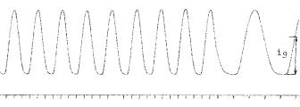
Figure 2.55 illustrates the spontaneous mechanical activity of a piece of rabbit small intestine, placed in a thermic physiological bath, and attached to a sensitive isotonic transducer. These contractions, as shown in the figure, are usually regular in amplitude, frequency and pattern, allowing the recording of rather long strips. In this particular case, there was a pacemaker activity of about 20 periods/min. Such intrinsic automaticity may vanish or may be enhanced (for example, by pharmacological stimulation) decreasing in the oral-caudal direction (Alvarez Law). The latter means that always a sample from either the duodenum or the jejunum or the ileum will show a higher spontaneous frequency than a sample from lower regions, as, say, the large intestine (ascending, transverse or descending colon).
Figure 2.55. RABBIT ILEUM ACTIVITY. Spontaneous contractions of a small intestine sample (ileum) immersed in an adequate physiological solution, temperature controlled, and with gentle air bubbling. The maximum force of contraction was about 1.75 g and very regular. The pattern was also regular and quasi-sinusoidal. Lower marks are 1 s apart. To the right of the record, paper speed was increased to better visualize the waveform. An isotonic transducer attached to one end of the sample while the other end was anchored to the holder longitudinally detected these contractions. Thus, they cannot be identified either as segmental or peristaltic. Records obtained by the author at the Department of Bioengineering, UNT, 1980.
Chapter 2. Source: Physiological Systems and Levels |
145 |
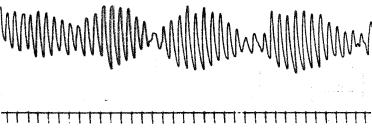
Figure 2.56. BEATING EFFECT FROM A RABBIT ILEUM SAMPLE: The sample was about 2 cm in length; thus, it probably caught two pacemakers with slightly different intrinsic frequencies (recall Alvarez Law: frequency decreases towards the tail, hence, the caudal portion of the segment should show a slightly lower activity than the upper one). The net result was the beating effect as manifested by the slow modulation. Time marks are 5 s apart. The carrier had a frequency of about 15 cycles/min while the beat had a very low 1.5 cycles/min. Records obtained by the author at the Department of Bioengineering, UNT, 1980.
Observe the sinusoidal-like waveform of the record shown in Figure 2.55, a rather interesting feature that can be explored a little further. When the sample is too short (say, less than 1 cm), no activity is detected because no pacemaker site is in it; conversely, if the sample is longer than 1 cm and about 2 cm, a waveform resembling the beating effect of two near sinusoidal frequencies can be produced (Figure 2.56). This phenomenon is typical of coupled systems, which can be modeled with a second order differential equation. If the sample is even longer, then more than two pacemakers may be picked up and a complex waveform will be the result.
Historical tip: Bayliss and Starling, in l899–l901, formulated the “Law of the Intestine” (not to be confused with Alvarez Law) to provide an explanation for peristalsis. They found that the response of the small intestine to a local stimulus consisted of contraction of the muscularis externa layer immediately above, and relaxation immediately below the point of stimulation. It was attributed to a reflex that involved the myenteric plexus and was independent of the external innervations of the intestine. Cannon, in l911, called it a “myenteric” reflex, as referred to above; the biphasic wave of relaxation and contraction was found to pass over the muscular layer in an aboral direction for short distances from the point of stimulation. Later workers, Alvarez among them, in l940, and Bozler, in l949, improved these studies.
There are longitudinal and circular smooth muscles all along the intestinal walls. Contractions of the former and of the latter musculature during
146 |
Understanding the Human Machine |
peristalsis are out of phase by 90 degrees, according to Davenport. On distension of the lumen the longitudinal muscle contracts, followed by progressive contraction of the circular layer. The circular layer begins contraction when contraction of the longitudinal layer is half complete; contraction of the circular layer is complete when relaxation of the longitudinal is half complete. Peristalsis is defined rather superficially in many scientific and medical dictionaries. The consensus appears to be that it is a vermiform or a progressive, wave-like movement in tubular organs, consisting of alternating waves of relaxation and contraction in the muscular coat by means of which the contents are propelled. More details can be found in the website med.plig.org/index.html (look for The Pyloric Sphincteric Cylinder in Health and Disease, by A.D. Keet).
Contraction of a smooth muscle cell is associated with change in potential of the cell membrane; the potential depends on the distribution of electrolytes between the cell and the extracellular space, much like in skeletal muscle. Two main types of spontaneously arising changes in the membrane potential may be detected, i.e., slow potential variations and spike potentials. The latter are superimposed on the former and are associated with mechanical activity. Thus, the electrophysiology of the gastrointestinal tract appears as another research field still with many unresolved problems and unknowns.
2.5.3. Secretions
Many substances are secreted into the alimentary canal. Some are hormones (which are internal secretions, that is, they get into the circulatory stream). The first to be found is saliva. The salivary glands in the mouth produce about 1,200 mL/day of saliva (an external secretion) at a pH of 6 to 7, that is, from acid to neutral.
The stomach secretes a hormone, gastrin, whose principal effect is to stimulate the production of pepsine (an enzime) and of gastric juice, both from the stomach itself. They are exocrine secretions, i.e., they do not get into blood. Gastric juice contains a variety of substances, it shows a very low pH (from 1 to 3.5, which means high acidity, due to the predominance of hydrochloric acid, HCl), and is secreted by parietal cells at a rate of about 3,000 mL/day. Such high acid level should damage the gastric wall, however, the surface membrane of the mucosal cells and the tight junction between cells seem to act as a protective barrier. Sub-
Chapter 2. Source: Physiological Systems and Levels |
147 |
stances that tend to break the barrier (such as aspirin or vinegar) may lead to gastric irritation, from mild to severe. Vagal activity also stimulates acid secretion from the stomach cells. This perhaps explains the rather common and so-called heartburns (upper gastric burning sensation) reported by worried or overstressed persons who may show an increased tone of the vagi.
As many as 33% to 44% of Americans experience heartburn at least once a month and up to 13% have heartburn each day. The likelihood of having heartburn increases with age and among women who are pregnant. Having heartburn every once in a while is something almost everyone experiences, but if it occurs 2 or more days per week, it can be a sign of a
more serious problem called gastroesophageal reflux disease.
The small intestine secretes two hormones, secretin and cholecystokininpancreozymin (CCK-PZ). They both act on the exocrine pancreatic function stimulating the secretion of pancreatic juice (1,200 mL/day, pH = 8.0 to 8.3, rich in enzymes to break up carbohydrates, proteins and lipids). Besides, CCK-PZ stimulates the contraction of the gall bladder to inject bile (produced by the liver) into the duodenum. Bile is temporarily stored in the gall bladder. There is a production of about 700 mL of bile per day at a constant pH of 7.8, that is, it is on the alcaline side.
Secretin was the first hormone ever found, discovered by Bayliss and Starling, in 1902. It causes the secretion of a watery, alcaline pancreatic juice. Its action on the duct cells of the pancreas is mediated by cyclic AMP. This hormone is produced by cells located deep in the glands of the mucosa of the upper portion of the small intestine.
Suggested reading exercise: Search for information about cyclic AMP (adenosine monophosphate). Find the definition of enzyme. The are extremely important substances.
A stimulating factor was isolated in crude form from hog intestines by William Maddock Bayliss and Ernest Henry Starling (the latter is the same of the law of the heart), very early in the XXth Century. These scientists, realizing the messenger role of a chemical substance, coined a new word — hormone (which means “I move” in Greek) — and called it secretin. It turned out, however, to be rather difficult to achieve its isolation from gut mucosa in pure form. The pure hormone was finally obtained about fifty years later by Jorpes and Mutt of the Karolinska Institute, in Stockholm in 1961. The same group was also successful in determining the sequence of the twenty-seven amino acids constituting the peptide chain of secretin.