

Micro-Nano Technology for Genomics and Proteomics BioMEMs - Ozkan
.pdf102 |
MIHRIMAH OZKAN ET AL. |
[140]J. Wang, G. Rivas, X. Cai, E. Palecek, P. Nielsen, H. Shiraishi, N. Dontha, D. Luo, C. Parrado, M. Chicharro, P.A.M. Farias, F.S. Valera, D.H. Grant, M. Ozsoz, and M.N. Flair. Anal. Chim. Acta, 347:1, 1997.
[141]J.V.A. Watson, S.H. Nakeff, H. Chambers, and P. Smith. Cytometry, 6:310, 1985.
[142]H.J. Watts, D. Yeung, and H. Parkes. Anal. Chem., 67:4283, 1995.
[143]C.J. Weijer. Science, 300:96, 2003.
[144]R. Wegeroff. From Membrane to Mind, 1st ed., Thieme, Stuttgart, p. 10, 1997.
[145]E. Wilkins and P. Atanasov. Med. Eng. Phys., 18:273, 1996.
[146]B.C. Wheeler, J.M. Corey, G.J. Brewer, and D.W. Branch. J. Biomech. Eng., 121:73, 1999.
[147]F.S. Wouters, P.J. Verveer, and P.I.H. Bastiaens, Trends Cell Biol., 11(5):212, 2001.
[148]C. Wyart, C. Ybert, L. Bourdieu, C. Herr, C. Prinz, and D. Chattenay. J. Neurosci. Meth., 117(2):23, 1997.
[149]M.Yang, S. Prasad, X. Zhang, A. Morgan, M. Ozkan, and C.S. Ozkan. Sens. Mat. (accepted)
[150]G. Zeck and P. Fromherz. P. Nat’l. Acad. Sci. USA, 98(18):10457, 2001.
[151]J.R. Zysk, and W.R. Baumbach. Comb. Chem., 1:171, 1998.
4
Cell Physiometry Tools based on Dielectrophoresis
Ronald Pethig
School of Informatics, University of Wales, Dean Street, Bangor, Gwynedd LL57 1UT, UK
Keywords: Cell-based assays; cell separation; dielectrophoresis, microelectrodes.
Dielectrophoresis is a technique for moving cells and other particles using radiofrequency electric fields. The usefulness of this method depends on the ability to generate highly non-uniform electric fields using microelectrodes, and also on the intrinsic dielectric properties of the cells and their surrounding medium. Selective cell isolation or concentration can be achieved without the need for biochemical labels, dyes or other markers and tags, and the cells remain viable after this process. Changes in cell state, such as those associated with activation, apoptosis, differentiation, necrosis, as well as responses to chemical and physical agents for example, can be monitored by observing changes in dielectrophoretic behavior. The basic theories and experimental techniques of dielectrophoresis are described in this chapter, and a summary is given of our present understanding of how the dielectrophoretic behavior of cells relate to their physiological and physico-chemical properties.
4.1. INTRODUCTION
Although dielectrophoresis (DEP) can be considered to be a mature subject (with publications extending back more than 50 years), it has until recently mainly been a topic of interest for a relatively small number of engineers and biophysicists. There is now, however, a growing interest in exploiting the modes of interaction of electrokinetic forces with biological particles (such as cells, bacteria, viruses, proteins and DNA) with the objective of addressing key opportunities in medical diagnostics, drug discovery, cell therapeutics
104 |
RONALD PETHIG |
and biothreat defence. It has been demonstrated that DEP is capable of selectively isolating, concentrating, or purifying target bioparticles when present in complex mixtures. Examples include the isolation of stem cells, cancer cells and bacteria from blood for therapy or further analysis. DEP also lends itself readily to miniaturization and automation, either as stand-alone microdevices or as the means for rapid and efficient sample collection and preparation.
The purpose of this chapter is to provide, for interdisciplinary biomedical scientists, a top level description of how dielectrophoresis (DEP) can be used to manipulate and characterize cells and other bioparticles. The term manipulate signifies the selective separation, fractionation, enrichment, concentration, assembly or positioning of cells. Although the method may be used as part of a process to achieve genetic manipulation (e.g., positioning cells prior to electrofusion or electroporation) DEP acting alone cannot perform this function. The term characterize refers to those physico-chemical or physiological properties of bioparticles that have so far been found amenable to DEP investigation. An objective here is to outline our understanding of how the DEP properties of cells may relate to their cellular states of function, viability and differentiation, or serve as a means of phenotyping.
In line with the objective to give a top level description, a comprehensive review of dielectroophoresis is not attempted here. The relevant methodologies and theories are stripped down to bare essentials. Readers interested in further details can usefully refer to monographs [41, 47, 59, 66, 79] and recent reviews [25, 42, 68, 69]. The methods used in the microfabrication of devices are not described at all here, but many of the referenced papers contain such details. Finally, as the term dielectrophoresis implies, we are dealing with cells being carried as a result of their dielectric properties. There is an extensive literature on the dielectric properties of cells and other biological materials (e.g., tissue, axons, bacteria, DNA, proteins, amino acids) extending back to the early 1900’s. The quickest way into this literature is through the monographs of Cole [14], Hasted [31], Grant et al [28], Pethig [66], Takashima [96] and Grimnes & Martinsen [29].
4.2. DIELECTROPHORESIS
Dielectrophoresis (DEP) is defined [78, 79] as the translational motion of neutral matter caused by polarization effects in a nonuniform electric field. This effect was observed by the ancient Greeks (e.g., Thales of Miletus, 600 B.C.) in the action of ‘animated’ amber attracting small particles. Polishing a piece of amber with cloth creates electric charges on its surface, and the resulting electric field can in turn polarize nearby dielectric particles into small electrets. These electrified particles (characterized by an induced electric dipole moment) can then be attracted to regions of high electric field strength on the amber surface. This is analogous to magnetophoresis, commonly observed as the attraction of iron filings to a magnet.
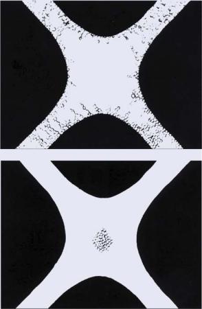
The dielectrophoretic collection of particles at electrode edges is shown in figure 4.1(a). An effect not possible by magnetophoretic manipulation of iron filings, namely their repulsion from a magnet followed by concentration in an aqueous medium, is shown in figure 4.1(b). The ability to attract or repel particles from electrodes is an important aspect of DEP. The translational forces producing these effects arise from the interaction of the
CELL PHYSIOMETRY TOOLS BASED ON DIELECTROPHORESIS |
105 |
(a)
(b)
FIGURE 4.1. Top (a): Yeast cells collecting at electrode edges under the influence of positive dielectrophoresis. Bottom (b): Yeast cells directed away from electrode edges, into a field cage, under the action of negative dielectrophoresis. The polynomial microelectrodes used here [33] are based on isomotive electrodes used to characterize macroscopic objects [80].
particle’s dipole moment m (permanent or induced) with the nonuniform electric field. This force is given by:
F = (m. )E |
(4.1) |
where is the grad vector operator defining the gradient of the local electric field E (rms or D.C. value). The dipole moment m induced in a particle, of volume v, is given by:
m = pv E |
(4.2) |
where p is the effective polarizability (per unit volume) of the particle, generally referred to as the Clausius-Mosotti factor (e.g., [17, 90]).

106 |
RONALD PETHIG |
From (4.1) and (4.2) we can write the translational (DEP) force as:
F = pv(E. )E = |
1 |
|
2 pv |E|2 |
(4.3) |
This well known result in electrostatics (e.g., [1], p. 91) can be interpreted as the energy which must be expended to withdraw the particle from the local field E into a region where there is no field. Eqn. 4.3 provides several important facts, namely that the DEP force is zero if the field is uniform (i.e., for E = 0), and the force depends on:
The polarizability of the particle
The effective volume of the particle
The square of the applied electric field magnitude
The geometry of the electrodes producing the nonuniform field
The polarizability of a particle is a sensitive function of its physico-chemical properties and structure. This means that particles can be selectively manipulated as a result of their own intrinsic properties, without the need for biochemical labels, beads, dyes or other markers and tags. The dependence on particle volume indicates that we are dealing with a ponderomotive effect—with all other factors remaining constant the larger the object the greater will be the DEP force acting on it. Indeed, for objects the size of a pebble or pencil, the DEP force can be measured using a conventional weighing balance [60, 80]. The term effective volume reminds us that the dominant polarizabilities of small particles (e.g., proteins, DNA, viruses) are often dominated by relaxations of their electrical double layers whose total volume can be comparable to or greater than the particle. The dependence on the square of the field emphasizes that the DEP force is independent of the polarity of the applied field—dielectrophoresis can thus be observed using either A.C. or D.C. fields. The range of frequencies that can be employed is large, extending from around 100 Hz or less, up through radio wave frequencies and well into the microwave region (100 MHz and above).
The strong dependence on electrode geometry arises because the factor (E. )E in eqn. 4.3 has dimensions of Volt2/m3. This instructs us that the same DEP force can be produced using a smaller applied field if the electrode dimensions are scaled down accordingly. For example, a hundred-times smaller field can produce the same effect if the electrodes are scaled down one thousand-fold (e.g., ten-fold reduction in size in each dimension). This has important implications for minimizing Joule heating effects when using DEP to characterize and manipulate cells and other biological particles in aqueous media. This was the reason, in our laboratory, to explore the use of microelectrode arrays fabricated by photolithography [81] and then excimer laser micromachining [72]. As discussed by Pethig and Markx [69] a value for the factor (E. )E of around 1013 V2/m3 is required to produce a significant DEP force, and this can be achieved using microelectrodes with applied voltages of less than 10V, corresponding to an applied field of less than 10 kV/m.
Care has been taken above to emphasize the expression ‘applied field’ rather than ‘applied voltage’. Electrode polarization effects commonly influence dielectric experiments, and are manifested as voltage drops across electrical double layers that form at electrodesolution interfaces. Electrode polarization therefore reduces the effective field in the bulk solution, and along with it a reduction of the DEP force. Electroosmotic fluid flow is

CELL PHYSIOMETRY TOOLS BASED ON DIELECTROPHORESIS |
107 |
also induced at the electrode-solution interface, and this can either impede or enhance the desired DEP manipulation of particles. The electrode polarization impedance has a
ω−β β =
frequency dependence of , where 0.7 0.9 (e.g., [4]) and so its effect increases with decreasing frequency. As a rough working rule, for solutions of conductivity 100 mS/m and lower, electrode polarization effects become apparent at frequencies below around 10 kHz. This can either be corrected by determining the magnitude of the electrode polarization (e.g., [9]) or the effect can be reduced by the well known method of applying platinum black to the electrodes (e.g., [37]). Electroosmotic fluid flow effects become increasingly important as the dimensional scale of a DEP device is reduced below the micron range (e.g., [8, 59, 82]).
4.3. DIELECTRIC POLARIZABILITY OF BIOPARTICLES
Some biological particles (e.g., proteins, peptides) possess permanent dipole moments, whereas others (e.g., DNA, RNA) do not, but can have large dipoles induced in them as a result of counter-ion displacement effects [69]. Other bioparticles (e.g., viruses, bacteria, cells, yeast, parasites) are assumed to possess no permanent dipole moment, and the accepted method of understanding their dielectric and dielectrophoretic behavior is to treat them as particles having a multilayered (multi-shell) structure. Each concentric layer represents a constituent component, such as a membrane, cell wall, cytoplasm, nucleus, vacuole, etc. [45, 47, 67, 86, 95].
When cells are subjected to an electric field, charges are induced to appear at the interfaces defining their gross structure. The induced charge density can be calculated [67] as typically some three decade orders of magnitude smaller than the uniformly distributed net surface charge that exists ( −1µC. cm−2) on the surface of cells and micro-organisms. The important fact is that the induced charge is not uniformly distributed about the cell surface, but is distributed so as to form an effective dipole moment. An alternative approach is to quantify this effect in terms of the electromagnetic momentum balance via the Maxwell stress tensor ([1], pp. 104–108; [87]). The effective dipole moment approach leads to the same phenomenological results and is more ‘user friendly’. Jones [47] provides an excellent treatment of the effective dipole moment method for understanding DEP and related phenomena.
4.4. DYNAMICS OF INTERFACIAL POLARIZATION
One of the important factors controlling the frequency-dependent behavior of DEP is related to the dynamics of the interfacial charging effect. This effect is known as MaxwellWagner interfacial polarization, formulated by Maxwell [56] and Wagner [100]. The induced charge is generated with a characteristic relaxation time τ given by:
τ |
= |
εp + 2εm |
ε |
o |
(4.4) |
|
σp + 2σm |
|
|||
where εo is the permittivity of free space, and εm and σm are the relative permittivity and conductivity of the suspending medium, respectively. The parameters εp and σp are the

108 |
RONALD PETHIG |
effective relative permittivity and conductivity of the particle, respectively. The term effective is used to signify that a heterogeneous (multi-shell) particle may be replaced conceptually with one having homogeneous smeared-out bulk properties, such that substitution of one particle with the other would not alter the electric field in the surrounding medium. For red blood cells, suspended in an aqueous electrolyte, eqn. 4.4 yields values for τ of about 0.1 µs, signifying the presence of a dielectric dispersion centered at a frequency (2πτ)−1 of about 2 MHz.
Various formulations of the polarization dynamics are given in the literature, and one that leads to simple insights into frequency-dependent behavior gives the following expression for the interfacial (Maxwell-Wagner) polarizability p(mw) of a spherical particle [44]:
|
|
ω2τ 2 |
|
ε |
εm |
|
|
|
|
|
1 |
|
|
σp |
σm |
|
|
||
p(mw) = εoεm |
|
|
|
|
|
p − |
|
+ |
|
|
|
|
|
|
|
− |
|
(4.5) |
|
1 |
+ |
ω2 |
τ 2 |
εp 2εm |
1 |
+ |
ω2 |
τ 2 |
σp |
2σm |
|||||||||
|
|
|
|
|
|
+ |
|
|
|
|
|
|
|
|
+ |
|
|
||
where ω is the radian frequency (2πf) of the applied field. For low values of the frequency (ωτ 1) eqn. 4.5 reduces to:
p(mw) ≈ εoεm |
σpp −2σm |
(4.6) |
|
|
σ |
σm |
|
+
and at high frequencies (ωτ 1) we have:
p(mw) ≈ εoεm |
εpp −2εm |
(4.7) |
|
|
ε |
εm |
|
+
The low frequency DEP force arising from interfacial polarization thus depends on the conductive properties of the particle and suspending medium, whilst at high frequencies the permittivity values are important. At intermediate frequencies, both the conductive and dielectric properties of the medium and particle dictate the magnitude and polarity of the DEP force. The low frequency polarization stabilizes when continuity of electric current densities are established across the various interfaces that define the particle structure and its immediate environment. This involves the movement of free ions around and through the particle, and thus the particle size and its effective surface area are important factors. At a high frequency, which in the time-domain corresponds to the initial dielectric response of the particle to an imposed electric field, the system strives to attain continuity of so-called displacement flux densities. This involves perturbations of bound charges at the molecular scale, and so is not controlled by the physical size of the particle. It is important to remember that other polarizations (e.g., double layer relaxations, considered in the next section) can contribute significantly to the low frequency DEP response of particles. Eqn. 4.5 does not encompass such additional polarizations.
If the polarizability factor p(mw) is positive (i.e., the particle is more polarizable than the surrounding medium) then the DEP force is positive and the particle is directed towards high field regions at electrode edges (For the range of frequencies and electrode dimensions commonly used in DEP applications, the field maxima are always located at electrode surfaces). If p(mw) has a negative value, the DEP force is negative and the particle is directed towards field minima away from the electrode edges. These two effects are shown in

CELL PHYSIOMETRY TOOLS BASED ON DIELECTROPHORESIS |
109 |
Figure 4.1. The concept shown in this figure, of directing and assembling colloidal particles into field cages under the action of negative DEP, has been significantly extended using 3-D electrode arrays [19, 21, 61, 88]. The continuous separation of particle mixtures can be achieved using synchronized pulses of fluid flow and electrode energization to selectively move particles into and out of positive and negative DEP traps [71] or using DEP combined with field flow fractionation [7, 37, 84] or electrokinesis [15]. Extruded quadrupolar traps [99], ‘zipper electrodes’ [32], high-density electrode arrays [16] and cell manipulation on a CMOS chip [51] have been described.
In DEP studies of mammalian cells (e.g., blood cells, cancer cells, stem cells) the suspending medium commonly takes the form of a low conductivity (10 180 mS/m) electrolyte containing sufficient concentrations of sugars (e.g., mannitol, sucrose, dextrose) to raise the osmolarity to the normal physiological level of around 280 mOs/kg. For viable cells the plasma membrane acts as an electrical insulator to passive ion conduction, and thus for frequencies below about 25 kHz the cell will appear as an insulating object suspended in a conducting medium. This corresponds to the situation σp < σm, so that the polarizability p(mw) given by eqn. 4.6 will be negative and viable cells will exhibit negative DEP at the lower frequencies. Physiological electrolytes typically have a conductivity and relative permittivity of the order 1.4 S/m and 79, respectively. For frequencies below around 1 MHz, the relative permittivity of the cytoplasm will be greater than 79 because of the presence of solvated polar molecules (proteins, peptides, amino-acids) and interfacial polarizations of membrane surfaces (e.g., endoplasmic reticulum, nucleus, mitochondria). We therefore commonly have the condition εp > εm, so that p(mw) given by eqn. 4.7 will be positive and the cells will exhibit positive DEP at frequencies above around 100 kHz. Positive DEP is enhanced by the fact that for frequencies above 100 kHz the electrical field penetrates into the cytoplasm. Electronic engineers will recognize this as capacitive coupling between the suspending medium and cytoplasm, where the effective capacitance of the plasma membrane shorts out the membrane resistance. The effective conductivity of the cytoplasm will be less than that ( 1.4 S/m) of a pure physiological strength electrolyte because of the presence of insulating bodies and structures (e.g., protein cytoskeleton, lipid membranes). Values for σp in the range 0.1 0.5 S/m are commonly deduced for viable cells above 100 kHz, so that depending on the choice of suspending medium conductivity we can achieve the condition σp > σm. From this we deduce that a transition from negative to positive DEP may occur as the frequency is increased above 10 kHz. The frequency value, commonly referred to as the DEP cross-over frequency fxo, of such a transition occurs when p(mw) is zero. From eqn. 4.5 this frequency is given by:
fxo = |
2π εo |
|
|
(εp |
− εm )(εp |
+ |
2εm ) |
(4.8) |
|
1 |
|
|
(σm |
σp )(σp |
|
2σm ) |
|
|
|
|
|
|
− |
+ |
|
|
The same result was derived by Jones & Kallio [46], using phasor notation to describe the induced dipole moment, in their investigation of the cut-off frequency for DEP levitation of particles. For viable cells with intact membranes of high electrical resistance the value for fxo is given to a good approximation by:
fxo = |
√2 |
(4.9) |
2πr Cmem σm |
110 |
RONALD PETHIG |
where r is the cell radius and Cmem is the membrane capacitance [74]. This dependence on the cell radius r and medium conductivity σm reflects the fact that the interfacial polarization at the lower frequencies is controlled by the movement of free ions around and through the particle. The concept of a membrane capacitance Cmem is introduced to account for the build-up of charge at the outer and inner membrane surfaces. We can think of the membrane as a very thin dielectric material separating two conducting mediums—namely the suspending electrolyte and the cytoplasm. This is somewhat similar to a conventional electrical capacitor formed by a dielectric held between two metal plates. The way in which the induced charges distribute themselves on the outer and inner membrane surfaces defines the magnitude and polarity of the resultant induced dipole moment m given in equations (4.1) and (4.2), and hence also the dielectrophoretic response of the cell. As for the case of a conventional electrical capacitor, where the magnitude of the induced charge is proportional to the electrode surface area, the effective surface area of the membrane will influence the value of Cmem in equation (4.9). The presence of microvilli or membrane folds will therefore influence the value of Cmem .
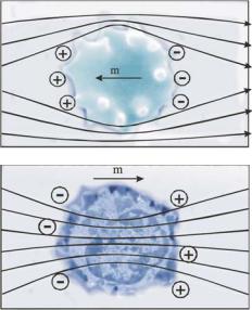
The situation for frequencies either side of fxo is shown in figure 4.2. At low frequencies (f < fxo) a mammalian cell with an intact, viable, plasma membrane will appear as an electrically insulating particle. The imposed electric field, and resulting ionic conduction flow, will skirt around the cell membrane and seek more conductive paths in the surrounding
FIGURE 4.2. Top: At low frequencies a mammalian cell with an intact, viable, plasma membrane appears as an electrically insulating particle. The applied electric field, and resulting ionic currents, skirt around the cell membrane to seek more conductive paths in the surrounding electrolyte. Induced charges will appear at the membrane-electrolyte interface to produce a dipole moment opposing the applied field. The cell will exhibit negative DEP. Bottom: At high frequencies the cell appears as a more polarizable volume than the surrounding electrolyte, the field will penetrate the cell interior, and the induced dipole moment will be oriented in the same sense as the field. The cell will exhibit positive DEP.
CELL PHYSIOMETRY TOOLS BASED ON DIELECTROPHORESIS |
111 |
electrolyte. Induced charges will appear at the cell surface to produce a dipole moment m opposing the applied field. The cell will exhibit negative DEP. At high frequencies (f > fxo) the field will penetrate the cell interior and the cell will appear to be more polarizable than the surrounding electrolyte. The induced dipole moment will be oriented in the same sense as the field, and the cell will exhibit positive DEP.
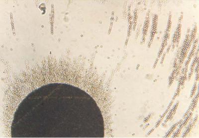
At the cross-over frequency fxo the induced dipole moment is zero—in other words the cell appears to be transparent to the applied field and no charges are induced on or within the cell. A very small change in either the cell state or physico-chemical properties of its membrane can result in a measurable change of fxo (e.g., [24]). (A dramatic change will occur if the membrane becomes degraded and no longer acts as an insulting barrier to passive ion flow. Ions will leak out of the cell and the condition σp > σm will no longer hold at high frequencies). A PhysioNetics instrument has been developed [74, 77] to measure such changes for individual cells in a cell suspension. This information can be used to refine protocols for separating different cell types from each other, or to monitor the effects of chemical agents added to a cell culture, for example. An early example of how knowledge of the fxo values for different cell types can lead to their separation by DEP was demonstrated by Gascoyne et al [23] for mixtures of erythrocytes and erythroleukeia cells. More recently, Huang et al [40] have demonstrated that DEP separation significantly improves the accuracy of gene expression profiling by purifying out cells of interest in a complex cell population. Basically, by performing DEP at a frequency between the fxo values of two cell types, one type will be repelled from the electrodes by negative DEP and the other type trapped at the electrodes by positive DEP. Examples of this are shown in figures 4.3–4.5. Particles
FIGURE 4.3. Bacteria (Micrococcus lysodeikticus) separated from blood cells by dielectrophoresis. The bacteria collect by positive DEP at the electrode, whilst erythrocytes are repelled under the action of negative DEP (unpublished work, related to Wang et al [104]).