

Kluwer - Handbook of Biomedical Image Analysis Vol
.3.pdf96 |
Angelini, Jin, and Laine |
Mathematical Methods in Biomedical Image Analysis, pp. 44–51, Kauai,
HI, USA, 2001.
[60]Vemuri, B. and Chen, Y., Joint image registration and segmentation, In: Geometric Level Set Methods in Imaging, Vision and Graphics, N. P. S. Osher, eds., Springer Verlag, New York, pp. 251–269, 2003.
[61]Tsai, A., Yezzi-Jr., A., Wells, W., III, Tempany, C., Tucker, D., Fan, A., Grimson, W. E., and Willsky, A., Model-based curve evolution technique for image segmentation, In: Proceedings of Conference on Computer Vision and Pattern Recognition, pp. 463–468, 2001.
[62]Cremers, D., Schnorr, C., and Weickert, J., Diffusion-snakes: combining statistical shape knowledge and image information in a variational framework, In: Proceedings of Workshop on Variational and Level Set Methods in Computer Vision, pp. 137–144, Vancouver, BC, Canada, 2001.
[63]Paragios, N., A level set approach for shape-driven segmentation and tracking of the left ventricle, IEEE Transactions on Medical Imaging, Vol. 22, pp. 773–776, 2003.
[64]Wang, Y. and Staib, L., Boundary finding with correspondence using statistical shape models, In: Proceedings of IEEE Conference on Computer Vision and Pattern Recognition, Santa Barbara, CA, USA, 1998.
[65]Staib, L. H. and Duncan, J. S., Model-based deformable surface finding for medical images, IEEE Transactions on Medical Imaging, Vol. 15, pp. 720–731, 1996.
[66]Chen, Y., Thiruvenkadam, S., Tagare, H. D., Huang, F., Wilson, D., and Geiser, E. A., On the incorporation of shape priors into geometric active contours, In: IEEE Workshop on Variational and Level Set Methods in Computer Vision, Vancouver, BC, Canada, 2001.
[67]Chen, Y., Huang, F., Tagare, H. D., Murali, R., Wilson, D., and Geiser,
E. A., Using prior shape and intensity profile in medical image
segmentation, In: Proceedings of IEEE ICCV, pp. 1117–1124, 2003.
[68]Chen, Y., Thiruvenkadam, S., Tagare, H. D., Huang, F., Wilson, D., and Geiser, E. A., On the incorporation of shape priors into geometric
Segmentation of Medical Images |
97 |
active contours, In: Proceedings of IEEE Workshop on Variational and Level Set Methods in Computer Vision, Vancouver, BC, Canada, 2001.
[69]Paragios, N., Rousson, M., and Ramesh, V., Knowledge-based registration & segmentation of the left ventricle: a level set approach, In: Proceedings of IEEE Workshop on Applications of Computer Vision, 2002.
[70]Paragios, N., A variational approach for the segmentation of the left ventricle in MR cardiac images, In: Proceedings of IEEE Workshop on Variational and Level Set Methods in Computer Vision, Vancouver, BC, Canada, 2001.
[71]Xu, C. and Prince, J. L., Snakes, shapes and gradient vector Flow, IEEE Transactions on Image Processing, pp. 359–369, 1998.
[72]Han, X., Xu, C., and Prince, J., Topology preserving geometric deformable models for brain reconstruction, In: Geometric Level Set Methods in Imaging, Vision and Graphics, N. P. S., Osher, ed., New York: Springer Verlag, 2003.
[73]Kwan, R. K.-S., Evans, A. C., and Pike, G. B., An extensible MRI simulator for post-processing evaluation, In: Proceedings of Visualization in Biomedical Computing, Hamburg, Germany, 1996.
[74]Collins, D. L., Zijdenbos, A. P., Kollokian, V., Sled, J. G., Kabani, N. J., Holmes, C. J., and Evans, A. C., Design and construction of a realistic digital brain phantom, IEEE Transactions on Medical Imaging, Vol. 17, pp. 463–468, 1998.
[75]Cocosco, C. A., Kollokian, V., Kwan, R. K.-S., Evans, A. C. and v., no. 4, part 2/4, S425, May 1997., BrainWeb: online interface to a 3d MRI simulated brain database, in the Proceedings of III International Conference on Functional Mapping of the Human Brain, pp. S425, Copenhagen, Denmark, 1997.
[76]Kwan, R. K.-S., Evans, A. C., and Pike, G. B., MRI simulation-based evaluation of image-processing and classification methods, IEEE Transactions on Medical Imaging, Vol. 18, pp. 1085–1097, 1999.
98 |
Angelini, Jin, and Laine |
[77]Mykkanen, J., Tohka, J., and Ruotsalainen, U., Automated delineation of brain stuctures with snakes in PET, In Physiological Imaging of the Brain with PET, A. Press, ed., 2001, pp. 39–43.
[78]Ofili, E. O. and Nanda, N. C., Three-dimensional and four-dimensional echocardiography, Ultrasound Medical Biology, Vol. 20, 1994.
[79]Fenster, A. and Downey, D. B., Three-Dimensional Ultrasound Imaging, In: Handbook of Medical Imaging. Volume1. Physics and Psychophysics, Vol. 1, H. L. K. Jacob Beutel, Richard L. Metter, ed., Bellingham, WA, USA.: SPIEThe International Society of Optical Engineering, 2000, pp. 463–510.
[80]Rankin, R. N., Fenster, A., Downey, D. B., Munk, P. L., Levin, M. F., and Vellet, A. D., Three-dimensional sonographic reconstruction: technique and diagnostic applications, American Journal of Radiology, Vol. 161,
pp.695–702, 1993.
[81]Belohlavek, M., Foley, D. A., Gerber, T. C., Kinter, T. M., Greenleaf, J. F., and Seward, J. B., Threeand four-dimensional cardiovascular ultrasound imaging: a new era for echocardiography, In: Mayo Clinic Proceedings, Vol. 68, pp. 221–240, 1993.
[82]Stetten, G., Ota, T., Ohazama, C., Fleishman, C., Castelucci, J., Oxaal, J., Ryan, T., Kisslo, J., and Von Ramm, O. T., Real-time 3D ultrasound: a new look at the heart, Journal of Cardiovascular Diagnosis Procedures, Vol. 15, pp. 73–84, 1998.
[83]Smith, S. W., Pavy, J. H. G., Miglin, J. A., and Von Ramm, O. T., Improved real-time volumetric ultrasonic imaging, Ultrasonic Imaging, Vol. 14,
pp.186–211, 1992.
[84]Von Ramm, O. T., Pavy, J. H. G., Smith, S. W., and Kisslo, J., Realtime, three-dimensional echocardiography: the first human images, Circulation, Vol. 84, pp. 685, 1991.
[85]Ramm, O. T. Von and Smith, S. W., Real time volumetric ultrasound imaging system, Journal of Digital Imaging, Vol. 3, pp. 261–266, 1990.
Segmentation of Medical Images |
99 |
[86]Ota, T., Fleishman, C. E., Strub, M., Stetten, G., Ohazama, C. J., Ramm,
O.T. Von, and Kisslo, J., Real-time, three-dimensional echocardiography: Feasibility of dynamic right ventricular volume measurement with saline contrast., American Heart Journal, Vol. 137, pp. 958–66, 1999.
[87]Kisslo, J., Firek, B., Ota, T., Kang, D. H., Fleishman, C. E., Stetten, G., Li, J., Ohazama, C. J., Adams, D., Landolfo, C., Ryan, T., and Ramm,
O.T. Von, Real-time volumetric echocardiography: the technology and the possibilities, Echocardiography, Vol. 17, pp. 773–779, 2000.
[88]Morsy, A. A., Stetten, G. D., and Ramm, O. T. Von, Detection and quantification of true 3D motion components of the myocardium using 3D speckle tracking in volumetric ultrasound scans: simulations and initial experimental results, In: Proceedings of Medical Imaging 1997: Physiology and Function from Multidimensional Images, pp. 346–353, 1997.
[89]Ota, T., Fleishman, C. E., Ohazama, C. J., Stetten, G., Lewis, C. W., Glower, D. D., Li, J., Ryan, T., Kisslo, J., and Ramm, O. T. Von, Measurement of left ventricular volume by real-time, three-dimensional echocardiography in dogs, Circulation, Vol. 94, pp. 379, 1990.
[90]Shattuck, D. P., Weinshenker, M. D., Smith, S. W., and Ramm, O. T. Von, Exploroscan: A parallel processing technique for high speed ultrasound imaging with linear phased arrays, The Journal of the Acoustical Society of America, Vol. 75, pp. 1273–1282, 1984.
[91]Shiota, T., Jones, M., Chikada, M., Fleishman, C., Castellucci, J., Cotter, B., DeMaria, A., Ramm, O. T. Von, Kisslo, J., Ryan, T., and Sahn, D., Real-time three dimensional echocardiography for determining right ventricular stroke volume in an animal model of chronic right ventricular volume overload, Circulation, Vol. 97, pp. 1897–1900, 1998.
[92]Masson, P. and Pieczynski, W., SEM algorithm and unsupervised statistical segmentation of satellite images, IEEE Transactions on Geoscience and Remote Sensing, Vol. 31, pp. 618–633, 1993.
[93]M. B. I. Centre: Montreal´ Neurological Institute, McGill University, Montreal,´ Canada.
100 |
Angelini, Jin, and Laine |
[94]Handbook of Medical Informatics. Heidelberg: Springer-Verlag, 2002.
[95]Han, X., Xu, C., Rettmann, M., and Prince, J., Automatic segmentation editing for cortical surface reconstruction, In: Proceedings of SPIE Medical Imaging, pp. 194–203, San Diego, CA, USA, 2001.
[96]Lorensen, W. E. and Cline, H. E., Marching cubes: A high resolution 3D surface construction algorithm, Computer Graphics, Vol. 21,
pp.163–169, 1987.
[97]Resnick, S., Goldszal, A., Davatzikos, C., Golski, S., Kraut, M., Metter, E., Bryan, R., and Zonderman, A., One-year changes in MRI brain volumes in older adults, Cerebral Cortex, Vol. 10, pp. 464–472, 2000.
[98]Pham, D. L. and Prince, J. L., Adaptive fuzzy segmentation of magnetic resonance images, IEEE Transactions on Medical Imaging, Vol. 18,
pp.737–752, 1999.
[99]Han, X., Xu, C., Braga-Neto, U., and Prince, J., Topology correction in brain cortex segmentation using a multiscale graph-based algorithm, IEEE Transactions on Medical Imaging, Vol. 21, pp. 109–121, 2002.
[100]Xu, C., Pham, D. L., Rettmann, M. E., Yu, D. N., and Prince, J. L., Reconstruction of the human cerebral cortex from magnetic resonance images, IEEE Transactions on Medical Imaging, Vol. 18, pp. 467–480, 1999.
[101]M. G. H. Center for Morphometric Analysis, Brain Segmentation Repository.
[102]Rajapakse, J. C. and Fruggel, F., Segmentation of MR images with intensity inhomogeneities, Image and Vision Computing, Vol. 16,
pp.165–180, 1998.
[103]Schultz, R. T. and Chakraborty, A., Magnetic resonance image analysis, In: Handbook of Human Brain Function: Neuroimaging, E. Bigler, ed., Plenum, New York, 1996.
[104]Pakkernenberg, B. and Gundersen, H. J., Neocortical neuron number in humans: effect of sex and age, Journal of Comparative Anatomy, Vol. 384, 1997.
Segmentation of Medical Images |
101 |
[105]Corsi, C., Saracino, G., Sarti, A., and Lamberti, C., Left ventricular volume estimation for real-time three-dimensional echocardiography, IEEE Transactions on Medical Imaging, Vol. 21, pp. 1202–1208, 2002.
[106]Angelini, E., Laine, A., Takuma, S., Holmes, J., and Homma, S., LV volume quantification via spatio-temporal analysis of real-time 3D echocardiography, IEEE Transactions on Medical Imaging, Vol. 20, pp. 457–469, 2001.
[107]Udupa, J., LeBlanc, V., Schmidt, H., Imielinska, C., Saha, P., Grevera, G., Zhuge, Y., Molholt, P., Jin, Y., and Currie, L., A methodology for evaluating image segmentation algorithm, In: Proceedings of SPIE Conference on Medical Imaging, pp. 266–277, San Diego CA, USA, 2002.

Chapter 3
Three-Dimensional Rigid and Non-Rigid Image Registration for the Pelvis and Prostate
Baowei Fei,1 Jasjit Suri,1 and David L Wilson1,2
3.1 Introduction
Several important applications require registration of images of the pelvis and prostate [1, 2]. First, comparison of registered MR images acquired before and immediately after therapies can be used to determine whether a tumor is adequately treated. This is particularly helpful in instances where the edematous response to treatment can be confused with a highly perfused tumor. Second, registration of serial examinations can be used to follow regression/progression of tumor. Third, registration of functional, biochemical images such as single photon emission computed tomography (SPECT), positron emission tomography (PET), and MR spectroscopy with anatomical MR images is useful for detecting and localizing cancer. Fourth, incorporating the functional, biochemical images into the interventional magnetic resonance imaging (iMRI) paradigm will aid image-guided treatments. Fifth, on a low-field magnet system during iMRI treatments where fast imaging is important, it might be highly desirable to register high quality MR image from a conventional MR scanner to the live-time iMRI images [3, 5].
There are challenges for registration in the pelvis and prostate that might reduce the effectiveness of automatic voxel-based registration. First, the
1 Department of Biomedical Engineering, Case Western Reserve University, Cleveland, OH,
44106, USA
2 Department of Radiology, University Hospitals of Cleveland & Case Western Reserve
University, Cleveland, OH 44106, USA
103
104 Fei, Suri, and Wilson
abdomen has irregular boundaries, unlike the head to which registration has been most often applied. Second, the normal prostate is a small organ that when healthy measures only about 38.0 mm in its widest dimension transversely across the base [6]. Third, different patient positions such as legs flat and raised significantly change the legs in lower portions of image volumes as well as cause movement and deformation of internal organs in the pelvis. Fourth, the prostate might move relative to the pelvic bones due to changes in bladder and rectal filling [7, 8]. The alignment of the pelvic bones, a most prominent anatomical feature in MR grayscale images, does not necessarily mean that the prostate is aligned. In addition, it is more difficult to evaluate pelvic and/or prostate registration because no external markers are available.
Many reports describe methods and evaluations for registration in the brain [9]. A few describe results for the pelvis or prostate. For example, manual registration has been used where an operator cues on segmented vascular structures [10] or other anatomical landmarks [11, 13]. Others have used automated 3D schemes that match contours of bones and sometimes other structures that are extracted using manual or interactive segmentation [14, 16]. Manual segmentation has also been used to create surfaces for automatic registration [17, 18]. All of these methods require either segmentation or visual identification of structures. Voxel based methods, particularly those based upon mutual information, are robust, require no segmentation that can be prone to error, are suitable for multimodality registration, are highly accurate for brain registration [19], and are suitable for abdominal registration [20]. For registration of brain and other organs, registration accuracy has been assessed using fiducial markers [21, 22] and anatomical landmarks [23, 25].
The next section will describe a three-dimensional mutual information rigid body registration algorithm with special features for MRI volumes of the pelvis and prostate. Section 3.3 describes a three-dimensional nonrigid registration algorithm that is based upon independent optimization of many interactively placed control points using mutual information and a thin plate spline transformation. Detailed implementation, comparisons with rigid body method, and discussions are reported at the end of the section.

Three-Dimensional Registration Methods |
105 |
3.2Three-Dimensional Rigid Body Registration Algorithm with Special Features
3.2.1 Similarity Measurements
Two similarity measures, mutual information and correlation coefficient (CC), are used in the registration. Suppose one volume R is the reference, and the other F is floating. Their mutual information MI(R,F) is given below [19]:
MI(R, F) = r, f |
pRF (r, f ) log |
pRF (r, f ) |
pR(r) · pF ( f ) |
The joint probability pRF (r, f ) and the marginal probabilities pR(r) of the reference image and pF ( f ) of the floating image, can be estimated from the normalized joint and marginal intensity histogram, respectively. The correlation coefficient CC(R, F) is given below [26]:
(R(r) − R(r))(F( f ) − F( f ))
CC(R, F) = (R(r) − R(r))2 (F( f ) − F( f ))2
Here R(r), F( f ) denote the average intensities of the reference and floating volumes and the summation includes all voxels within the overlap of both volumes.
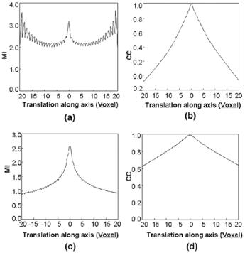
In Fig. 3.1, we compare the two similarity measures at different resolutions. Plotted are MI and CC values as a function of translation along the transverse axis where the origin is the optimal transformation. For images at a resolution of 1/4 voxels along a linear dimension, the CC curves are much smoother than MI, which is noisy and contains many local maximums as shown in Fig. 3.1a. In addition, there is a false global maximum in Fig. 3.1a at 18 voxels. At full resolution, Fig. 3.1c shows that MI is much more peaked than CC, but there is high frequency noise in the MI curves far from the optimum that give rise to local maximums that must be avoided. From these figures, we infer that CC is better at low resolution and that MI is better at full resolution when one is close
106 |
Fei, Suri, and Wilson |
|
|
− |
|
|
− |
|
|
|
|
||||
− − − |
− |
− |
− − |
|||
− |
− |
− |
− |
− − − − |
Figure 3.1: MI and CC similarity functions are plotted to show their relative advantages for registration at different resolutions. Two high-resolution MRI volumes were registered to obtain the optimal parameters. We then computed similarity values as a function of translation along the transverse axis. MI is plotted in (a) and (c); CC is plotted in (b) and (d). Graphs on the top, (a) and (b), are at a resolution of 1/4 voxels along a linear dimension, giving a distance between voxel centers of ≈5.5 mm. MI gives a noisy plot having many local maximums, and a false global maximum occurs at 18 voxels. Graphs on the bottom are obtained at full resolution. MI has a much sharper peak than CC, which is relatively flat. The voxel size is 1.4 mm. Images are from volunteer V2 in the diagnostic and reference conditions.
to the optimum value. As described in Section 3.2.2, our registration algorithm makes use of these features.
3.2.2 Registration Algorithm with Special Features
The algorithm shown in Fig. 3.2 include special features to improve robustness
for registration of MR prostate images. We use a multiresolution approach and