

Kluwer - Handbook of Biomedical Image Analysis Vol
.3.pdf86 |
Angelini, Jin, and Laine |
performed on nine RT3D cases. The correlation coefficient was 0.87 when compared to MRI measurement. The main limitation of this study is the absence of testing on normal physiological ventricular volume in the range [40–150 ml] for which the behavior of the level set segmentation can be significantly different as these volumes are much smaller.
2.4.3.5Segmentation of RT3D Ultrasound with Implicit Deformable Models Without Gradients
This research was published by Angelini, Holmes, Laine and Homma in [43].
Method: This study focused on the same clinical problem as the previous study for segmentation of echocardiographic RT3D ultrasound data. The proposed method uses the homogeneity-based implicit deformable model proposed by Chan and Vese in [40] as an extension of the Mumford-Shah segmentation functional. Motivations for selection of this method include robustness with arbitrary initialization of the object anywhere in the image, topology adaptation for multiobject segmentation (for potential cosegmentation of ventricles and atria, for example), self-adaptation of the deformation flow to inward and outward flows. Minor modifications of the method were performed to adapt the design to the specificity of the 3D ultrasound data. The homogeneity terms from Eq.
(2.28) were weighted by the mean intensity value as:
I − c0 2 I − c1 2
E (C) = d + d . (2.43)
insideC outsideC
A similar approach was followed by Lin et al. [44] for segmentation of 3D echocardiographic data where they normalized the homogeneity term by the variance of the data inside and outside the object segmented, after pre-processing with multiscale Gaussian filtering.
Parameters were set to υ = 0 (no constant inflation force was used), µ = 1,
λ1 = 0.25, λ2 = 0 (no homogeneity constraint on the outside of the ventricle to reduce the effect of the noisy myocardium texture), x = y = z = 1,
t = min( x, y, z)/|υ + µ + λ1 + λ2| (to respect CFL condition with explicit numerical scheme). The system was let to deform over 20 iterations.
Experiments: A clinical study was performed on 10 patients with pulmonary hypertension to segment both right and left ventricular volumes. A 2D parametric deformable model and a 3D level set deformable model illustrated in
Segmentation of Medical Images |
87 |
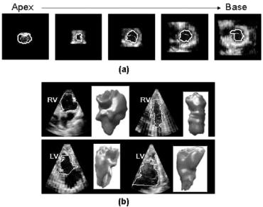
Figure 2.6 were used for segmentation of the ultrasound data after denoising with a spatio-temporal brushlet expansion [106]. The model was initialized with a cone in which dimensions were defined manually on slices at the base and apex. Manual tracing on ultrasound data was performed by an expert clinician. MRI data were also acquired on the patients and manually traced by a second expert. Absolute errors of measures were computed for RV and LV ejectionfraction. Mean-error values and standard deviation over the 10 cases for the two ventricles were equal to [Mean Std Max Min]: [8.6% 5.7% 17.8% 0.3%] for manual tracing on ultrasound vs. MRI, [4.9% 4.1% 12.21% 0.2%] for 2D parametric deformable model vs. MRI, [4.6% 4.2% 13.9% 0.8%] for 3D level set deformable model vs. MRI. Improvement of correlation measurements with deformable models (with good statistical significance) was reported when compared to MRI as well as better accuracy with a Bland-Altman analysis. The study concluded that errors of EF measurements using deformable models were within
Figure 2.6: Segmentation of right and left ventricular volumes with a 3D implicit deformable model on RT3D ultrasound data. (a) Initialization of the segmentation with a cone shape surface (dashed line) and final position of the contour (continuous line) on the endocardial surface. (b) Illustration of diversity of right and left ventricular shapes and sizes extracted for the clinical study reported in [43].
88 |
Angelini, Jin, and Laine |
the range of interand intraobserver variability for both ventricles and compared favorably to similar studies performed by other groups using RT3D ultrasound for quantification of cardiac function. Manual tracing measures were significantly less reliable with large standard deviation of errors and low correlation coefficients. Finally, the 3D level set deformable model achieved the highest degree of accuracy, which can be explained by a more accurate segmentation of small and distorted ventricular shapes when integrating the third spatial dimension.
2.5 Conclusion
Level set methods for segmentation and registration of medical images have been the focus of intense research for the past decade producing very promising results. Major advantages of the method include its robustness to noisy conditions, its aptitude in extracting curved objects with complex topology and its clean numerical framework of multidimensional implementation. Despite their success, these methods still need to be refined to address two limitations:
1.Computation time needs to be further reduced, for viability of the method in clinical application where interactivity (and therefore close to real-time computation) is critical. This optimization will have to handle the constant increase in data size observed in medical imaging applications with improvements of spatial resolution, temporal resolution and now the introduction of combo scanners such as PET/CT machines.
2.Robustness to variation in image quality and organ anatomy needs to be studied. Unfortunately, the methods described in this chapter were only rarely validated in clinical studies. On the other hand, it is well known that these methods require tuning of their parameters to adapt to the nature of the image data to segment. In that optic, it is therefore critical to evaluate robustness of the performance on a set of data that covers the range of quality encountered in clinical practice for a particular examination. For methods based on shape models, it is also critical to test the method on a variety of abnormal (e.g., disease) cases that differ from the average anatomy that they typically represent. Such validation for medical application should always clearly specify the context of the problem at
Segmentation of Medical Images |
89 |
hand in terms of anatomy of interest (e.g., endocardial surface of myocardium muscle), imaging modality (e.g., three-dimensional real-time ultrasound) and clinical application targeted (e.g., quantification of volume). Only in this context can a segmentation method be really tuned, tested, and validated for clinical application [107].
Questions
1.Define the principle idea of the level set framework for segmentation of an image given an initial contour C and a speed function V > 0. What are the advantages of using an implicit formulation of the problem?
2.What are the limitations associated with gradient-based speed terms? Why is it especially problematic with medical images?
3.What is a regularization term? What is its main functionality? Give some examples.
4.What is the entropy principle for implementation of a level set deformable model with finite difference? In what case does it apply?
5.Explain the concept of speed extension for image-based speed terms? Why is it necessary? Propose a simple algorithm to implement it.
6.Is the standard level set framework preserving the distance function? Why is this an important concept for segmentation applications?
7.Why is there a need for reinitialization of the distance function?
8.Outline in a flowchart the structure of an iterative level set segmentation algorithm using a gradient-based speed term. Use a convergence criteria (without detailing it) to stop the iterations.
9.Design a level set segmentation algorithm for extraction of the endocardial and epicardial surfaces of the left ventricle from an MRI volume? What are the properties of the data that can be used to define the speed function? Is there a way to perform simultaneous segmentation of the two surfaces?

90 |
Angelini, Jin, and Laine |
Bibliography
[1]McInerney, T. and Terzopoulos, D., Deformable models in medical image analysis: A survey, Medical Image Analysis, Vol. 1, pp. 91–108, 1996.
[2]Kass, M., Witkin, A., and Terzopoulos, D., Snakes: Active contour models, International Journal of Computer Vision, Vol. 1, pp. 321–331, 1987.
[3]Metaxas, D. N., Physics-based deformable models. Applications to computer vision, graphics and medical imaging, 1997.
[4]Xu, C., Pham, D., and Prince, J., Image segmentation using deformable models., In: Handbook of Medical Imaging, Vol. 2: SPIE, 2000, pp. 129– 174.
[5]McInerney, T. and Terzopoulos, D., Topologically adaptable snakes, In: Proceedings of 5th International Conference on Computer Vision,
pp.840–845, 1995.
[6]Durikovic, R., Kaneda, K., and Yamashita, H., Dynamic contour: a texture approach and contour operations, The Visual Computer, Vol. 11,
pp.277–289, 1995.
[7]Osher, S. and Sethian, J. A., Fronts propagating with curvaturedependent speed: Algorithms based on Hamilton-Jacobi formulations, Journal of Computational Physics, Vol. 79, pp. 12–49, 1988.
[8]Malladi, R., Sethian, J. A., and Vemuri, B. C., Shape modeling with front propagation: A level set approach, IEEE Transactions on Pattern Analysis and Machine Intelligence, Vol. 17, pp. 158–175, 1995.
[9]Suri, J. S., Liu, K., Singh, S., Laxminarayan, S. N., Zeng, X., and Reden, L., Shape recovery algorithms using level sets in 2D/3D medical imagery: a state-of-the-art review, IEEE Transactions on Information Technology in Biomedicine, Vol. 6, pp. 8 -28, 2002.
[10]Suri, J. S. and Liu, K., Level set regularizers for shape recovery in medical images, In: Proceedings of IEEE Symposium on Computer-Based Medical Systems, pp. 369–374, Bethesda, MD, USA, 2001.
Segmentation of Medical Images |
91 |
[11]Montagnat, J., Delingette, H., and Ayache, N., A review of deformable surfaces: topology, geometry and deformation, Image and Vision Computing, Vol. 19, pp. 1023–1040, 2001.
[12]Caselles, V., Catte, F., Coll, T., and Dibos, F., A geometric model for active contours, Numerische Mathematik, Vol. 66, pp. 1–31, 1993.
[13]Sethian, J., Numerical algorithms for propagating interfaces: HamiltonJacobi equation and conservation laws, Journal of Differential Geometry, Vol. 31, pp. 131–161, 1990.
[14]Sethian, J. A., Level Set Methods and Fast Marching Methods: Evolving interfaces in computational geometry, fluid mechanics, computer vision, and materials science, Cambridge University Press, 1999.
[15]Kichenassamy, S., Kumar, A., Olver, P., Tannenbaum, A., and Yezzi, A., Gradient flows and geometric active contour models, In: Proceedings of IEEE International Conference in Computer Vision, pp. 810–815, Cambridge, MA, USA, 1995.
[16]Caselles, V., Kimmel, R., and Sapiro, G., Geodesic active contours, In: Proceedings of International Conference on Computer Vision, pp. 694– 699, Cambridge, MA, USA, 1995.
[17]Yezzi-Jr, A., Kichenassamy, S., Kumar, A., Olver, P., and Tannenbaum, A., A geometric snake model for segmentation of medical imagery, IEEE Transactions on Medical Imaging, Vol. 16, pp. 199–209, 1997.
[18]Malladi, R., Kimmel, R., Adalsteinsson, D., Sapiro, G., Caselles, V., and Sethian, J. A., A geometric approach to segmentation and analysis of 3D medical images, In: Proceedings of Workshop on Mathematical Methods in Biomedical Image Analysis, pp. 244–252, 1996.
[19]Niessen, W. J., Romeny, B. M. t. H., and Viergever, M. A., Geodesic deformable models for medical image analysis, IEEE Transactions on Medical Imaging, Vol. 17, pp. 634 - 641, 1998.
[20]Siddiqi, K., Lauziere, Y. B., Tannenbaum, A., and Zucker, S. W., Area and Length Minimizing Flows for Shape Segmentation, IEEE Transactions on Image Processing, Vol. 7, pp. 433–443, 1998.
92 |
Angelini, Jin, and Laine |
[21]Deschamps, T., Curve and Shape Extraction with Minimal Path and Level-Sets techniques. Applications to 3D Medical Imaging, In: Mathematiques´ Appliquees´ et Traitements d’Images. Paris, France: Universite´ de Paris Dauphine, 2001.
[22]Yezzi-Jr., A., Tsai, A., and Willsky, A., A statistical approach to snakes for bimodal and trimodal imagery, In: Proceedings of IEEE International Conference on Computer Vision, pp. 898–903, Kerkyra, Greece, 1999.
[23]Paragios, N. and Deriche, R., Geodesic active contours and level sets for the detection and tracking of moving objects, IEEE Transactions on Pattern Analysis and Machine Intelligence, Vol. 22, pp. 266–280, 2000.
[24]Sarti, A. and Malladi, R., A Geometric Level Set Model for Ultrasounds Analysis, University of California, Berkeley, 1999.
[25]Jin, Y., Laine, A., and Imielinska, C., An adaptive speed term based on homogeneity for level-set segmentation, In: Proceedings of SPIE Medical Imaging Conference, pp. 383–390, San Diego, CA, USA, 2002.
[26]Saha, P. K., Udupa, J. K., and Odhner, D., Scale-based fuzzy connected image segmentation: theory, algorithms, and validation, Computer Vision and Image Understanding, Vol. 77, pp. 145–174, 2000.
[27]Sarti, A., Malladi, R., and Sethian, J. A., Subjective surfaces: a geometric model for boundary completion, International Journal of Computer Vision, Vol. 46, pp. 201–221, 2002.
[28]Suri, J. S., Leaking prevention in fast level sets using fuzzy models: an application in MR brain, In: Proceedings of Information Technology Applications in Biomedicine, pp. 220–225, Arlington, VA, USA, 2000.
[29]Suri, J. S., Leaking prevention in fast level sets using fuzzy models: an application in MR brain, In: Proceedings of IEEE EMBS International Conference on Information Technology Applications in Biomedicine, pp. 220–225, Arlington, VA, USA, 2000.
[30]Baillard, C., Barillot, C., and Bouthemy, P., Robust Adaptive Segmentation of 3D Medical Images with Level Sets, INRIA, Rennes, France, Research Report Nov. 2000.
Segmentation of Medical Images |
93 |
[31]Leventon, M. E., Grimson, W. E. L., and Faugeras, O., Statistical shape influence in geodesic active contours, In: Proceedings of IEEE Conference on Computer Vision and Pattern Recognition, pp. 316–325, 2000.
[32]Leventon, M., Grimson, E., Faugeras, O., Wells, S., and Kikinis, R., Knowledge-based segmentation of medical images, In: Geometric Level Set Methods in Imaging, Vision and Graphics, Osher, N. P. S., ed., New York: Springer Verlag, 2003.
[33]Zeng, X., Staib, L. H., Schultz, R. T., and Duncan, J. S., Segmentation and measurement of the cortex from 3D MR images using coupledsurfaces propagation, IEEE Transactions on Medical Imaging, Vol. 18,
pp.927–937, 1999.
[34]Gomes, J. and Faugeras, O., Segmentation of the inner and outer surfaces of the human cortex: an approach based on partial differential equations, In: Proceedings of 22nd Annual International Conference of the IEEE Engineering in Medicine and Biology Society, pp. 1764–1774, Chicago, IL, USA, 2000.
[35]Barles, G., Soner, H. M., and Souganidis, P. E., Front propagation and phase field theory, SIAM Journal of Control and Optimization, Vol. 31,
pp.439–469, 1993.
[36]Mumford, D. and Shah, J., Boundary detection by minimizing functional, In: Proceedings of International Conference on Computer Vision and Pattern Recognition, pp. 22–26, San Francisco, CA, USA, 1985.
[37]Chan, T. F. and Vese, L. A., Active contour and segmentation models using geometric PDE’s for medical imaging, In Geometric Methods in Bio-Medical Image Processing, Mathematics and Visualization, R. Malladi, ed., Springer, 2002.
[38]Chan, T. F. and Vese, L. A., A level set algorithm for minimizing the Mumford-Shah functional in image processing, In: Proceedings of IEEE Workshop on Variational and Level Set Methods in Computer Vision,
pp.161–168, Vancouver, BC, Canada, 2001.
[39]Chan, T. F. and Vese, L. A., An efficient variational multiphase motion for the Mumford-Shah segmentation model, In: Proceedings of 34th
94 |
Angelini, Jin, and Laine |
Asilomar Conference on Signals, Systems and Computers, pp. 490–494,
Pacific Grove, CA, USA, 2000.
[40]Chan, T. F. and Vese, L. A., Active contours without edges, IEEE Transactions on Image Processing, Vol. 10, pp. 266–277, 2001.
[41]Vese, L. A. and Chan, T. F., A multiphase level set framework for image segmentation using the Mumford and Shah model, University of California, Los Angeles, CA, USA, Computational and Applied Mathematics Report 01-25, 2001.
[42]Tsai, A., Yezzi-Jr, A., and Willsky, A. S., Curve evolution implementation of the Mumford-Shah functional for image segmentation, denoising, interpolation, and magnification, IEEE Transactions on Image Processing, Vol. 10, pp. 1169–1186, 2001.
[43]Angelini, E. D., Holmes, J., Laine, A. F., and Homma, S., Segmentation of real-time 3D cardiac ultrasound with implicit deformable models without gradients, In: Proceedings of International Symposium on Image and Signal Processing and Analysis, pp. 711–716, Rome, Italy, 2003.
[44]Lin, N., Yu, W., and Duncan, J. S., Combinative multiscale level set framework for echocardiographic image segmentation, Medical Image Analysis, Vol. 7, pp. 529–537, 2003.
[45]Chan, T. F., Sandberg, B. Y., and Vese, L. A., Active contours without edges for vector-valued images, Journal of Visual Communication and Image Representation, Vol. 11, pp. 130–141, 2000.
[46]Maintz, J. B. and Viergever, M. A., A survey of medical image registration, Medical Image Analysis, Vol. 2, pp. 1–36, 1998.
[47]Ginneken, B. v., Frangi, A. F., Staal, J. J., Romeny, B. M. t. H., and Viergever, M. A., Active shape model segmentation with optimal features, IEEE Transactions on Medical Imaging, Vol. 21, pp. 924–933, 2002.
[48]Frangi, A. F., Rueckert, D., Schnabel, J. A., and Niessen, W. J., Automatic construction of multiple-object three-dimensional statistical shape models: application to cardiac modeling, IEEE Transactions on Medical Imaging, Vol. 21, pp. 1151–1166, 2002.
Segmentation of Medical Images |
95 |
[49]Taylor, C. J., Cootes, T. F., Hill, A., and Haslam, J., Medical image segmentation using active shape models, In: Medical Imaging, M. H. K. L. Beolchi, ed., IOS Press, Amsterdam, 1994.
[50]Angelini, E. D. and Ciaccio, E. J., Optimized region finding and edge detection of knee cartilage surfaces from magnetic resonance images, Annals of Biomedical Engineering, Vol. 31, pp. 336–345, 2003.
[51]Medical Image Registration: CRC Press, 2001.
[52]Vemuri, B. C., Ye, J., Chen, Y., and Leonard, C. M., Image registration via level-set motion: Applications to atlas-based segmentation, Medical Image Analysis, Vol. 7, pp. 1–20, 2003.
[53]Yahia, H. M., Huot, E. G., Herlin, I. L., and Cohen, I., Geodesic distance evolution of surfaces: a new method for matching surfaces, In: Proceedings of IEEE CVPR, pp. 663–668, Hilton Head Island, SC, USA, 2000.
[54]Cohen, I. and Herlin, I., Curve matching using geodesic paths, In: Proceedings of IEEE CVPR, pp. 741–746, Santa Barbara, CA, USA, 1998.
[55]Lavallee, S. and Szeliski, R., Recovering the position and orientation of free-form objects from image contours using 3D distance maps, IEEE Transactions on Pattern Analysis and Machine Intelligence, Vol. 17, pp. 378–390, 1995.
[56]Rousson, M. and Paragios, N., Shape priors for level set representations, In: Proceedings of European Conference on Computer Vision, pp. 78–93, Copenhagen, Denmark, 2002.
[57]Paragios, N., Rousson, M., and Ramesh, V., Matching distance functions: A shape-to-area variational approach for global-to-local registration, In: Proceedings of European Conference in Computer Vision, Copenhagen, Denmark, 2002.
[58]Paragios, N. and Rousson, M., Shape analysis towards model-based segmentation, In: Geometric Level Set Methods in Imaging, Vision and Graphics, N. P. S. Osher, ed., Springer Verlag, New York, 2003.
[59]Yezzi, A., Zollei, L., and Kapur, T., A variational framework for joint segmentation and registration, In: Proceedings of IEEE Workshop on