
книги студ / Color Atlas of Pathophysiology (S Silbernagl et al, Thieme 2000)
.pdf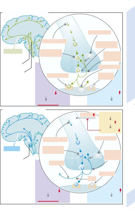
A. Noradrenergic Transmission |
|
|
|
|
|
|
|
Tyrosine |
|
|
|
|
|
Dopa |
|
|
|
|
|
MAO inhibitor |
|
|
|
|
|
Dopamin |
|
|
|
|
Synthesis inhibitor |
Stimulation |
|
|
|
|
|
|
|
|
|
|
|
Norepinephrine |
of release |
|
|
|
|
|
|
|
|
Norepinephrine |
Inhibition of |
Break- |
|
|
|
|
storage |
down MAO |
Inhibition |
|
|
|
|
|
of re-uptake |
Depression |
|
Tegmentum |
1 |
|
|
|
|
|
|
COMT inhibitor |
|
||
Locus |
|
|
|
||
|
|
|
|
||
coeruleus |
Receptor blocker |
Receptor |
|
||
|
2 |
||||
|
|
Break- |
agonists |
10.27 |
|
|
|
down |
|
|
|
|
|
|
|
|
|
|
Norepinephrine |
Receptor COMT |
Norepinephrine |
|
Plate |
|
effect |
|
effect |
|
|
|
Deterioration |
|
Improvement |
|
|
B. Serotonergic Transmission |
|
|
|
|
|
|
|
|
Uptake |
|
Glucose supply |
|
|
|
Tryptophan |
|
|
|
|
|
|
|
|
|
Insulin |
|
|
|
|
5-OH- |
|
Amino acids |
|
|
|
|
tryptophan |
|
|
|
|
|
|
|
|
|
|
|
|
Synthesis inhibitor |
|
MAO inhibitor |
|
|
|
|
|
|
|
|
|
|
Serotonin |
Inhibition |
Serotonin |
|
|
|
|
Break- |
|
|
|
|
||
|
of storage |
down |
|
Inhibition |
|
|
|
|
|
MAO |
|
|
|
Raphe nuclei |
|
|
of re-uptake |
|
||
|
|
|
|
|
|
|
1 |
Darkness |
|
|
|
|
|
|
Melatonin |
|
|
Agonists |
2 |
|
|
|
|
|
|
|
|
|
|
Light |
|
|
|
|
|
Serotonin effect |
Receptor |
Serotonin effect |
|
||
|
|
|
||||
|
Deterioration |
|
|
Improvement |
351 |
|
Silbernagl/Lang, Color Atlas of Pathophysiology © 2000 Thieme
All rights reserved. Usage subject to terms and conditions of license.

10 Neuromuscular and Sensory Systems
352
Schizophrenia
Schizophrenia is a disease with an increased familial incidence. It is characterized by delusions, hallucinations, socially inacceptable behavior and/or inadequate associations (socalled positive symptoms). Lack of motivation and of emotion also frequently occur (socalled negative symptoms). In some patients the positive symptoms predominate (type I), in others the negative ones (type II).
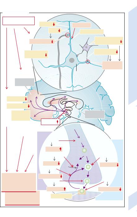
In schizophrenia there is reduced blood flow and glucose uptake especially in the prefrontal cortex and, in type II patients, also a decrease in the number of neurons (reduction in the amount of gray matter). In addition, abnormal migration of neurons during brain development is of pathophysiological significance (→A2).
Atrophy of the spiny dendrites of pyramidal cells has been found in the prefrontal cortex and the cingulate gyrus. The spiny dendrites contain glutamatergic synapses; their glutamatergic transmission is thus disturbed (→A1). In addition, in the affected areas the formation of GABA and/or the number of GABAergic neurons seems to be reduced, so that inhibition of pyramidal cells is reduced.
Special pathophysiological signficance is ascribed to dopamine: excessive availability of dopamine or dopamine agonists can produce symptoms of schizophrenia, and inhibitors of D2 dopamine receptors have been successfully used in the treatment of schizophrenia (see below). On the other hand, a reduction in D2 receptors has been found in the prefrontal cortex (→A1), and a reduction of D1 and D2 receptors correlates with negative symptoms of schizophrenia, such as lack of emotions. It is possible that the reduction in dopamine receptors is the result of an increased dopamine release and in itself has no pathogenetic effect.
Dopamine serves as a transmitter in several pathways (→B):
Dopaminergic pathways to the limbic (mesolimbic) system; and
to the cortex (mesocortical system) are probably essential in the development of schizophrenia.
In the tubuloinfundibular system dopamine controls the release of hypophyseal hormones
(especially inhibition of prolactin release; →p. 260ff.).
It controls motor activity in the nigrostriatal system (→p. 312ff.).
Release and action of dopamine are increased by several substances that promote the development of schizophrenia (→A3, left). Thus, the dopaminergic treatment of Parkinson’s disease can lead to symptoms of schizophrenia, which in turn can limit the treatment of Parkinson’s disease:
L-dopa leads to an increased formation and release of dopamine.
Monoamine oxidase inhibitors (MAO inhibitors) inhibit the breakdown of dopamine and thus increase its availability for release in the synaptic cleft.
Cocaine stimulates dopamine release in the synaptic cleft, too.
Amphetamine inhibits dopamine uptake in presynaptic nerve endings and thus at the same time raises the transmitter concentration in the synaptic cleft.
Conversely, antidopaminergic substances
can improve schizophrenia (→A3, right):
Some substances (e.g., phenothiazines, haloperidol) displace dopamine from receptors and thus have an antidopaminergic action.
Inhibition of the uptake of dopamine in the synaptic vesicle, for example, by reserpine, ultimately impairs the release of the transmitter in the synaptic cleft. However, reserpine is at present not used therapeutically.
The long-term use of dopamine antagonists in a patient with schizophrenia can lead to “tardive dyskinesia” as a result of their action on the striatum (→p. 314). This complication can limit the treatment of schizophrenia.
It is possible that serotonin also plays a role in producing schizophrenic symptoms. Excessive serotonin action can cause hallucinations,
and many antipsychotic drugs block 5-HT2 A receptors (→A1).
Silbernagl/Lang, Color Atlas of Pathophysiology © 2000 Thieme
All rights reserved. Usage subject to terms and conditions of license.
A. Schizophrenia |
|
|
|
|
|
Genetic and other factors |
|
|
|
|
|
|
Dendrites |
|
Hallucinogens |
|
|
|
|
|
|
|
|
1 |
Spines |
|
5-HT2A receptors |
|
|
|
|
|
|
||
Glutamate effect |
|
|
Inhibitory |
|
|
|
|
GABAergic neurons |
|
||
Dopamine |
|
|
GABA formation |
|
|
receptors (D1) |
|
|
|
Schizophrenia |
|
|
|
|
|
Reduced |
|
|
|
|
|
inhibition |
|
|
|
|
Pyramidal cells |
|
|
|
|
|
|
|
|
Prefrontal |
|
|
|
|
10.28 |
cortex |
|
|
|
|
|
2 |
|
|
|
|
|
|
|
|
|
Plate |
|
Circulation |
|
|
|
|
|
|
|
|
|
|
|
Number |
|
|
|
|
|
of neurons |
|
|
|
|
|
Abnormal |
|
|
Limbic |
|
|
neuron |
|
|
system |
|
|
migration |
Dopamine |
|
|
|
|
|
|
|
|
|
|
|
|
|
Tyrosine |
|
|
3 |
Symptoms increased |
|
|
|
|
|
by: |
|
|
|
|
|
L-Dopa |
|
Dopa |
Symptoms reduced |
|
|
|
|
|
|
|
|
Synthesis |
|
|
by: |
|
|
|
|
|
|
|
|
MAO inhibitors |
Dopamine |
Reserpine |
|
|
|
|
|
|
|
|
|
Breakdown |
|
Breakdown |
Storage |
|
|
MAO |
|
|
||
|
Amphetamine |
|
|
||
|
|
|
|
|
|
Positive symptoms: |
Re-uptake |
|
|
Phenothiazine, |
|
delusions, hallucinations, |
Cocaine |
|
|
haloperidol |
|
socially inacceptable |
|
|
|
|
|
|
|
|
|
|
|
behavior, |
|
|
|
|
|
inadequate associations |
Release |
|
|
Free receptors |
|
Negative symptoms: |
D2 receptors |
|
|
|
|
lack of motivation, |
|
|
|
|
353 |
lack of emotion |
|
|
|
|
|
Silbernagl/Lang, Color Atlas of Pathophysiology © 2000 Thieme
All rights reserved. Usage subject to terms and conditions of license.

10 Neuromuscular and Sensory Systems
354
Dependence, Addiction
Dependence or rather addiction is an acquired compulsion that dictates the behavior of those who are dependent or addicted. In drug dependence there is a great craving for the particular drug. For the dependent person, obtaining and supply of the drug become priorities over all other kinds of behavior. Among the most important of such drugs are nicotine, alcohol, opiates, and cocaine. There are, however, also many other drugs (especially sleeping pills [hypnotics] and analgesics) that can lead to dependence.
It is not only the supply of the particular drug that is important in the development of addiction, as only some of those who take a drug become dependent. Of great significance for the development of addictive behavior is a genetic disposition (→A). It has been shown that in those dependent on alcohol or cocaine, certain polymorphisms of the gene for the dopamine transporter (DAT-1) are especially common. Genetic defects of acohol dehydrogenase (ADH) or acetaldehyde dehydrogenase (ALDH) impair the breakdown of alcohol and thus increase its toxic effect. These enzyme defects therefore protect against alcohol dependence. The attempt has been made to achieve pharmacological inhibition of ALDH (with desulfiram) in order to force an increase in acetaldehyde and thus stop addictive behavior through the toxic effect of acetaldehyde (nausea, vomiting, hypotension). Because of the high risk and relatively limited success this approach has now been abandoned.
Another important factor in dependence is the social context (→A). Thus, a change in social environment can make it easier to give up drugs. Most of the soldiers, for example, who took drugs during the Vietnam War were not addicted after their return to the USA.
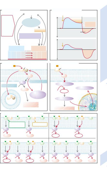
Frequently addicts develop a tolerance to the substance and the initial effect gradually weakens if drug intake continues (→A,B). If intake is suddenly discontinued, there is a reversal of effect (→B). Chronic intake weakens the effect of the drug and increases the reversal effect on discontinuance. If the addict wants to attain the same effect, the dosage has to be increased. When the drug is discontinued, withdrawal symptoms develop that
get worse the longer the drug addiction had lasted. Withdrawal symptoms lead to physical dependence in the addict. Psychological dependence is the result of the need for the positive effects of the drug and/or the fear of the neurobiological or psychological withdrawal symptoms (→A). The desire for the positive effects remains after the withdrawal symptoms have abated. Stress, among other factors, favors relapse.
Mesolimbic and mesocortical dopaminergic pathways (→A; see also p. 352) apparently play an important role in the development of dependence or addiction. By activating these pathways, for example, with alcohol or opiates, the addict tries to produce a feeling of wellbeing or euphoria or, conversely, to prevent dysphoria. It is possible that on withdrawing the substance the activity of the dopaminergic system is reduced or the target cells are less sensitive. Withdrawal symptoms can be attenuated by activating endorphinergic, GABAergic, dopaminergic, or serotoninergic receptors.
The cellular mechanisms of tolerance have been in part elucidated for opiates. Stimulation of the receptors leads to phosphorylation via G protein receptor kinases and thus to the inactivation of the receptor (→C). The receptors are also internalized. The effectiveness of receptor stimulation can also be reduced by influencing cellular signal transmission. The opiate receptor acts partly via inhibition of adenylylcyclase (AC), a decrease of cyclic adenosine monophosphate (cAMP) and reduced activation of protein kinase A (→D). Taking opiates thus at first diminishes cAMP formation (→E2). Chronic intake, however, raises the expression of adenylylcyclase by influencing cAMP-re- sponsive element-binding protein ([CREB] →p. 6ff.). As a result, even in the presence of opiates, cAMP is still formed (→E3). Subsequent withdrawal of opiates will, for example, via a massive increase in cAMP (→E4), lead to withdrawal symptoms.
Silbernagl/Lang, Color Atlas of Pathophysiology © 2000 Thieme
All rights reserved. Usage subject to terms and conditions of license.
A. Drug Usage |
|
B. Reversal of Drug Effect |
|
|||
|
|
Drug intake |
|
Primary intake |
Sudden |
|
Stress |
|
|
|
|
withdrawal |
|
|
|
|
|
|
||
Genetic |
|
Mesolimbic |
Effect negative positive negative positive |
Tolerance |
||
disposition |
|
|
|
|||
|
dopaminergic |
|
Effect |
|||
(DAT-1, |
|
|
||||
|
system |
|
reversal |
|||
ADH) |
|
|
||||
|
|
|
|
|||
Social |
|
Tolerance |
Withdrawal |
|
||
context |
|
|
symptoms |
|
||
(e. g. war) |
Endorphins, dopamine, |
Dependence, Addiction |
||||
|
||||||
|
serotonin, GABA |
|
||||
|
|
|
Renewed intake |
|||
|
Positive |
Withdrawal |
|
|||
|
effects: |
symptoms: |
|
|||
|
e. g. Euphoria |
Depression |
|
|||
Feeling of omnipotence |
Anxiety |
|
||||
|
Relaxation |
Nervousness |
|
|||
|
|
|
|
|||
C. Receptor Inactivation |
|
|
|
D. Signal Transduction |
|
|
Opiates |
Receptor |
Opiates |
Receptor |
10.29 |
|
||||
|
|
|
||
|
P |
|
|
Plate |
|
G |
|
Gi |
|
Phosphorylation |
Internalization |
Adenylylcyclase |
|
Adenylylcyclase
|
cAMP |
|
|
G protein |
|
P |
receptor kinase |
|
Proteinkinase A |
Cell |
|
|
|
nucleus |
Cellular effects
|
|
|
|
|
Cellular effects |
|
CREB |
|
|
|
|
|
|
|
|
|
|
||
E. Adaptation of Signal Transduction |
1 |
Primary intake |
|
2 |
|
||||
|
|
|
|
|
|
||||
|
AC-activating |
Opiate receptor |
|
|
|
|
|
||
|
receptor |
|
|
|
|
|
|||
|
|
|
|
|
|
|
|
|
|
AC |
|
AC |
|
|
AC |
|
AC |
|
|
cAMP |
Normal |
cAMP |
|
|
cAMP |
|
cAMP |
|
|
|
|
|
|
|
|
||||
Chronic intake |
|
|
3 |
|
Withdrawal |
4 |
|
||
AC |
AC |
AC |
AC |
|
AC |
AC |
AC |
AC |
|
cAMP |
cAMP |
cAMP |
cAMP |
cAMP |
cAMP |
cAMP |
cAMP |
355 |
|
|
|||||||||
Silbernagl/Lang, Color Atlas of Pathophysiology © 2000 Thieme
All rights reserved. Usage subject to terms and conditions of license.

10 Neuromuscular and Sensory Systems
356
Cerebrospinal Fluid, Blood–Brain Barrier
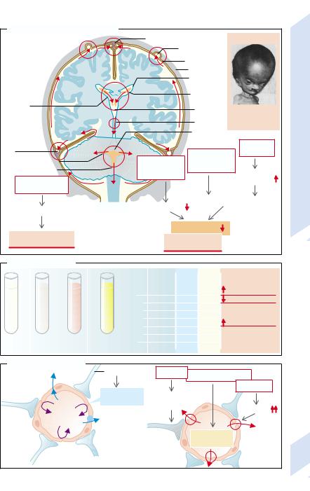
Cerebrospinal fluid (CSF) flow (→A). CSF is formed mainly in the choroid plexus of the lateral ventricles. It flows via the interventricular foramina (→A1) into the third ventricle and from there into the fourth ventricle via the aqueduct (→A2). It then circulates via the foramina of Luschka and Magendi (→A3) into the subarachnoid space and the arachnoid villi of the sinuses of the dura mater (Pacchionian bodies) and from there into the venous sinuses (→A4).
CSF flow may be slowed or interrupted at each of the named structures. This results in
CSF backward congestion (hydrocephalus) with raised pressure. Depending on the site of the obstruction, one distinguishes a communicating hydrocephalus, in which CSF flow between the ventricles is uninterrupted, from a non-communicating hydrocephalus, where the connections between the ventricles are obstructed.
Obstruction of the CSF channels, especially the aqueduct, can be the result of malformations, scars (as after an infection or bleeding), or tumors. The absorption of CSF in the arachnoid villi is impaired if drainage in the sinuses is obstructed (e.g., in thrombosis) or the systemic venous pressure is raised (e.g., in heart failure). Drainage can also be reduced after subarachnoid hemorrhage or meningitis as well as by a high protein concentration in CSF (tumors or infection), because the arachnoid villi can be obstructed by proteins. Lastly, absorption may be reduced for no obvious external reasons. An increase of the CSF space caused by primary cerebral atrophy is termed hydrocephalus e vacuo.
In congenital hydrocephalus the cranial bones may be separated because their sutures have not yet fused, resulting in an enlarged cranium (“water on the brain”, the literal meaning of the term hydrocephalus) (→A5). Once the bony sutures have fused, an excess of CSF causes an increased CSF pressure (→p. 358).
Composition of CSF (→B). The normal composition of CSF is approximately the same as that of serum. However, it has lower protein and protein-bound Ca2+ concentrations. The K+ concentration is also lower (about 1 mmol/ l). Changes in the composition of CSF are of
great diagnostic significance in certain brain diseases:
CSF is normally as clear as water and does not contain any erythrocytes and only very few leukocytes (< 4 per µL, largely lymphocytes). However, in infections (e.g., meningitis) leukocytes may pass into the CSF (→cloudy CSF), and after hemorrhage (e.g., a brain tumor) erythrocytes may be found in CSF ( reddish discoloration). A yellowish CSF may indicate the presence of blood pigments or bilirubin-binding plasma proteins.
The protein concentration in CSF is increased if there is no CSF absorption in the arachnoid villi or in infection (especially formation by immune competent cells).
The glucose concentration in CSF is decreased by tumors, acute bacterial infections, tuberculosis, fungal infections of the brain as well as defective glucose transport in rare cases.
Blood–brain barrier (→C). The endothelial cells of the blood capillaries in the brain (except for the posterior pituitary, area postrema, choroid plexus, and circumventricular organs) under the influence of astrocytes form dense tight junctions that prevent the passage of substances dissolved in blood (electrolytes, proteins) or of cells. In this way the extracellular milieu of the brain is separated from the blood, thus preventing nerve cells being exposed to electrolyte changes, transmitters, hormones, growth factors, and immune reactions. Under abnormal circumstances the tight junctions can be opened. This happens, for example, in brain tumors that contain no functional astrocytes. The blood–brain barrier may also be breached in hyperosmolarity (brought about by infusion of hypertonic mannitol solutions) or in bacterial meningitis.
The blood–brain barrier is not yet closed in newborns. As a result, in hyperbilirubinemia of the newborn bilirubin can reach the brain and damage nuclei (“Kerne”) in the brain stem (hence kernicterus). Damage to the basal ganglia may, for example, cause hyperkinesias (→p.134).
Silbernagl/Lang, Color Atlas of Pathophysiology © 2000 Thieme
All rights reserved. Usage subject to terms and conditions of license.
A. Cerebrospinal Fluid (CSF) Flow |
|
|
|
|
||
|
|
|
Sagittal sinus |
|
5 |
Barrier |
|
4 |
|
Arachnoid villi |
|
||
|
|
4 |
Dura mater |
|
||
|
|
|
|
Bone |
|
Blood–Brain |
|
|
|
|
Lateral |
|
|
|
|
1 |
|
ventricle |
|
|
|
|
|
Choroid |
|
||
Interventricular |
|
|
|
|
||
|
|
|
plexus |
|
||
foramina |
|
|
|
3rd |
|
|
|
|
|
Hydrocephalus |
Fluid, |
||
|
|
|
|
ventricle |
||
|
|
2 |
|
Aqueduct |
in newborn |
|
|
|
|
|
|||
|
|
|
|
4th ventricle |
|
Cerebrospinal |
Arachnoid |
|
|
|
with plexus |
Tumors, |
|
|
|
|
|
|||
villi |
4 |
3 |
|
Meningitis, |
infections |
|
Foramen of Luschkae |
Thrombosis, |
|
||||
|
|
subarachnoid |
|
|||
Foramen of Magendii |
|
|
sinus occlusion, |
hemorrhage, etc. |
||
|
|
cardiac failure |
|
Protein |
||
|
|
|
|
|||
Malformations, |
|
|
|
|
concentration |
10.30 |
scarring, tumors |
|
|
|
|
|
|
|
|
|
|
Obstruction of arachnoid villi |
||
|
|
|
Venous outflow |
Plate |
||
CSF flow obstructed |
|
|
|
|||
|
|
|
|
|||
|
|
|
|
|
|
|
|
|
|
CSF absorption |
|
|
|
Hydrocephalus |
|
|
Hydrocephalus |
|
|
|
(non-communicating) |
|
|
|
|
||
|
|
(communicating) |
|
|
||
|
|
|
|
|
||
B. CSF Composition |
|
|
|
|
|
|
|
|
|
|
Serum |
CSF |
Abnormal CSF |
|
|
|
|
normal |
normal |
Infections, |
|
|
|
Proteins g/L |
70 |
0.2 |
|
|
|
|
CSF obstruction |
|||
|
|
|
Glucose |
5 |
3 |
Tumors, infection |
|
|
mmol/L |
Na+ |
145 |
150 |
|
|
|
K+ |
4 |
3 |
Infections, |
|
|
|
Ca2+ |
2.5 |
1 |
CSF obstruction |
|
|
|
Mg2+ |
0.8 |
1 |
|
|
|
|
|
|
|||
Normal |
Leukocytes, Erythrocytes Blood pigments, |
Osm |
295 |
295 |
|
|
pH |
7.4 |
7.33 |
|
|||
|
proteins |
plasma proteins |
|
|||
C. Blood-Brain-Barrier |
Astrocytes |
|
|
|
Lipid-soluble |
Tumors |
Bacterial meningitis |
||
substances |
|
|
|
|
|
|
|
Infusions |
|
|
|
|
|
|
|
Closed |
|
Defective |
|
|
tight junctions |
|
||
|
astrocytes |
|
||
|
|
|
Osmolarity |
|
Electrolytes, |
Selective |
|
|
|
proteins, |
|
|
|
|
carrier |
|
|
|
|
cells |
|
|
|
|
|
|
|
|
Open |
|
|
|
|
tight junctions |
1 Normal cerebral capillary |
2 Abnormal |
357 |
||
|
|
|
||
Silbernagl/Lang, Color Atlas of Pathophysiology © 2000 Thieme
All rights reserved. Usage subject to terms and conditions of license.

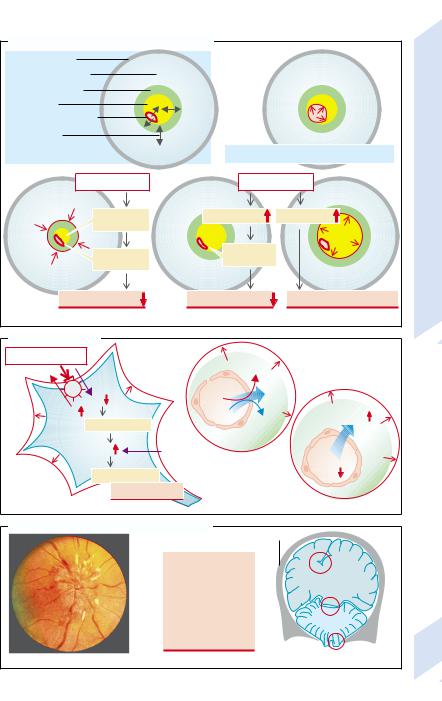
Cerebrospinal Fluid Pressure, Cerebral Edema
|
After the the cranial bone sutures have fused, |
|
|
the brain is confined within a rigid casing. Its |
|
|
volume cannot expand and any intracranial |
|
|
compartments can get larger only at the ex- |
|
Systems |
pense of other compartments (→A1). |
|
cord via the foramen magnum. The intravascu- |
||
|
The cerebrospinal fluid (CSF) space of the |
|
|
brain is open to the CSF space of the spinal |
|
Sensory |
lar space is momentarily increased with each |
|
systolic pulse wave, and synchronously with |
||
|
||
|
the pulse a small volume of CSF escapes |
|
and |
through the foramen magnum into the spinal |
|
CSF space, i.e., the intravascular space is in- |
||
|
||
Neuromuscular |
creased at the expense of the CSF space |
|
(→A2). |
||
|
||
|
Similarly, an increase in interstitial or intra- |
|
|
cellular volume at first occurs at the expense |
|
|
of the CSF space. Once this reserve is used up |
|
|
and the CSF space has collapsed, CSF pressure |
|
10 |
quickly rises and there is a marked decrease |
|
in cerebral perfusion (→A3). |
||
|
Several forms of cerebral edema are distin- |
|
|
guished (→B): |
|
|
Cytotoxic edemas enlarge the intracellular |
|
|
space as a result of cell swelling (→B1). |
|
|
Among causes are energy deficiency (e.g., due |
|
|
to hypoxia or ischemia). Impairment of Na+/ |
|
|
K+-ATPase raises the intracellular Na+ concen- |
|
|
tration and decreases intracellular K+ concen- |
|
|
tration. Subsequent depolarization leads to |
|
|
Cl– entry and cell swelling (→p.10). |
|
|
Reduction of extracellular osmolarity can |
|
|
also cause cell swelling, for example, in hypo- |
|
|
tonic hyperhydration (→p.122). |
|
|
Treatment of prolonged hypernatriemia de- |
|
|
mands caution. The glial cells and neurons |
|
|
compensate for the extracellular hyperosmo- |
|
|
larity by intracellular accumulation of osmo- |
|
|
lytes (e.g., inositol), a process that takes days. |
|
|
If the hypernatremia is corrected too quickly, |
|
|
the osmolytes are not removed quickly enough |
|
|
and the cells swell. |
|
|
Cerebral edemas of vascular origin occur |
|
|
when there is increased permeability of the |
|
|
cerebral capillaries. The resulting capillary fil- |
|
|
tration of proteins with osmotically obliged |
|
|
water (→B2) thus increases the interstitial |
|
358 |
space. Among causes of increased permeability |
|
are tumors, infections, abscesses, infarcts, |
||
|
bleeding, or poisoning (lead). |
Water can also accumulate in the interstitial space when the blood–brain barrier is intact but the osmolarity of the interstitial space is higher than that of blood, for example, if there is a rapid fall in the concentration of blood sugar (during treatment of diabetes mellitus), of urea (dialysis), or of Na+ (interstitial cerebral edema; →B3). In those conditions the increase of interstitial space may be accompanied by cell swelling.
CSF congestion also increases cerebral pressure (→p. 356). An acute disorder of CSF drainage causes a rise in pressure that, via narrowing of the vessel lumen, impairs cerebral perfusion (→A4). Chronic drainage abnormality, by bringing about the death of neurons, i.e., a decrease in intracellular space, will ultimately result in a decrease in cerebral mass (→B5).
Tumors and bleeding (→A3) take up intracranial volume at the expense of other compartments, especially the CSF space.
Symptoms of increased CSF pressure. Due to the increased cerebral pressure, lymph from the back of the eye can no longer flow toward the intracranial space via the lymphatic canal at the center of the optic nerve. Lymph thus collects at the exit of the optic nerve and causes bulging of the papilla (papilledema; →C1). Other consequences of increased CSF pressure are headache, nausea, vomiting, dizziness, impaired consciousness (e.g., due to decreased perfusion), bradycardia and arterial hypertension (through pressure on the brain stem), squinting (compression of the abducens nerve), and dilated pupils which are unresponsive to light (compression of the oculomotor nerve) (→C2). The pressure gradients bear an increasing risk of herniation of parts of the brain through the cerebellar tentorium (→C3a) or the foramen magnum (→C3b). The herniated parts compress the brain stem causing immediate death. If the increase in CSF pressure is unilateral, the cingulate gyrus may herniate under the falx cerebri (→C3), causing compression of the anterior cerebral vessels with corresponding deficits in cerebral function (→p. 360).
Silbernagl/Lang, Color Atlas of Pathophysiology © 2000 Thieme
All rights reserved. Usage subject to terms and conditions of license.
A. Volume Changes of Brain Compartments |
|
|
|
||
Skull |
|
|
|
|
|
Intracellular (~ 80 %) |
|
|
|
|
|
Interstitial (<10 %) |
|
|
|
|
|
CSF (~10 %) |
|
|
|
|
|
Intravascular (~1– 3 %) |
|
|
|
Pressure |
|
Exchange |
|
|
|
||
of metabolites |
|
|
|
||
1 Cranial volumes |
2 Pulse-synchronous vessel dilation |
||||
Fluid |
|||||
Cell swelling |
Outflow obstruction |
||||
Cerebrospinal |
|||||
CSF space |
CSF pressure |
CSF space |
|||
collapsed |
|
|
|
||
Vessel |
Vessel |
|
|
||
|
|
|
|||
narrowing |
|
|
|
||
narrowing |
|
|
10.31 |
||
|
|
|
|||
|
|
|
|
||
Cerebral perfusion |
Cerebral perfusion |
|
Death of neurons |
Plate |
|
3 Cell swelling |
4 Acute CSF obstruction |
5 |
Chronic CSF obstruction |
||
|
|||||
B. Cerebral Edema |
|
|
|
Energy deficiency |
|
|
|
|
|
Pr– |
|
ATP |
Blood |
H2O |
|
+ |
|
|
|
K |
|
|
|
Na+ |
|
Na+ |
Osm |
Depolarization |
|
||
|
|
|
|
|
Interstitial space |
|
|
Cl– |
2 Of vascular origin |
H2O |
|
|
|||
H2O entry |
|
|
Osm |
|
|
|
|
Cell swelling |
|
|
|
1 Cytotoxic cerebral edema |
|
|
3 Interstitial |
C. Effects of Increased Intracranial Pressure |
Skull |
|
|
||
Augenheilkunde. |
|
|
|
|
|
|
Headache |
|
c |
|
|
|
|
|
|
||
der |
|
Nausea |
|
|
|
|
Vomiting |
|
|
|
|
TaschenatlasF.HollwichPhoto: |
1987Thieme;Stuttgart:ed.3rd |
|
a |
|
|
Coma |
|
|
|||
|
|
|
b |
|
|
|
|
Bradycardia |
|
|
|
|
|
Hypertension |
|
Cerebellum |
|
|
|
Squint |
|
|
|
|
|
|
|
|
|
|
|
Fixed pupils |
|
|
|
1 Papilledema |
|
2 Additional effects |
3 |
Herniation |
359 |
|
|
||||
Silbernagl/Lang, Color Atlas of Pathophysiology © 2000 Thieme
All rights reserved. Usage subject to terms and conditions of license.

10 Neuromuscular and Sensory Systems
360
Disorders of Cerebral Blood Flow, Stroke
Complete cessation of cerebral blood flow causes loss of consciousness within 15 – 20 seconds (→p. 342) and irreversible brain damage after seven to 10 minutes (→A1). Occlusion of individual arteries results in deficits in circumscribed regions of the brain (stroke). The basic mechanism of damage is always energy deficiency caused by ischemia (e.g., atherosclerosis, embolism). Bleeding (due to trauma, vascular aneurysm, hypertension; → p. 208) also causes ischemia by compressing neighboring vessels.
By inhibiting Na+/K+-ATPase, energy deficiency causes the cellular accumulation of Na+ and Ca2+ as well as an increased extracellular concentration of K+, and thus depolarization. This results in the cellular accumulation of Cl–, cell swelling, and cell death (→A; see also p.10). It also promotes the release of glutamate, which accelerates cell death via the entry of Na+ and Ca2+.
Cell swelling, release of vasoconstrictor mediators, and occlusion of vessel lumina by granulocytes sometimes prevent reperfusion, despite the fact that the primary cause has been removed. Cell death leads to inflammation that also damages cells at the edge of the ischemic area (penumbra).
The symptoms are determined by the site of the impaired perfusion, i.e., the area supplied by the vessel (→B).
The frequent occlusion of the middle cerebral artery causes contralateral muscle weakness and spasticity as well as sensory deficits (hemianesthesia) by damage to the precentral and postcentral lateral gyri. Further consequences are ocular deviation (“déviation conjugée” due to damage of the visual motor area), hemianopsia (optic radiation), motor and sensory speech disorders (Broca and Wernicke speech areas of the dominant hemisphere), abnormalities of spatial perception, apraxia, and hemineglect (parietal lobe).
Occlusion of the anterior cerebral artery causes contralateral hemiparesis and sensory deficits (due to loss of the medial portion of the precentral and postcentral gyri), speech difficulties (due to damage of the supplementary motor area) as well as apraxia of the left arm, when the anterior corpus callosum, and thus
the connection from the dominant hemisphere to the right motor cortex, is impaired. Bilateral occlusion of the anterior cerebral artery leads to apathy as a result of damage to the limbic system.
Occlusion of the posterior cerebral artery leads to partial contralateral hemianopsia (primary visual cortex) and blindness in bilateral occlusion. In addition, there will be memory losses (lower temporal lobe).
Occlusion of the carotid or basilar artery can cause deficits in the supply area of the anterior and middle cerebral arteries. When the anterior choroid artery is occluded, the basal ganglia (hypokinesia), the internal capsule (hemiparesis), and optic tract (hemianopsia) are affected. Occlusion of the branches of the posterior communicating artery to the thalamus primarily causes sensory deficits
Complete occlusion of the basilar artery causes paralysis of all limbs (tetraplegia) and of the ocular muscles as well as coma (→p. 342). Occlusion of the branches of the basilar artery can cause infarctions in the cerebellum, mesencephalon, pons, and medulla oblongata. The effects depend on the site of damage:
–Dizziness, nystagmus, hemiataxia (cerebellum and its afferent pathways, vestibular nerve).
–Parkinson’s disease (substantia nigra), contralateral hemiplegia and tetraplegia (pyramidal tract).
–Loss of pain and temperature sensation (hypesthesia or anesthesia) in the ipsilateral half of the face and the contralateral limbs (trigeminal nerve [V] and spinothalamic tract).
–Hypacusis (auditory hypesthesia; cochlear nerve), ageusis (salivary tract nerve), singultus (reticular formation).
–Ipsilateral ptosis, miosis, and facial anhidrosis (Horner’s syndrome, in loss of sympathetic innervation).
–Paralysis of the soft palate and tachycardia (vagal nerve [X]). Tongue muscle paralysis
(hypoglossal nerve [XII]), drooping mouth (facial nerve [VII]), squinting (oculomotor nerve [III], abducens nerve [VI]).
–Pseudobulbar paralysis with global muscular paralysis (but consciousness maintained).
Silbernagl/Lang, Color Atlas of Pathophysiology © 2000 Thieme
All rights reserved. Usage subject to terms and conditions of license.
