

книги студ / Color Atlas of Pathophysiology (S Silbernagl et al, Thieme 2000)
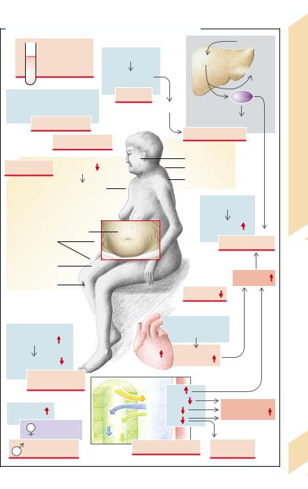
.pdfA. Antidiuretic Hormone (Vasopressin, ADH) Excess and Deficiency
|
|
|
|
Tumors, |
Alcohol, |
Autoimmune |
Pain, stress, |
|
|
|
|||
CNS damage, |
|
|
|
esp. small-cell |
cold |
disease |
hypothyroidism, |
|
|
|
bronchial carcinoma |
Genetic |
|
drugs |
|
|
|
|
defect |
|
|
|
|
|
|
||
|
|
|
|
|
|
|
|
|
|
|
Damage to |
|
Prolactin |
|
|
|
|
|
hypothalamus |
|
||
|
|
|
|
|
|
|
|
|
|
|
|
Renal defect |
|
Hormone, |
|
|
|
|
ADH |
|
|
|
|
|
|
|
|
|
ADH |
|
|
Pulmonary |
|
|
|
|
|
Adiuretic |
|
|
|
|
|
|
|
||
disease |
|
|
|
|
|
|
|
|
|
|
H2O |
|
|
H2O excess |
|
|
|
|
|
|
|
||
1 |
|
|
|
|
Polydipsia |
The |
|
|
|
|
|
H2O |
|||
|
|
|
|
|
|
|
|
Excretion |
|
Hypotonic |
|
NaCl |
|
9.3 |
|
|
|
|
|
Thirst |
|||
|
|
|
hyperhydration |
|
|
Plate |
|
|
|
|
|
|
|
Excretion |
|
|
|
|
|
|
|
|
|
Urolithiasis |
|
|
|
Hypertonic |
|
|
|
|
|
|
|
2 |
dehydration |
|
|
|
|
|
|
|
|
|
|
|
|
|
Cerebral |
|
Cell shrinkage |
|
|
|
|
|
edema |
|
|
||
B. Prolactin Excess |
|
|
|
|
|
|
|
Liver and renal failure |
|
Absence of dopaminergic inhibition |
Dopamine antagonists |
|
|||
Hypothyroidism |
|
|
Opiates, |
|
|
||
|
|
stress |
|
|
|||
|
|
|
|
|
|
|
|
|
|
|
|
|
Post-partum, |
|
|
TRH secretion |
|
|
estrogens |
|
|
||
|
|
|
|
|
|||
|
|
|
|
|
Tumor |
Inhibition |
|
|
|
|
|
|
|
of glucose |
|
|
|
|
|
|
|
absorption |
|
LH, FSH |
|
|
Prolactin |
|
|
||
|
|
|
|
|
|
||
|
|
|
|
|
Glucose |
|
|
Gonadal function |
|
|
|
|
|
|
|
|
|
|
|
|
Hyperglycemia |
|
|
|
|
|
|
Galactorrhea |
|
|
|
Amenorrhea, |
Androgen deficiency, |
(flow of milk) |
|
|
|||
|
loss of libido, |
|
|
|
|
||
estrogen deficiency |
|
|
|
|
|||
|
impotence |
|
Risk of diabetes mellitus developing |
261 |
|||
|
|
|
|
||||
|
|
|
|
|
|||
Silbernagl/Lang, Color Atlas of Pathophysiology © 2000 Thieme
All rights reserved. Usage subject to terms and conditions of license.

9 Hormones
262
Somatotropin
Somatotropin (growth hormone [GH]) is formed in the anterior lobe of the pituitary gland. It inhibits the uptake of glucose in fat and muscle cells and promotes lipolysis, gluconeogenesis, collagen synthesis, and the formation of erythropoietin (in part through the mediation of hepatic somatomedins or insulinlike growth factors [IGF], e.g., IGF-1). Somatotropin stimulates the enteric absorption of calcium and phosphates as well as the renal excretion of calcium. It also promotes bone growth (before the end of epiphyseal fusion and thus longitudinal growth) as well as soft tissue growth. Somatotropin promotes T-cell proliferation, interleukin 2 (IL-2) formation and the activity of natural killer cells, cytotoxic T cells, and macrophages. In this way it strengthens immune defense. Estrogens inhibit the formation of somatomedins and thus also reduce the effects of somatotropins.
The normally pulsatile liberation of somatotropin is regulated by the hypothalamic messenger substances somatoliberins (somatocrinin) and somatostatin (inhibitory). The release of somatotropin is stimulated by amino acids, hypoglycemia, glucagon, dopamine, and stress. Hyperglycemia, hyperlipidacidemia, obesity, and cold inhibit its release.
An excess of somatotropin is usually due to uncontrolled formation of the hormone, for example, by a pituitary adenoma or, in rare cases, by an ectopic tumor. Increased stimulation of hormone synthesis by somatoliberin is equally rare. Finally, uncontrolled therapeutic administration of somatotropin can also result in an iatrogenic excess of somatotropin (→A1).
Massive somatotropin excess before epiphyseal fusion is completed leads to gigantism (height up to 2.6 m). In adults it results in acromegaly (enlarged cheek bones, mandibula, feet and hands, and supraorbital bulge), cartilage hypertrophy with arthropathy and calcification of cartilage and intervertebral disks
(→A2). At the same time there is an increase in the size of soft tissues, for example, tongue, heart, liver, kidneys, thyroid, salivary glands, and skin (→A3). This increase in organ size can lead to further complications. If, for example, vascularization does not increase with myocardial hypertrophy, impaired coronary
oxygen delivery will result (angina pectoris;
→p. 218). Arterial hypertension occurs relatively frequently (in 30% of cases). Thickening of the skin is associated with increased sweat and sebum production. Compression of the median nerve can lead to carpal tunnel syndrome. Decreased glucose uptake in peripheral cells favors the development of hyperglycemia (→A4), in some cases of diabetes mellitus. Increased intestinal absorption results in calcium excess followed by hypercalciuria (→A5). The latter may cause precipitation of calcium salts in urine (nephrolithiasis;
→p.120). Somatotropin excess also promotes the development of tumors.
A somatotropin-producing pituitary tumor often causes enlargement of the sella turcica; pressure on the optic chiasma (→A6) can give rise to visual field defects (typically bitemporal hemianopia, as though the patient were wearing blinkers; →p. 326). Displacement of other endocrine cells can lead to gonadotropin deficiency, and thus to amenorrhea as well as loss of libido, and impotence (→A7). Conversely, somatotropin-producing tumors can also release other hormones, such as prolactin (→ p. 260).
Somatotropin deficiency can be genetically determined or due to damage of the hormoneproducing cells (e.g., tumor, hemorrhage, radiation), decreased hypothalamic stimulation, or an inibition of release (cortisol, hypothyroidism). The effect of somatotropin can also be weakened by estrogens. If somatotropin deficiency occurs before epiphyseal fusion, pituitary dwarfism will result. However, a deficiency that occurs after the completion of longitudinal growth will have no visible effect. Nevertheless, decreasing release of somatotropin in the elderly probably contributes to a weaken-
ing of the immune system.
Silbernagl/Lang, Color Atlas of Pathophysiology © 2000 Thieme
All rights reserved. Usage subject to terms and conditions of license.

A. Somatotropin Excess |
Magnetic resonance image kindly supplied by |
Santen, Düsseldorf Univ., Endocrinology Clinic |
|
|
|
|
|
|
Stress, |
|
|
||
|
|
NREM sleep, |
|
|||
|
|
hypoglycemia, |
|
|||
|
|
amino acids |
|
|||
|
|
|
|
|
||
Pituitary adenoma |
|
R. |
|
|
|
|
|
|
|
|
|
||
6 |
7 |
|
|
Rare: |
|
|
|
|
Ectopic adenoma |
|
|||
|
|
|
|
Somatotropin |
||
Compression of |
Gonadotropin |
|
|
|
||
optic chiasma |
deficiency |
|
|
|
|
|
Visual field |
Amenorrhea, |
|
|
|
|
|
defects |
loss of libido |
|
|
|
|
|
|
|
1 |
Iatrogenic |
|
||
|
and impotence |
|
9.4 |
|||
|
|
|
Somatotropin |
|
|
|
|
|
|
|
|
Plate |
|
|
|
|
|
Before epiphyseal fusion: |
||
|
|
|
|
|
Gigantism |
|
|
|
|
|
|
|
|
Glucose |
4 |
|
|
|
|
|
absorption |
|
|
|
|
|
|
reduced |
|
|
|
|
Hyperphosphatemia |
|
Hyperglycemia |
|
|
|
5 |
|
|
|
|
|
|
|
||
Visceromegaly |
|
|
2 |
|
|
|
|
|
|
|
|
|
|
|
Secretion of |
|
|
|
|
|
|
sweat and sebum |
|
Calciuria |
|
||
|
|
|
|
|
|
|
Carpal tunnel |
3 |
|
|
|
|
|
|
|
|
|
|
||
syndrome |
|
|
Acromegaly |
|
||
|
|
|
|
|
||
|
|
Macroglossia |
|
|
|
|
Risk of |
|
|
|
|
|
|
angina pectoris |
|
|
|
|
|
|
|
|
|
|
Enlargement of |
|
|
|
|
|
|
cheek bones, |
|
|
Hypertension |
|
|
|
hands and feet |
|
|
|
|
|
|
|
263 |
|
|
|
|
|
|
|
|
Silbernagl/Lang, Color Atlas of Pathophysiology © 2000 Thieme
All rights reserved. Usage subject to terms and conditions of license.

Adrenocortical Hormones: Enzyme Defects in Formation
9 Hormones
The most important adrenocortical hormones (corticoids) are the glucocorticoids and mineralocorticoids. Androgens, progestogens, and estrogens are also formed in the adrenal cortex.
All adrenocortical hormones (see also p. 272ff.) are formed from cholesterol. The transport of cholesterol to the mitochondria and subsequent transformation in pregnenolone can be impaired by a deficiency in steroidogenic acute regulatory protein (StAR). Several enzymes, which may be absent in genetic defects, are necessary for the formation of the various hormones.
Enzyme defects lead to decreased synthesis of enzyme products, and thus also of the hormones formed through their action. However, reduced glucocorticoid synthesis leads to disinhibition of the formation of corticoliberins
(CRH) and of corticotropin (adrenocorticotropic hormone [ACTH]). Corticotropin, in turn, stimulates the growth of the adrenal cortex, the release of cholesterol and the expression of several enzymes involved in the synthesis of adrenocorticoid hormones. As a result of this action, there is a rise in the concentration of enzyme substrates, their precursors, and metabolites as well as of steroids which are active preceding the enzyme defect in the metabolic chain. These steroids have partly hormonal effects, namely glucocorticoid (blue), mineralocorticoid (green), androgenic (red), progestogenic (orange), and estrogenic (violet) ones, as illustrated in Figs. 9.7 –9.10. Depending on what activity those products, substrates, precursors, and metabolites possess, there may thus be reduced (↓) or increased (↑) hormonal effects (see Table).
By using ACTH to stimulate adrenocorticoid hormone production, glucococorticoid production can be (practically) normalized, in spite of an enzyme defect. More frequently, though, the glucocorticoid action decreases (→ p. 270). If there is an excess of gestagenic metabolites, their weak antimineralocorticoid effect can trigger natriuresis (→ p. 276). Some enzyme defects increase concentrations of androgenic metabolites, with the corresponding consequences for sexual development (→ p. 272f.). If there is a 3β-hydroxydehydro- genase defect (→ A3), then insufficient amounts of androgens are formed for normal male sexual development to take place; too many androgens are formed for normal female sexual development. Limiting the production of the sexual hormones in the adrenal cortex does not, however, generally impair sexual development, since the sexual hormones are normally mainly formed in the gonads.
The most common enzyme defect is a deficiency of 21β-hydroxylase (cytochrome P450c21). Such a deficiency impairs transformation of progesterone into 11-desoxycorti- costerone and of 17-hydroxyprogesterone into 11-desoxycortisol (→ A5). Depending on the extent to which enzyme activity is impaired, there will be a moderate to severe cortisol deficiency. Increased formation of androstendion and testosterone leads to virilization of girls and premature development of male sex characteristics (incomplete precocious puberty) in boys (adrenogenital syndrome; see also p. 272). These effects can already be detected at birth, since the excess androgens are formed intrauterinely.
|
Enzym Defect |
Androgenic |
Glucocorticoid |
Mineralcorticoid |
|
(→ A1 –8) |
Action |
Action |
Action |
|
|
|
|
|
|
20,22-Desmolase (P450scc, StAR) |
↓ |
↓ |
↓ |
|
17α-Hydroxylase (P450c17) |
↓ |
↓ |
↑ |
|
3β-Hydroxydehydrogenase |
↑ (!) ↓ (") |
↓ |
↓ |
|
17-Reductase |
↓ |
– |
– |
|
21β-Hydroxylase (P450c21) |
↑ |
↓ |
↓ |
264 |
11β-Hydroxylase (P450c11) |
↑ |
↓ |
↑ |
18-Hydroxylase (P450c11AS) |
– |
– |
↓ |
|
|
18-Methyloxidase (P450c11AS) |
– |
– |
↓ |
|
|
|
|
|
Silbernagl/Lang, Color Atlas of Pathophysiology © 2000 Thieme
All rights reserved. Usage subject to terms and conditions of license.

A. Enzyme Defects in Formation of Adrenocortical Hormones
ACTH |
|
|
|
|
|
|
CRH |
|
|
|
|
|
|
|
|
|
|
|
|
|
|
|
|
12 18 |
|
StAR |
|
Enzymes: |
|
|
|
|
11 |
17 |
1 |
20,22-Desmolase |
|||
2 |
|
1 19 |
9 |
13 |
16 |
|
2 |
17α-Hydroxylase |
|
|
|
10 |
|
814 15 |
|
3 |
3β-Hydroxydehydrogenase |
||
|
3 |
|
|
|
|||||
HO |
|
|
|
7 |
|
|
4 |
17-Reductase |
|
|
|
5 |
|
|
|
||||
|
4 |
|
Cholesterol |
|
|||||
|
|
|
|
|
5 |
21β-Hydroxylase |
|||
|
|
|
|
|
|
|
|
||
1 |
|
|
|
|
|
|
|
6 |
11β-Hydroxylase |
|
|
|
|
CH3 |
|
CH3 |
7 |
18-Hydroxylase |
|
|
|
|
|
|
8 18-Methyloxidase |
||||
C O
HO
Pregnenolone
3
CH3
C O
O
Progesterone
5
CH2OH
C O
O 11-Desoxy- corticosterone
6
CH2OH
C O
HO
11
O
Corticosterone
7
|
|
C |
O |
|
|
O |
|
|
17 |
OH |
|
|
|
|
|
|
|
|
|
|
|
HO |
17-Hydroxy- |
HO |
|
Dehydro- |
|
|
|
|
|
|||
2 |
|
pregnenolone |
|
epiandrosterone |
||
|
3 |
|
3 |
|
|
|
|
|
|
|
|
||
|
|
CH3 |
|
|
|
|
|
|
C |
O |
|
|
O |
|
|
|
OH |
|
|
|
|
|
17 |
|
|
|
|
|
|
|
|
|
|
|
|
O |
17-Hydroxy- |
O |
|
|
|
|
|
|
Androstenedione |
|||
2 |
|
progesterone |
|
|||
|
5 |
|
|
|
|
|
|
|
|
|
|
|
|
|
|
CH2OH |
|
|
|
|
|
|
C |
O |
|
|
|
|
|
11 |
OH |
|
|
HO |
|
|
|
|
|
||
|
O |
|
|
|
|
|
|
|
11-Desoxycortisol |
|
|
|
|
|
|
6 |
|
6 |
|
|
|
|
CH2OH |
|
|
|
|
|
|
C |
O |
|
OH |
O |
|
|
HO |
OH |
|
||
|
|
11 |
|
|
|
|
|
|
|
|
|
|
|
|
O |
|
|
O |
11-Hydroxy- |
|
|
|
Cortisol |
|
|||
|
|
|
androstenedione |
|||
OH
|
HO |
Dihydroxy- |
|
|
|
4 |
|
androstene |
|
3 |
|
|
|
|
|
|
OH |
|
O |
|
4 |
|
Testosterone |
|
|
|
|
|
O |
Estrone
OH
HO
Estradiol
OH |
CH2OH |
CH2OH |
|
|
|
HO |
H2C C O |
HO |
OHC C |
O |
Hormonal effects: |
androgenic |
|
|
|
|
|
|||
|
|
|
|
|
|
glucocorticoid |
gestagenic |
O |
|
|
O |
|
|
mineralocorticoid |
estrogenic |
|
18-Hydroxy- |
|
|
|
|
||
|
|
|
Aldosterone |
|
|
||
|
corticosterone 8 |
|
|
|
|||
Plate 9.5 Adrenocortical Hormones
265
Silbernagl/Lang, Color Atlas of Pathophysiology © 2000 Thieme
All rights reserved. Usage subject to terms and conditions of license.

9 Hormones
266
Adrenocorticoid Hormones: Causes of Abnormal Release
The glucocorticoids serve, in the first instance, in the adaptation of metabolism, circulation, blood, immune system, etc. to stress, i.e., severe physical and psychological threat. The mineralocorticoids act on mineral and water balance (for mechanism of action see p. 268) by aiding in the renal retention of Na+ and the elimination of K+ and other ions.
The release of glucocorticoids (e.g., cortisol) is regulated by ACTH from the pituitary gland, which is, in turn, under the control of corticoliberin (corticotropin-releasing hormone [CRH]) from the hypothalamus (→ A). The most important stimulus for the release of CRH, and thus of ACTH and cortisol, is stress. Other stimuli are epinephrine, ADH, histamine, pyrogens, pain, fall in blood pressure, and hypoglycemia (→ A1). The organism’s diurnal rhythm also plays a role: release of cortisol is highest in the early morning hours and then slowly falls during the day (→ A2). The release is inhibited by morphine.
An excess of glucocorticoids often has an iatrogenic cause (therapeutic administration of glucocorticoids for immunosuppression; → A4), but it can also be the result of a hor- mone-producing tumor in the adrenal gland or other organs (especially small-cell bronchial carcinoma; → A3) (Cushing’s disease; → p. 268). The cause may be an excess stimulation of the adrenal by ACTH (secondary Cushing syndromes, for example, due to a pituitary tumor, other causes of CRH release, or ectopic formation of ACTH or, rarely, of CRH).
An important stimulus for the release of the mineralocorticoid aldosterone is angiotensin II, which is formed in increased amounts via the renin–angiotensin system when the renal perfusion pressure is reduced (→ A5). Aldosterone release is also stimulated by ADH, whose secretion is stimulated by angiotensin II. Aldosterone release is increased by hyperkalemia, but decreased by dopamine and the atrial natriuretic factor (ANF).
A selective excess of mineralocorticoids in the majority of cases occurs in the form of secondary hyperaldosteronism caused by increased renin release. In hypovolemia (e.g in dehydration) the increased release of aldosterone is adequate for controlling volume, but
usually too high for K+ balance. If hypovolemia occurs, the resulting “intertwining” of the regulatory circuits for plasma volume and potassium (→ p. 258) regularly leads to hypokalemia. Even if blood volume is normal or increased, renal perfusion may be impaired and thus renin release increased in a number of renal diseases. If the pumping action of the heart is reduced (→ p. 224), or in peripheral vasodilation (e.g., in sepsis or liver failure; → p.118) the blood pressure can be maintained only by massive activation of the sympathetic system, resulting in renal vasoconstriction, renin release, and hyperaldosteronism. Another cause may be an aldosterone-producing tumor in the adrenal (Conn’s syndrome). Furthermore, a defect of 11β-hydroxysteroid dehydrogenase (→ p. 212) may result in an increased mineralocorticoid effect. The enzyme is normally formed in the target cells of aldosterone and inactivates cortisol. This fits into the mineralocorticoid receptor and its mineralocorticoid action is normally stopped only by enzymatic inactivation. Because its concentration in blood is more than a hundred times higher than that of aldosterone, cortisol will cause a massive mineralocorticoid effect if 11β-hydroxysteroid dehydrogenase is defective. In a rare genetic defect (glucocorticoid remediable hyperaldosteronism), the expression of aldosterone producing enzymes is driven by an ACTH-sensitive promotor, leading to enhanced aldosterone production, whenever ACTH is high. Treatment of the patients with glucocorticoids suppresses ACTH release and thus hyperaldosteronism. In yet another rare genetic disease the mineralocorticoid receptor is sensitive to progesterone, leading to pseudohyperaldosteronism which exacerbates in pregnancy.
A deficiency of adrenal hormones (→ B) can be the consequence of adrenal insufficiency (Addison’s disease;→ p. 270; e.g., in genetic defects, autoimmune adrenal disease, tuberculosis, metastases, surgical removal) or of enzyme defects in adrenal hormone synthesis (→ p. 264). In addition, there may be insufficient stimulation by ACTH, as in damage to the pituitary gland or hypothalamus. Aldosterone release can also be reduced as a result of hypokalemia or decreased angiotensin II formation.
Silbernagl/Lang, Color Atlas of Pathophysiology © 2000 Thieme
All rights reserved. Usage subject to terms and conditions of license.

A. Causes of Cortisol and Aldosterone Excess |
|
|
|
|
|
|
|
|
|
||
Psychological and |
ADH, epinephrine, |
Pain, drop in blood |
|
|
|
||||||
physical stress |
histamine, pyrogens |
pressure, hypoglycemia |
|
|
|||||||
|
1 |
|
|
|
|
|
|
|
|
|
|
|
|
Corticoliberin |
|
|
|
Morphine |
|
|
|
|
|
|
Pituitary |
(CRH) |
|
|
|
Diurnal rhythm of |
|
AdrenocorticoidHormones |
|||
|
|
|
|
|
|
||||||
3 |
adenoma |
|
|
|
|
plasma glucocorticoids |
|||||
|
|
|
|
|
|
|
|
|
|
||
Bronchial carcinoma |
|
|
|
|
|
|
|
|
2 |
||
and others |
|
|
|
|
|
|
|
|
|
|
|
|
Corticotropin |
|
|
|
|
|
|
|
|
||
|
|
(ACTH) |
|
|
|
|
|
|
|
|
|
|
|
|
20 |
0 |
4 |
8 |
12 |
16 |
20 |
24 h |
|
|
|
|
Concentration |
|
|
|
Relative hypovolemia |
||||
|
|
Enzyme defects |
|
|
|
9.6 |
|||||
Therap. cortisol |
|
|
Angiotensinogen |
|
|
||||||
|
|
|
|
|
|
||||||
administration |
|
|
|
|
|
|
|
|
Renin |
Plate |
|
|
|
|
|
|
Angiotensin I |
|
|
|
|||
|
|
Adrenals |
|
|
Angiotensin ll |
|
|
5 |
|
||
|
|
|
|
|
|
|
|
||||
|
|
|
|
|
|
|
ADH |
Hyperkalemia |
|
||
|
|
|
|
|
|
|
|
|
|||
4 |
|
|
|
|
|
|
Dopamine, ANF |
|
|||
|
Tumors or |
|
|
|
|
|
|
|
|
|
|
|
|
hyperplasia |
|
|
|
|
|
|
|
|
|
Cortisol |
|
Aldosterone |
|
|
|
|
|
|
|||
B. Causes of Cortisol and Aldosterone Deficiency |
|
|
|
|
|
|
|
|
|||
|
|
Damage to |
|
Damage to pituitary |
hypothalamus |
||
Hypervolemia |
|||
|
|
||
|
|
Angiotensinogen |
|
Enzyme defects |
ACTH |
Renin |
|
|
|
||
|
|
Angiotensin I |
|
Adrenal insufficiency |
|
|
|
(e. g. inflammations, |
|
Angiotensin ll |
|
surgical removal) |
|
||
Hypokalemia
Cortisol |
Aldosterone |
267 |
Silbernagl/Lang, Color Atlas of Pathophysiology © 2000 Thieme
All rights reserved. Usage subject to terms and conditions of license.

9 Hormones
268
Excess Adrenocorticoid Hormones: Cushing’s Disease
Glucocorticoids (especially cortisol) stimulate gluconeogenesis in the liver and inhibit glucose uptake in peripheral cells. They also stimulate lipolysis, the breakdown of proteins in the periphery, and the formation of plasma proteins (e.g., angiotensinogen) in the liver. They promote the formation of erythrocytes, thrombocytes, and neutrophil granulocytes (neutrophils). At the same time they reduce the number of eosinophil granulocytes (eosinophils) and basophil granulocytes (basophils), lymphocytes, and monocytes. They also, via the formation of the proteins lipocortin and vasocortin, suppress the release of histamine, interleukins, and lymphokines. By inhibiting phospholipase A2 they suppress the formation of prostaglandins and leukotrienes. They diminish antibody formation and thus act as immunosuppressives. Glucocorticoids suppress inflammation by inhibiting connective tissue proliferation, but at the same time impede collagen synthesis and repair. They stimulate the secretion of acids and pepsin in the stomach and slow down mucus production. In addition, they decrease the plasma levels of calcium and phosphate, in part by inhibiting calcitriol formation. They also sensitize blood vessels and the heart to catecholamines, partly by inhibiting prostaglandin synthesis, stimulate the release of norepinephrine, and increase the excitability of the nervous system.
Mineralocorticoids (especially aldosterone) further renal retention of Na+ and water. They thus facilitate a rise in blood pressure. They also stimulate renal elimination of K+, Mg2+, and H+ and simultaneously the intracellular uptake of potassium. However, at high plasma levels cortisol also exerts a significant mineralocorticoid effect, even though it is largely inactivated in the target cells of the mineralocorticoids (→p. 266). Dehydro-epiandrosterone
(DHEA), the precursor of the steroid sex hormones, is also formed in the adrenals, in addition to mineralocorticoids and glucocorticoids.
The metabolic effects of glucocorticoid excess favor the development of diabetes mellitus (→p. 286ff.), i.e., steroid diabetes, in which the release of insulin is increased (→A2). The free fatty acids formed by stimulated lipolysis are utilized in the liver to generate very low
density lipoproteins (VLDL) which are passed into the blood (→A3). In addition, the liver forms ketone bodies from fatty acids. A redistribution of fat tissue occurs due to differing sensitivities of peripheral fatty tissue for glucocorticoids and insulin. This results in centripetal fat stores, rounded or moon faces and fat deposits in the neck (“buffalo” hump), while the limbs are noticeably thin. Peripheral protein breakdown (→A5) leads to muscle wasting, osteoporosis (loss of bone matrix), striae (breakdown of subcutaneous connective tissue), and purpura (increased vascular fragility). Because repair is impeded, wound healing is delayed. The effect on bone is aggravated by CaHPO4 deficiency and in children results in delayed growth. The effects on blood lead to polycythemia (→A1), thrombocytosis, and raised coagulability (→A6). Weakened immune defenses encourage infections (→A4). Sensitization of the circulation to catecholamines causes, among other things, an increase in cardiac contractility as well as peripheral vasoconstriction, and thus leads to hypertension (→A7), which, together with hyperlipidemia and raised coagulability of blood, promotes the development of atherosclerosis, thrombosis, and vascular occlusions (→A6). Due to stimulation of hydrochloric acid and pepsin secretion and the inhibition of mucus secretion in the stomach, gastric and/or duodenal (peptic) ulcers develop (→A8). The effects on the nervous system can trigger an endocrine psychogenic syndrome.
An increased mineralcorticoid effect causes hypervolemia, which in turn leads to hypertension; it also causes hypokalemia, hypomagnesemia, and alkalosis, which in turn lead to increased neuromuscular excitability (→A10). The effects are, among others, abnormal action potential formation and conduction in the heart.
An excess of androgens (→A9) can lead to masculinization and amenorrhea (virilism) in women, and to an accelerated onset of sexual characteristics in male children (incomplete precocious puberty; →p. 272).
Silbernagl/Lang, Color Atlas of Pathophysiology © 2000 Thieme
All rights reserved. Usage subject to terms and conditions of license.
A. Effects and Symptoms of Adrenocortical Hormone Excess |
|
|
||||||
|
|
|
|
|
|
|
Lipolysis |
|
|
|
Polycythemia, |
Gluconeogenesis |
|
|
|
||
|
|
leukocytosis, |
Free fatty acids |
|
||||
|
|
eosinopenia |
|
|
||||
|
|
1 |
|
Ketone bodies |
||||
|
|
|
|
|
Hyperglycemia |
|
|
|
|
|
|
|
|
2 |
|
|
|
|
Lymphopenia, |
|
|
Diabetes |
|
VLDL |
|
|
|
|
|
|
|
|
|||
|
inhibition of immune defenses |
Insulin |
|
|
||||
|
4 |
Susceptibility |
|
3 |
Lipogenesis |
|
||
|
|
|
|
Disease |
||||
|
|
to infection |
|
Redistribution |
||||
|
|
|
|
|
|
|||
|
|
|
Neuropsychological |
of fat tissue |
||||
|
|
|
disorders |
|
|
|
||
|
Protein |
|
|
|
Moon face |
Cushing’s |
||
|
|
CaHPO4 |
‘Buffalo’ hump |
|||||
|
breakdown |
|
||||||
|
|
|
|
|
|
|||
|
|
|
|
|
|
Truncal obesity |
||
|
|
|
|
Osteoporosis |
|
|
9.7 |
|
|
|
5 |
|
|
|
Thrombocytosis |
||
|
|
|
|
|
|
Plate |
||
|
|
|
|
|
|
|
|
|
|
|
Muscular |
|
Striae |
|
Clotting |
|
|
|
|
weakness: |
|
6 |
|
|
||
|
|
|
|
|
|
|
||
|
|
thin limbs |
|
|
|
|
Atherosclerosis |
|
|
|
Purpura |
|
|
|
|
|
|
|
|
Delayed |
|
|
|
|
Blood |
|
|
|
|
|
|
in children: |
pressure |
||
|
wound healing |
|
|
|
||||
|
|
|
|
|
|
|||
|
|
|
|
|
|
Growth |
|
|
|
|
|
|
|
|
Sensitization to |
|
|
Gastric acid and |
|
|
|
catecholamines |
|
|
||
|
|
|
|
7 |
|
|||
pepsin secretion |
|
|
Cardiac |
|
||||
|
|
|
|
|
|
|
||
|
|
|
|
|
output |
Peripheral |
|
|
|
Mucus secretion |
|
|
resistance |
|
|
||
|
|
|
|
|
|
|||
8 |
|
Gastric and |
|
|
|
|
|
|
|
|
|
Transport in |
|
|
|
||
|
|
duodenal ulcers |
|
|
|
|||
|
|
distal nephron |
Na+ |
|
|
|||
|
|
|
|
|
|
10 |
||
|
|
|
|
|
Na+ |
Mg2+ |
Neuromuscular |
|
Androgens |
9 |
|
|
H+ |
||||
|
|
excitability |
|
|||||
|
|
|
|
H+ |
K+ |
|
||
|
|
Hirsutism, |
|
|
|
|||
|
|
|
+ |
|
|
|
||
|
|
amenorrhea |
K |
|
|
|
||
|
|
|
|
|
|
|||
|
|
Precocious |
|
Electrolyte disturbances |
Renal damage |
|
||
|
|
pseudopuberty |
|
|
|
269 |
||
Silbernagl/Lang, Color Atlas of Pathophysiology © 2000 Thieme
All rights reserved. Usage subject to terms and conditions of license.

9 Hormones
270
Deficiency of Adrenocorticoid Hormones: Addison’s Disease
For the effects of the adrenocorticoid hormones, see p. 268.
Glucocorticoid deficiency frequently leads to hypoglycemia as a result of disinhibited glycolysis and reduced gluconeogenesis (→A1). This is especially marked in secondary deficiency of adrenocorticoid hormones due to pituitary insufficiency, because it is associated with decreased somatotropin secretion, the hyperglycemic effect of which will be absent (→p. 262). The hypoglycemia activates the sympathetic nervous system and inhibits the release of insulin, and thus also of its influence on lipolysis and protein breakdown. The reduced lipolytic and proteolytic action of cortisol is more than compensated by a decreased insulin and an increased epinephrine effect. Lipolysis and protein breakdown are thus stimulated. Further effects of the raised epinephrine release are tachycardia and sweating (→A2). The reduced sensitivity to catecholamines of the heart and blood vessels leads to a fall in blood pressure despite the release of epinephrine. Due to the diminished secretion of hydrochloric acid, pathogens that have been swallowed will be less effectively killed in the stomach and more commonly cause gastrointestinal infections (→ A6). Diarrhea and vomiting occur with corresponding loss of water and electrolytes. The lack in glucocorticoid effect on blood-forming cells results in anemia, neutropenia, eosinophilia, and lymphocytosis
(→A4). Other symptoms are fatigue and weakness. Furthermore, depression is caused by the lack of glucocorticoid action on the brain. However, while cortisol deficiency persists, sensitivity of the target cells is raised and they thus delay the onset of symptoms.
In primary adrenocorticoid insufficiency
(Addison’s disease) the diminished negative feedback from cortisol leads to a massive rise in the synthesis of pro-opiomelanocortin
(POMC), the precursor of ACTH. This increases formation not only of ACTH, but also of α-me- lanotropin (α-MSH or melanocortin). α-MSH as well as ACTH itself cause brown discoloration of the skin (→ A3), because of which Addison’s disease has been called “bronze disease”. If one adrenal cortex is absent, ACTH causes hypertrophy of the intact adrenal cor-
tex. If both adrenals are absent, ACTH can even cause the ectopic formation of adrenocorticoid hormones, but this is usually inadequate. In secondary adrenocorticoid insufficiency skin pigmentation is decreased because of a lack of α-MSH and ACTH.
Mineralocorticoid deficiency leads to renal salt loss and renal retention of K+, Mg2+, and H+ (→A5). Na+ reabsorption in the sweat glands and gut is also impaired. This results in salt deficiency, hypotonic dehydration, hypovolemia, drop in blood pressure, and in the increase of intracellular volume (→ p.122ff.). This can lead to a decrease in renal perfusion and glomerular filtration rate, causing an increase of plasma creatinine concentration. Also, due to the impaired renal perfusion the release of renin and angiotensin I–II will be raised. As angiotensin II stimulates ADH release and ADH leads to renal water retention the release of angiotensin II contributes to hypoosmolarity. The retention of K+, Mg2+, and H+ leads to reduced neuromuscular excitability as well as abnormalities of action potential formation and conduction in the heart due to hyperkalemia, hypermagnesemia, and acidosis (→A8 and p.124ff.). In combined mineralocorticoid and glucocorticoid deficiency, the increased fat and protein breakdown and loss of fluid cause weight loss, and arterial hypotension and anemia reduce physical fitness.
A lack of androgens manifests itself especially in sparse pubic hair as well as muscle wasting and loss of libido (→ A7). However, lack of adrenal androgens is of no consequence in men, as long as testosterone production in the testes is normal.
Acute worsening of the symptoms leads to Addisonian crisis with extreme weakness, fall in blood pressure, tachycardia, diarrhea, hypoglycemia, hyponatremia, hyperkalemia, and oliguria. It is frequently the consequence of an infection that normally, but not in patients with Addison’s disease, leads to an increase in cortisol release.
Silbernagl/Lang, Color Atlas of Pathophysiology © 2000 Thieme
All rights reserved. Usage subject to terms and conditions of license.