

Post-text assignments
1 Translate the following:
1 Each of the 5 senses consists of organs with specialized cells that have receptors for specific stimuli.
2 The subjects go through an initial period of great confusion, but subsequently they perceive the images as right side up.
3 In the dark, a substance produced by the rod cells increases the sensitivity of the eye so that it is possible to detect very dim light.
4 The human ear can perceive frequencies from 16 cycles per second, which is a very deep bass, to 28,000 cycles per second, which is a very high pitch.
5 Generally, the taste buds close to the tip of the tongue are sensitive to sweet tastes, whereas those in the back of the tongue are sensitive to bitter tastes.
6 The nose also has a structure called the vomeronasal organ whose function has not been determined, but which is suspected of being sensitive to pheromones that influence the reproductive cycle.
7 Hairs on the skin magnify the sensitivity and act as an early warning system for the body.
2 Answer the questions:
1 What is called the «blind spot» of the eye?
2 Why do birds have a higher visual acuity compared to humans?
3 What is the function of otoliths?
4 What is called a «perfect pitch»?
5 What sensations taste buds are able to detect?
6 Who have a greater number of taste buds – men or women?
7 What sensations the smell receptors are sensitive to?
8 What four kinds of touch sensations can be identified?
Unit 17
Pre-text assignment
Learn the key words and phrases:
ciliary body, conjuctiva, aqueous humor, iris, lens, pupil, rhodopsin.
The Human Eye
Cornea and sclera
The eye is made of three coats, or tunics. The outermost coat consists of the cornea and the sclera; the middle coat contains the main blood supply to the eye and consists of the choroid, the ciliary body, and the iris. The innermost layer is the retina. The sclera, or the white of the eye, is composed of tough fibrous tissue. On the exposed area of the eye the scleral surface is covered with a mucous membrane called the conjunctiva. This protects the eye from becoming dry. The cornea, a part of the sclera, is the transparent window of the eye through which light passes. The focusing of light begins in the cornea. Behind the cornea is a watery fluid called the aqueous humor. This fluid fills a curved, crescent-shaped space, thick in the center and thinner toward the edges. The cornea and the aqueous humor together make an outer lens that refracts, or bends, light and directs it toward the center of the eye.
Iris
Behind the aqueous humor is a colored ring called the iris. The color of the iris is inherited and does not affect vision. The iris is like a muscular curtain that opens and closes. It controls the amount of light entering the eye through the pupil, an opening in the iris. The pupil looks like a black spot. Light from everything a person sees must go through the pupil. When more or less light is needed to see better, the pupil becomes larger or smaller through the movement of the muscle in the iris. The aqueous humor flows through the pupil into a small space between the iris and the lens. A simple way to see how the pupils respond to light is to stand in front of a mirror with the eyes closed, covered by the hands for about ten seconds. When the hands are removed and the eyes opened, the pupils begin to get smaller, or contract, in response to the light. When light is reduced, pupils expand; when it is increased, they contract. The choroid is a layer of blood vessels and connective tissue squeezed between the sclera and the retina. It supplies nutrients to the eye. The ciliary body is a muscular structure that changes the shape of the lens.
Lens
Behind the pupil and iris are the crystalline lens and the ciliary muscle. The muscle holds the lens in place and changes its shape. The lens is a colorless, nearly transparent double convex structure, similar to an ordinary magnifying glass. Its only function is to focus light rays onto the retina. The lens is made of elongated cells that have no blood supply. These cells obtain nutrients from the surrounding fluids the aqueous humor in front and the vitreous body, a clear jelly, behind. The shape of the lens essentially that of a flattened globe can be changed by the movement of the ciliary muscles surrounding it. Hence, the eye can focus clearly on objects at widely varying distances. The ability of the lens to adjust from a distant to a near focus is called accommodation.
By contracting, the ciliary muscle pushes the lens to make it thicker in the middle. By relaxing, the muscle pulls the lens and flattens it. To see objects clearly when they are close to the eyes the lens is squeezed together and thickened. To see distant objects clearly it is flattened. For people with normal vision, the relaxed ciliary muscle flattens the lens enough to bring objects into sharp focus if they are 6 meters or more from the eye. To see closer objects clearly, the ciliary muscle must contract in order to thicken the lens. Young children can see objects clearly at distances as close as 6.4 centimeters. After about age 45 most people must have objects farther and farther away in order to see them clearly. The lens becomes less elastic as a person grows older.
Retina
The retina is a soft, transparent layer of nervous tissue made up of millions of light receptors. The retina is connected to the brain by the optic nerve. All of the structures needed to focus light onto the retina and to nourish it are housed in the eye, which is primarily a supporting shell for the retina. When light enters the eye it passes through the lens and focuses an image onto the retina. The retina has several layers, one of which contains special cells named for their
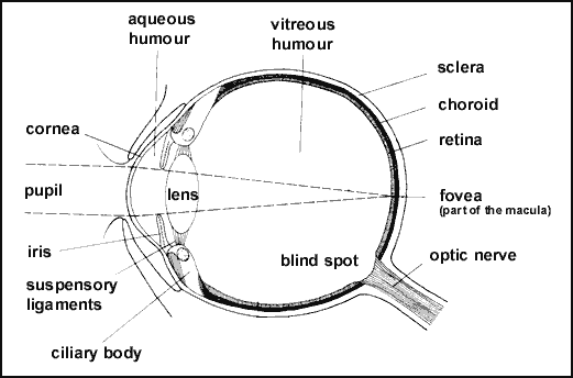
Figure 7 – The Eye
shapes rods and cones. Light-sensitive chemicals in the rods and cones react to specific wavelengths of light and trigger nerve impulses. These impulses are carried through the optic nerve to the visual center in the brain. Here they are interpreted, and sight occurs. Light must pass through the covering layers of the retina to reach the layer of rods and cones. There are about 75 to 150 million rods and about 7 million cones in the human retina. Rods do not detect lines, points, or color. They perceive only light and dark tones in an image. The sensitive rods can distinguish outlines or silhouettes of objects in almost complete darkness. They make it possible for people to see in darkness or at night. Cones are the keenest of the retina's receptor cells. They detect the fine lines and points of an image. The cones, for example, make it possible to read these words. There are three types of cones that receive color sensations. One type absorbs light best in wavelengths of blue-violet and another in wavelengths of green; a third is sensitive to wavelengths of yellow and red.