

Borchers Andrea Ann (ed.) Handbook of Signs & Symptoms 2015
.pdfAppendicitis. With appendicitis, a life-threatening disorder, pain initially occurs in the epigastric or umbilical region. Anorexia, nausea, or vomiting may occur after the onset of pain. Pain localizes at McBurney’s point in the right lower quadrant and is accompanied by abdominal rigidity, increasing tenderness (especially over McBurney’s point), rebound tenderness, and retractive respirations. Later signs and symptoms include malaise, constipation (or diarrhea), low-grade fever, and tachycardia.
Cholecystitis. Severe pain in the right upper quadrant may arise suddenly or increase gradually over several hours, usually after meals. It may radiate to the right shoulder, chest, or back. Accompanying the pain are anorexia, nausea, vomiting, fever, abdominal rigidity, tenderness, pallor, and diaphoresis. Murphy’s sign (inspiratory arrest elicited when the examiner palpates the right upper quadrant as the patient takes a deep breath) is common.
Cholelithiasis. Patients may suffer sudden, severe, and paroxysmal pain in the right upper quadrant lasting several minutes to several hours. The pain may radiate to the epigastrium, back, or shoulder blades. The pain is accompanied by anorexia, nausea, vomiting (sometimes bilious), diaphoresis, restlessness, and abdominal tenderness with guarding over the gallbladder or biliary duct. The patient may also experience fatty food intolerance and frequent indigestion.
Cirrhosis. Dull abdominal aching occurs early and is usually accompanied by anorexia, indigestion, nausea, vomiting, constipation, or diarrhea. Subsequent right upper quadrant pain worsens when the patient sits up or leans forward. Associated signs include fever, ascites, leg edema, weight gain, hepatomegaly, jaundice, severe pruritus, bleeding tendencies, palmar erythema, and spider angiomas. Gynecomastia and testicular atrophy may also be present.
Crohn’s disease. An acute attack in Crohn’s disease causes severe cramping pain in the lower abdomen, typically preceded by weeks or months of milder cramping pain. Crohn’s disease may also cause diarrhea, hyperactive bowel sounds, dehydration, weight loss, fever, abdominal tenderness with guarding, and possibly a palpable mass in a lower quadrant. Abdominal pain is commonly relieved by defecation. Milder chronic signs and symptoms include right lower quadrant pain with diarrhea, steatorrhea, and weight loss. Complications include perirectal or vaginal fistulas.
Diverticulitis. Mild cases of diverticulitis usually produce intermittent, diffuse left lower quadrant pain, which is sometimes relieved by defecation or passage of flatus and worsened by eating. Other signs and symptoms include nausea, constipation or diarrhea, a low-grade fever and, in many cases, a palpable abdominal mass that’s usually tender, firm, and fixed. Rupture causes severe left lower quadrant pain, abdominal rigidity and, possibly, signs and symptoms of sepsis and shock (high fever, chills, and hypotension).
Duodenal ulcer. Localized abdominal pain — described as steady, gnawing, burning, aching, or hunger-like — may occur high in the midepigastrium, slightly off center, usually on the right. The pain usually doesn’t radiate unless pancreatic penetration occurs. It typically begins 2 to 4 hours after a meal and may cause nocturnal awakening. Ingestion of food or antacids brings relief until the cycle starts again, but it may also produce weight gain. Other symptoms include changes in bowel habits and heartburn or retrosternal burning.
Ectopic pregnancy. Lower abdominal pain may be sharp, dull, or cramping and constant or intermittent in ectopic pregnancy, a potentially life-threatening disorder. Vaginal bleeding, nausea, and vomiting may occur, along with urinary frequency, a tender adnexal mass, and a 1- to 2-month history of amenorrhea. Rupture of the fallopian tube produces sharp lower abdominal pain, which may radiate to the shoulders and neck and become extreme with cervical or adnexal

palpation. Signs of shock (such as pallor, tachycardia, and hypotension) may also appear. Endometriosis. Constant, severe pain in the lower abdomen usually begins 5 to 7 days before the start of menses and may be aggravated by defecation. Depending on the location of the ectopic tissue, the pain may be accompanied by constipation, abdominal tenderness, dysmenorrhea, dyspareunia, and deep sacral pain.
Escherichia coli O157:H7. E. coli O157:H7 is an aerobic, gram-negative bacillus that causes food-borne illness. Most strains of E. coli are harmless and are part of normal intestinal flora of healthy humans and animals. However, E. coli O157:H7, one of hundreds of strains of the bacterium, is capable of producing a powerful toxin and can cause severe illness. Eating undercooked beef or other foods contaminated with the bacteria causes the disease. Signs and symptoms include watery or bloody diarrhea, nausea, vomiting, fever, and abdominal cramps. In children younger than age 5 and in elderly patients, hemolytic-uremic syndrome may develop, and this may ultimately lead to acute renal failure.
Gastric ulcer. Diffuse, gnawing, burning pain in the left upper quadrant or epigastric area commonly occurs 1 to 2 hours after meals and may be relieved by ingestion of food or antacids. Vague bloating and nausea after eating are common. Indigestion, weight change, anorexia, and episodes of GI bleeding also occur.
Gastritis. With acute gastritis, the patient experiences a rapid onset of abdominal pain that can range from mild epigastric discomfort to burning pain in the left upper quadrant. Other typical features include belching, fever, malaise, anorexia, nausea, bloody or coffee-ground vomitus, and melena. However, significant bleeding is unusual, unless the patient has hemorrhagic gastritis.
Gastroenteritis. Cramping or colicky abdominal pain, which can be diffuse, originates in the left upper quadrant and radiates or migrates to the other quadrants, usually in a peristaltic manner. It’s accompanied by diarrhea, hyperactive bowel sounds, headache, myalgia, nausea, and vomiting.
Heart failure. Right upper quadrant pain commonly accompanies heart failure’s hallmarks: jugular vein distention, dyspnea, tachycardia, and peripheral edema. Other findings include nausea, vomiting, ascites, productive cough, crackles, cool extremities, and cyanotic nail beds. Clinical signs are numerous and vary according to the stage of the disease and amount of cardiovascular impairment.
Hepatitis. Liver enlargement from any type of hepatitis causes discomfort or dull pain and tenderness in the right upper quadrant. Associated signs and symptoms may include dark urine, clay-colored stools, nausea, vomiting, anorexia, jaundice, malaise, and pruritus.
Intestinal obstruction. Short episodes of intense, colicky, cramping pain alternate with painfree intervals in an intestinal obstruction, a life-threatening disorder. Accompanying signs and symptoms may include abdominal distention, tenderness, and guarding; visible peristaltic waves; high-pitched, tinkling, or hyperactive sounds proximal to the obstruction and hypoactive or absent sounds distally; obstipation; and pain-induced agitation. In jejunal and duodenal obstruction, nausea and bilious vomiting occur early. In distal smallor large-bowel obstruction, nausea and vomiting are commonly feculent. Complete obstruction produces absent bowel sounds. Late-stage obstruction produces signs of hypovolemic shock, such as hypotension and tachycardia.
Irritable bowel syndrome. Lower abdominal cramping or pain is aggravated by ingestion of coarse or raw foods and may be alleviated by defecation or passage of flatus. Related findings

include abdominal tenderness, diurnal diarrhea alternating with constipation or normal bowel function, and small stools with visible mucus. Dyspepsia, nausea, and abdominal distention with a feeling of incomplete evacuation may also occur. Stress, anxiety, and emotional lability intensify the symptoms.
Listeriosis. A serious infection, listeriosis is caused by eating food contaminated with the bacterium Listeria monocytogenes. This food-borne illness primarily affects pregnant women, neonates, and those with weakened immune systems. Signs and symptoms include fever, myalgia, abdominal pain, nausea, vomiting, and diarrhea. If the infection spreads to the nervous system, meningitis may develop. Signs and symptoms include fever, headache, nuchal rigidity, and change in the level of consciousness.
 GENDER CUE
Listeriosis infection during pregnancy may lead to premature delivery, infection of the neonate, or stillbirth.
Mesenteric artery ischemia. Always suspect mesenteric artery ischemia in patients older than age 50 with chronic heart failure, cardiac arrhythmia, cardiovascular infarct, or hypotension who develop sudden, severe abdominal pain after 2 to 3 days of colicky periumbilical pain and diarrhea. Initially, the abdomen is soft and tender with decreased bowel sounds. Associated findings include vomiting, anorexia, alternating periods of diarrhea and constipation and, in late stages, extreme abdominal tenderness with rigidity, tachycardia, tachypnea, absent bowel sounds, and cool, clammy skin.
Norovirus. The highly contagious Norovirus infection is transmitted by the fecal-oral route. An acute onset of GI irritation, with abdominal pain or cramping, occurs in patients affected with Noroviruses. Other symptoms include vomiting, uncontrolled stool seepage, watery nonbloody diarrhea, and nausea. Less common symptoms include low-grade fever, headache, chills, muscle aches, and generalized fatigue. Typically, Norovirus symptoms last from 1 to 5 days without lasting effects.
Ovarian cyst. Torsion or hemorrhage causes pain and tenderness in the right or left lower quadrant. Sharp and severe if the patient suddenly stands or stoops, the pain becomes brief and intermittent if the torsion self-corrects or dull and diffuse after several hours if it doesn’t. Pain is accompanied by slight fever, mild nausea and vomiting, abdominal tenderness, a palpable abdominal mass and, possibly, amenorrhea. Abdominal distention may occur if the patient has a large cyst. Peritoneal irritation, or rupture and ensuing peritonitis, causes high fever and severe nausea and vomiting.
Pancreatitis. Life-threatening acute pancreatitis produces fulminating, continuous upper abdominal pain that may radiate to both flanks and to the back. To relieve this pain, the patient may bend forward, draw his knees to his chest, or move restlessly about. Early findings include abdominal tenderness, nausea, vomiting, fever, pallor, tachycardia and, in some patients, abdominal rigidity, rebound tenderness, and hypoactive bowel sounds. Turner’s sign (ecchymosis of the abdomen or flank) or Cullen’s sign (a bluish tinge around the umbilicus) signals hemorrhagic pancreatitis. Jaundice may occur as inflammation subsides.
Chronic pancreatitis produces severe left upper quadrant or epigastric pain that radiates to the

back. Abdominal tenderness, a midepigastric mass, jaundice, fever, and splenomegaly may occur. Steatorrhea, weight loss, maldigestion, and diabetes mellitus are common.
Pelvic inflammatory disease. Pain in the right or left lower quadrant ranges from vague discomfort worsened by movement to deep, severe, and progressive pain. Sometimes, metrorrhagia precedes or accompanies the onset of pain. Extreme pain accompanies cervical or adnexal palpation. Associated findings include abdominal tenderness, a palpable abdominal or pelvic mass, fever, occasional chills, nausea, vomiting, urinary discomfort, and abnormal vaginal bleeding or purulent vaginal discharge.
Perforated ulcer. With perforated ulcer, a life-threatening disorder, sudden, severe, and prostrating epigastric pain may radiate through the abdomen to the back or right shoulder. Other signs and symptoms include boardlike abdominal rigidity, tenderness with guarding, generalized rebound tenderness, absent bowel sounds, grunting and shallow respirations and, in many cases, fever, tachycardia, hypotension, and syncope.
Peritonitis. With peritonitis, a life-threatening disorder, sudden and severe pain can be diffuse or localized in the area of the underlying disorder; movement worsens the pain. The degree of abdominal tenderness usually varies according to the extent of disease. Typical findings include fever; chills; nausea; vomiting; hypoactive or absent bowel sounds; abdominal tenderness, distention, and rigidity; rebound tenderness and guarding; hyperalgesia; tachycardia; hypotension; tachypnea; and positive psoas and obturator signs.
Prostatitis. Vague abdominal pain or discomfort in the lower abdomen, groin, perineum, or rectum may develop with prostatitis. Other findings include dysuria, urinary frequency and urgency, fever, chills, low back pain, myalgia, arthralgia, and nocturia. Scrotal pain, penile pain, and pain on ejaculation may occur in chronic cases.
Pyelonephritis (acute). Progressive lower quadrant pain in one or both sides, flank pain, and CVA tenderness characterize this disorder. Pain may radiate to the lower midabdomen or to the groin. Additional signs and symptoms include abdominal and back tenderness, high fever, shaking chills, nausea, vomiting, and urinary frequency and urgency.
Renal calculi. Depending on the location of calculi, severe abdominal or back pain may occur. However, the classic symptom is severe, colicky pain that travels from the CVA to the flank, suprapubic region, and external genitalia. The pain may be excruciating or dull and constant. Pain-induced agitation, nausea, vomiting, abdominal distention, fever, chills, hypertension, and urinary urgency with hematuria and dysuria may occur.
Sickle cell crisis. Sudden, severe abdominal pain may accompany chest, back, hand, or foot pain. Associated signs and symptoms include weakness, aching joints, dyspnea, and scleral jaundice.
Smallpox (variola major). Worldwide eradication of smallpox was achieved in 1977; the United States and Russia have the only known storage sites for the virus. The virus is considered a potential agent for biological warfare. Initial signs and symptoms include high fever, malaise, prostration, severe headache, backache, and abdominal pain. A maculopapular rash develops on the mucosa of the mouth, pharynx, face, and forearms and then spreads to the trunk and legs. Within 2 days, the rash becomes vesicular and later pustular. The lesions develop at the same time, appear identical, and are more prominent on the face and extremities. The pustules are round, firm, and embedded in the skin. After 8 to 9 days, the pustules form a crust, and later, the scab separates from the skin, leaving a pitted scar. In fatal cases, death results from encephalitis, extensive bleeding, or secondary infection.

Splenic infarction. Fulminating pain in the left upper quadrant occurs along with chest pain that may worsen on inspiration. Pain usually radiates to the left shoulder with splinting of the left diaphragm, abdominal guarding and, occasionally, a splenic friction rub.
Ulcerative colitis. Ulcerative colitis may begin with vague abdominal discomfort that leads to cramping lower abdominal pain. As the disorder progresses, pain may become steady and diffuse, increasing with movement and coughing. The most common symptom — recurrent and possibly severe diarrhea with blood, pus, and mucus — may relieve the pain. The abdomen may feel soft, squashy, and extremely tender. High-pitched, infrequent bowel sounds may accompany nausea, vomiting, anorexia, weight loss, and mild, intermittent fever.
Other Causes
Drugs. Salicylates and nonsteroidal anti-inflammatory drugs commonly cause burning, gnawing pain in the left upper quadrant or epigastric area, along with nausea and vomiting.
Special Considerations
Help the patient find a comfortable position to ease his distress. He should lie in a supine position, with his head flat on the table, arms at his sides, and knees slightly flexed to relax the abdominal muscles. Monitor him closely because abdominal pain can signal a life-threatening disorder. Especially important indications include tachycardia, hypotension, clammy skin, abdominal rigidity, rebound tenderness, a change in the pain’s location or intensity, or sudden relief from the pain.
Withhold analgesics from the patient because they may mask symptoms. Also withhold food and fluids because surgery may be needed. Prepare for I.V. infusion and insertion of a nasogastric or other intestinal tube. Peritoneal lavage or abdominal paracentesis may be required.
You may have to prepare the patient for a diagnostic procedure, which may include a pelvic and rectal examination; blood, urine, and stool tests; X-rays; barium studies; ultrasonography; endoscopy; and biopsy.
Patient Counseling
Explain the diagnostic tests the patient will need, which foods and fluids he shouldn’t have, the need to report any changes in bowel habits, and how to position himself to alleviate symptoms.
Pediatric Pointers
Because a child typically has difficulty describing abdominal pain, you should pay close attention to nonverbal cues, such as wincing, lethargy, or unusual positioning (such as a sidelying position with knees flexed to the abdomen). Observing the child while he coughs, walks, or climbs may offer some diagnostic clues. Also, remember that a parent’s description of the child’s complaints is a subjective interpretation of what the parent believes is wrong.
In children, abdominal pain can signal a disorder with greater severity or different associated signs than in adults. Appendicitis, for example, has higher rupture and mortality rates in children, and vomiting may be the only other sign. Acute pyelonephritis may cause abdominal pain, vomiting, and diarrhea, but not the classic urologic signs found in adults. Peptic ulcer, which is becoming increasingly common in teenagers, causes nocturnal pain and colic that, unlike peptic ulcer in adults,
may not be relieved by food.
Abdominal pain in children can also result from lactose intolerance, allergic-tension-fatigue syndrome, volvulus, Meckel’s diverticulum, intussusception, mesenteric adenitis, diabetes mellitus, juvenile rheumatoid arthritis, and many uncommon disorders such as heavy metal poisoning. Remember, too, that a child’s complaint of abdominal pain may reflect an emotional need, such as a wish to avoid school or to gain adult attention.
Geriatric Pointers
Advanced age may decrease the manifestations of acute abdominal disease. Pain may be less severe, fever less pronounced, and signs of peritoneal inflammation diminished or absent.
REFERENCES
McCutcheon, T. (2013). The ileus and oddities after colorectal surgery. Gastroenterology Nursing, 36(5), 368–375.
Saccamano, S., & Ferrara, L. (2013). Evaluation of acute abdominal pain. The Nurse Practitioner: The American Journal of Primary Health Care, 38(11), 46–53.
Abdominal Rigidity
(See Also Abdominal Distention, Abdominal Mass, Abdominal Pain) [Abdominal muscle spasm, involuntary guarding]
Detected by palpation, abdominal rigidity refers to abnormal muscle tension or inflexibility of the abdomen. Rigidity may be voluntary or involuntary. Voluntary rigidity reflects the patient’s fear or nervousness upon palpation; involuntary rigidity reflects potentially life-threatening peritoneal irritation or inflammation. (See Recognizing Voluntary Rigidity, page 26.)
Involuntary rigidity most commonly results from GI disorders, but may also result from pulmonary and vascular disorders and from the effects of insect toxins. Usually, it’s accompanied by fever; nausea; vomiting; and abdominal tenderness, distention, and pain.
 EMERGENCY INTERVENTIONS
After palpating abdominal rigidity, quickly take the patient’s vital signs. Even though the patient may not appear gravely ill or have markedly abnormal vital signs, abdominal rigidity calls for emergency interventions.
Prepare to administer oxygen and to insert an I.V. line for fluid and blood replacement. The patient may require drugs to support blood pressure. Also prepare him for catheterization, and monitor intake and output.
A nasogastric tube may have to be inserted to relieve abdominal distention. Because emergency surgery may be necessary, the patient should be prepared for laboratory tests and X-rays.
History and Physical Examination

If the patient’s condition allows further assessment, take a brief history. Find out when the abdominal rigidity began. Is it associated with abdominal pain? If so, did the pain begin at the same time? Determine whether the abdominal rigidity is localized or generalized. Is it always present? Has its site changed or remained constant? Next, ask about aggravating or alleviating factors, such as position changes, coughing, vomiting, elimination, and walking.
Explore other signs and symptoms. Inspect the abdomen for peristaltic waves, which may be visible in very thin patients. Also, check for a visibly distended bowel loop. Next, auscultate bowel sounds. Perform light palpation to locate the rigidity and determine its severity. Avoid deep palpation, which may exacerbate abdominal pain. Finally, check for poor skin turgor and dry mucous membranes, which indicate dehydration.
Medical Causes
Abdominal aortic aneurysm (dissecting). Mild to moderate abdominal rigidity occurs with abdominal aortic aneurysm, a life-threatening disorder. Typically, it’s accompanied by constant upper abdominal pain that may radiate to the lower back. The pain may worsen when the patient lies down and may be relieved when he leans forward or sits up. Before rupture, the aneurysm may produce a pulsating mass in the epigastrium, accompanied by a systolic bruit over the aorta. However, the mass stops pulsating after rupture. Associated signs and symptoms include mottled skin below the waist, absent femoral and pedal pulses, lower blood pressure in the legs than in the arms, and mild to moderate tenderness with guarding. Significant blood loss causes signs of shock, such as tachycardia, tachypnea, and cool, clammy skin.
 EXAMINATION TIP Recognizing Voluntary Rigidity
Distinguishing voluntary from involuntary abdominal rigidity is a must for accurate assessment. Review this comparison so that you can quickly tell the two apart.
VOLUNTARY RIGIDITY
Usually symmetrical
More rigid on inspiration (expiration causes muscle relaxation)
Eased by relaxation techniques, such as positioning the patient comfortably and talking to him in a calm, soothing manner
Painless when the patient sits up using his abdominal muscles alone
INVOLUNTARY RIGIDITY
Usually asymmetrical
Equally rigid on inspiration and expiration
Unaffected by relaxation techniques
Painful when the patient sits up using his abdominal muscles alone

Insect toxins. Insect stings and bites, especially black widow spider bites, release toxins that can produce generalized, cramping abdominal pain, usually accompanied by rigidity. These toxins may also cause a low-grade fever, nausea, vomiting, tremors, and burning sensations in the hands and feet. Some patients develop increased salivation, hypertension, paresis, and hyperactive reflexes. Children commonly are restless, have an expiratory grunt, and keep their legs flexed.
Mesenteric artery ischemia. A life-threatening disorder, mesenteric artery ischemia is characterized by 2 to 3 days of persistent, low-grade abdominal pain and diarrhea leading to sudden, severe abdominal pain and rigidity. Rigidity occurs in the central or periumbilical region and is accompanied by severe abdominal tenderness, fever, and signs of shock, such as tachycardia and hypotension. Other findings may include vomiting, anorexia, and diarrhea or constipation. Always suspect this disorder in patients older than age 50 who have a history of heart failure, arrhythmia, cardiovascular infarct, or hypotension.
Peritonitis. Depending on the cause of peritonitis, abdominal rigidity may be localized or generalized. For example, if an inflamed appendix causes local peritonitis, rigidity may be localized in the right lower quadrant. If a perforated ulcer causes widespread peritonitis, rigidity may be generalized and, in severe cases, boardlike.
Peritonitis also causes sudden and severe abdominal pain that can be localized or generalized. In addition, it can produce abdominal tenderness and distention, rebound tenderness, guarding, hyperalgesia, hypoactive or absent bowel sounds, nausea, and vomiting. Usually, the patient also displays fever, chills, tachycardia, tachypnea, and hypotension.
Special Considerations
Continue to monitor the patient closely for signs of shock. Position him as comfortably as possible. The patient should lie in a supine position, with his head flat on the table, arms at his sides, and knees slightly flexed to relax the abdominal muscles. Because analgesics may mask symptoms, withhold them until a tentative diagnosis has been made. Because emergency surgery may be required, withhold food and fluids and administer an I.V. antibiotic. Prepare the patient for diagnostic tests, which may include blood, urine, and stool studies; chest and abdominal X-rays; a computed tomography scan; magnetic resonance imaging; peritoneal lavage; and gastroscopy or colonoscopy. A pelvic or rectal examination may also be done.
Patient Counseling
Explain diagnostic tests or surgery the patient will need, and give him instruction on measures to reduce anxiety.
Pediatric Pointers
Voluntary rigidity may be difficult to distinguish from involuntary rigidity if associated pain makes the child restless, tense, or apprehensive. However, in any child with suspected involuntary rigidity, your priority is early detection of dehydration and shock, which can rapidly become life threatening.
Abdominal rigidity in the child can stem from gastric perforation, hypertrophic pyloric stenosis, duodenal obstruction, meconium ileus, intussusception, cystic fibrosis, celiac disease, and appendicitis.
Geriatric Pointers
Advanced age and impaired cognition decrease pain perception and intensity. Weakening of abdominal muscles may decrease muscle spasms and rigidity.
REFERENCES
Boeckxstaens, G. E., & de Jonge, W. J. (2009). Neuroimmune mechanisms in post-operative ileus. Gut, 58(9), 1300–1311.
Iyer, S., Saunders, W. B. , & Stemkowski S. (2009). Economic burden of postoperative ileus associated with colectomy in the United States. Journal of Managed Care Pharmacy, 15(6), 485–494.
Accessory Muscle Use
When breathing requires extra effort, the accessory muscles — the sternocleidomastoid, scalene, pectoralis major, trapezius, internal intercostals, and abdominal muscles — stabilize the thorax during respiration. Some accessory muscle use normally takes place during such activities as singing, talking, coughing, defecating, and exercising. (See Accessory Muscles: Locations and Functions, page 28.) However, more pronounced use of these muscles may signal acute respiratory distress, diaphragmatic weakness, or fatigue. It may also result from chronic respiratory disease. Typically, the extent of accessory muscle use reflects the severity of the underlying cause.
 EMERGENCY INTERVENTIONS
If the patient displays increased accessory muscle use, immediately look for signs of acute respiratory distress. These include a decreased level of consciousness, shortness of breath when speaking, tachypnea, intercostal and sternal retractions, cyanosis, adventitious breath sounds (such as wheezing or stridor), diaphoresis, nasal flaring, and extreme apprehension or agitation. Quickly auscultate for abnormal, diminished, or absent breath sounds. Check for airway obstruction, and, if detected, attempt to restore airway patency. Insert an airway, or intubate the patient. Then, begin suctioning and manual or mechanical ventilation. Assess oxygen saturation using pulse oximetry, if available. Administer oxygen; if the patient has chronic obstructive pulmonary disease (COPD), use only a low flow rate for mild COPD exacerbations. You may need to use a high flow rate initially, but be attentive to the patient’s respiratory drive. Giving a patient with COPD too much oxygen may decrease his respiratory drive. An I.V. line may be required.
History and Physical Examination
If the patient’s condition allows, examine him more closely. Ask him about the onset, duration, and severity of associated signs and symptoms, such as dyspnea, chest pain, cough, or fever.
Explore his medical history, focusing on respiratory disorders, such as infection or COPD. Ask about cardiac disorders, such as heart failure, which may lead to pulmonary edema; also, inquire about neuromuscular disorders, such as amyotrophic lateral sclerosis, which may affect respiratory muscle function. Note a history of allergies or asthma. Because collagen vascular diseases can cause diffuse infiltrative lung disease, ask about such conditions as rheumatoid arthritis and lupus erythematosus.
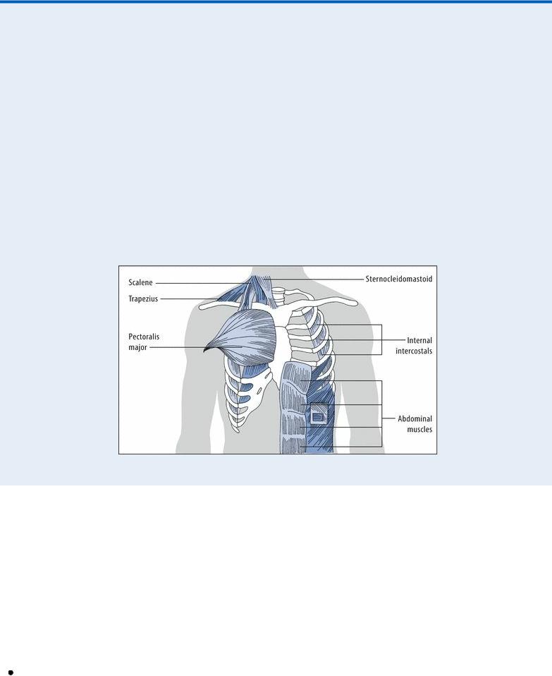
Accessory Muscles: Locations and Functions
Physical exertion and pulmonary disease usually increase the work of breathing, taxing the diaphragm and external intercostal muscles. When this happens, accessory muscles provide the extra effort needed to maintain respirations. The upper accessory muscles assist with inspiration, whereas the upper chest, sternum, internal intercostal, and abdominal muscles assist with expiration.
With inspiration, the scalene muscles elevate, fix, and expand the upper chest. The sternocleidomastoid muscles raise the sternum, expanding the chest’s anteroposterior and longitudinal dimensions. The pectoralis major elevates the chest, increasing its anteroposterior size, and the trapezius raises the thoracic cage.
With expiration, the internal intercostals depress the ribs, decreasing the chest size. The abdominal muscles pull the lower chest down, depress the lower ribs, and compress the abdominal contents, which exerts pressure on the chest.
Ask about recent trauma, especially to the spine or chest. Find out if the patient has recently undergone pulmonary function tests or received respiratory therapy. Ask about smoking and occupational exposure to chemical fumes or mineral dusts such as asbestos. Explore the family history for such disorders as cystic fibrosis and neurofibromatosis, which can cause diffuse infiltrative lung disease.
Perform a detailed chest examination, noting an abnormal respiratory rate, pattern, or depth. Assess the color, temperature, and turgor of the patient’s skin, and check for clubbing.
Medical Causes
Acute respiratory distress syndrome (ARDS). In ARDS, a life-threatening disorder, accessory muscle use increases in response to hypoxia. It’s accompanied by intercostal, supracostal, and sternal retractions on inspiration and by grunting on expiration. Other characteristics include tachypnea, dyspnea, diaphoresis, diffuse crackles, and a cough with pink, frothy sputum.