

Understanding the Human Machine - A Primer for Bioengineering - Max E. Valentinuzzi
.pdf8 |
Understanding the Human Machine |
favoring the intellectual act of creation. It is like taking photographs of a landscape from different positions. Some may show aspects that others may not. The creative act, by connecting previously unrelated dimensions of experience, enables the scientist to attain a higher level of mental evolution. It is an act of liberation, the defeat of the habit by originality. This is a theory put forward by Arthur Koestler —an Austrian journalist (who tragically died some years ago), also philosopher and thinker— in his book entitled The Act of Creation (1964). Koestler starts with a Theory of Humor: A joke would lead to laugh when two reference frames collide. An expert joke-teller should carefully prepare the first frame to suddenly exit into a totally unexpected situation, causing the collision and, thus, triggering laughter; in short, the stronger the collision, the higher the laugh intensity. Bioengineering, by its very definition and nature, favors all kinds and sorts of intellectual collisions. It brings together the improbable and the apparently unrelated, biology and mathematics, physiology and physics, medicine and engineering. Thus, through such cross-fertilization, it supplies a good culture broth or scenario for comprehension, discovery and invention. Is this not another strong attractor for the youngsters, enlightened by the Lampas Poesis?
Question for the curious inquisitive mind: What is an attractor? Where does this term come from? Does it have applications in biology?
1.6. Guiding Philosophy of this Book: The Recording Channel
‘Verba volant, scripta manent’, which means words fly, scriptures remain.
Johannes Gutenberg’s printing machine (1398–1468) with movable types represented an essential step forward in the advancement of culture for it permitted permanent and easily obtained records of the human thought. By digging into historical accounts, Koestler found out that this invention sprang out as a bisociation or collision product of two unrelated practical concepts, namely, the wine-press (well-known in those days) and a coin or lead seal which, owing to pressure, would leave a trace on paper.
Chapter 1. Introduction |
9 |
Before 1847, the physiological message given off by an animal or by man himself could not be graphically registered. Stephen Hales had to tell the ups and downs of the blood column (as it slowly and blandly coagulated), in a lengthy description, when he measured arterial blood pressure for the first time from a non-anesthetized mare, back in 1728. James Hope, in 1830, called for the presence of witnesses to certify (thus, “record”) his description of the heart sounds, to prove their valvular nature, during experiments made in donkeys.
Suggested study subjects: Find out about Hales’ and Hopes’ doings. Questions: When did anesthesia show up? What was its origin? Any collision of frames? How important was it in the development of medicine? Do you see any possible engineering inputs to these subjects?
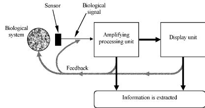
To pick up biological signals is essential because they carry, if not all, most of the information needed to understand the behavior of the system and, eventually, to take a decision (say, a therapy). Thus, the recording channel appears as a second system (a technological one) to be implemented, coupled to the biological system. This book takes it as its blueprint. Figure 1.1 explains its parts: The biological system, either as
Figure 1.1. THE RECORDING CHANNEL. The biological system produces signals, which are picked up by specific sensors. They are amplified, processed and displayed. Eventually, feedback signals modify different input levels according to information obtained from the different outputs.
10 |
Understanding the Human Machine |
an individual, organ or cell, is the source producing signals (Chapter 2), live signals, which are picked up by specific sensors. There are very many biological signals (Chapter 3), however, their nature can always be summarized in a few kinds: electrical, mechanical, chemical and thermal. Accordingly, electrodes and transducers represent a key critical bottleneck (Chapter 4), permanently opened to research and new developments. Most of the time, if not always, the signal requires amplification and processing. This basic unit needs to fulfill unique characteristics not covered by the amplifier usually applied in other fields (Chapter 5). Once the signal is faithfully displayed (by and large, there is always some kind of visualization, on paper, oscilloscope screen or monitor), we have to read it to obtain a meaningful interpretation. This is, in simple words, all what signal analysis is about (Chapter 6). But do not get too excited about the content of the chapter, for we merely want to offer you a lead. You will need specific courses to go deeper. The information produced by the analysis of the biological signals, hopefully, will offer a variety of messages leading to one or more decisions: the signal pick up must be improved, signal processing has to be changed, stimulation of some kind is advisable to isolate a given response, a therapy is instituted, a relationship is uncovered or discovered, a mathematical model can be set, thus starting a fruitful feedback loop (Chapter 7). Thus, feedback is meant here in a very wide sense and context. The final chapter will try to put things together, if possible, to jet you off into higher levels with the help of your inspiration, sweat and, perhaps, some light from the Seven Lamps.
1.7. Objectives
Set always the goal. It will be easier to find the way. Be a Pathfinder!
Leslie A. Geddes has been one of the outstanding contributors to Bioengineering. When I was a graduate student at Baylor College of Medicine, he told me: “Start the sentence with an infinitive. It means you are going to act. The sentence should be short and concise. If it is too long, either
Chapter 1. Introduction |
11 |
you do not know well what you want to do or you have to split the objective in two or three.” Let me follow the advice:
The general objective of this book is to offer an overview of Bioengineering. This is why it is simply an introduction at most, and more likely a primer, that is, a take off platform.
The specific objectives are:
1.To describe somewhat critically the basic sections of Bioengineering. The rest springs up from them.
2.To give a historical feeling. It teaches humility, it may show the way and avoids repetitions.
3.To show you how much you can learn by yourself. It is extremely difficult, but let us try. Your personal effort is indispensable.
In other words, after finishing it (perhaps a full semester work course), you should have the feeling of having caught almost everything and yet realizing that the true meat and potatoes and full taste of it is still to come, for the book is aimed at the undergraduate bioengineering student. Some mathematics is used, say, at the level of calculus, differential equations, and the concept of transform.
In the meantime, learn and put into practice as soon as possible the ABC of science (Bishop, 1997). The authors, believe me, are also trying hard in this respect. Unfortunately, the human being (scientists and engineers are also human beings) is not always too willing to comply with these nice suggestions:
Accept criticism graciously Become focused and organized
Cope with the burgeoning scientific literature Develop critical thinking and logical sequence of ideas Experience satisfaction and joy from your endeavors Finish tasks undertaken
Give the best at all times
Hone communication skills in writing and speaking.
Even though the word BIOENGINEERING may have conflicting meanings when its detailed coverage is required, as briefly referred to above, it is widely and loosely used in the daily language, probably because it is
12 |
Understanding the Human Machine |
shorter and powerfully says what the métier is. Therefore, we will stick to it in the text. From here on, it will not be capitalized, as the names of other disciplines will not either.
Chapter 2
Source: Physiological Systems and Levels
Know the ground thou set thy foot
Physiology, normal and pathological, is seminal to bioengineering. It is the ground we move about, source of our knowledge and recipient of our efforts. True, a bioengineer is first an engineer, but he/she must also be a physiologist and has to be fluent with its terminology. Otherwise, the bioengineer will not be able to communicate with his fellow physiologists, physicians, veterinarians and biologists. Thus, the objective of this chapter is to teach SOME physiology. It is assumed you know a little already and, more important, that you will study quite a bit in the near future. A few systems were left out (like the reproductive and bone systems), without meaning they are not important. At this level, it will not hurt. Those included are summarily and incompletely treated and probably will be criticized by the specialist or the prickly mind. Remember, it is a bird’s eye view (bioengineers like to fly). The approach, however, intends to be from the eyes of an engineer also, thus stressing the block diagram and even introducing some simple mathematical models. Descriptions are minimal. If necessary, the student will have to complement and supplement with any of the many available good physiology textbooks.
2.1. Organism in a Block Diagram
With the Great Engineer’s Forgiveness
To reduce a higher animal, or man, to a mere collection of interconnected functional blocks is a daring and even disrespectful task! However, and
13
14 |
Understanding the Human Machine |
very humbly, we just want to see what the main systems are, how they relate, and what perspective we may obtain from it. This is exactly what an engineer does when approaching an unknown equipment. Figure 2.1 underlines the two biological purposes: (1) maintenance or survival of the individual through maintenance of the tissues, and (2) maintenance or survival of the species by means of the reproductive system.
For the first essential task, the tissues take up oxygen and nutrients while releasing carbon dioxide and metabolites. These are passive transports occurring from and to the circulatory stream, via an exchanger, E1, represented by the peripheral capillary network closely associated to the tissue cells which, in the end, are the final users or customers. A second exchanger, E2, relating the pulmonary capillary bed to alveolar walls (in turn, connected to the external terrestrial atmosphere), allows the unloading of excess CO2 and the re-oxygenation of blood, also passive processes. Passive means that substances move along concentration gradients without utilizing any energy. Blood replenishes its nutrient load via a third exchanger, E3, divided in two sections (hepatic and intestinal) and
|
|
|
|
|
|
|
|
|
|
|
|
|
|
|
|
|
|
|
|
Inputs |
|
|
|
Nervous S. |
|
|
|
Endocrine S. |
|
|
|
|
Outputs |
||||||
|
|
|
|
|
|
|
|
|
|
|
|
|
|
|
|
|
|||
|
|
|
|
|
Coordination and Integration |
|
|
|
|
||||||||||
|
|
|
|
|
|
|
|
|
|
|
|
|
|
|
|
|
|
|
|
|
|
|
|
|
|
|
|
|
|
|
|
|
|
|
|
|
|
|
|
|
|
|
|
|
|
Ventilation to Outside |
|
|
|
|
|
|
|
||||||
|
|
|
|
|
|
|
|
|
|
|
|
|
|
|
|
|
|
|
|
|
|
|
|
|
|
|
|
|
|
|
|
|
|
|
|
|
|
|
|
Food Ingesta |
|
|
Respiratory S. |
|
|
|
|
|
|
|
|||||||||
|
|
|
|
|
|
|
|
|
|
|
|
|
|
|
|
|
|
|
|
|
|
|
|
|
E3 |
|
|
|
|
E2 |
E4 |
|
|
|
|
|
|||
|
|
|
|
|
|
|
|
|
|
|
|
|
|
|
|
|
|||
Gastrointestinal S. |
|
Cardiovascular S. |
|
|
|
Renal S. |
|||||||||||||
|
|
|
|
|
|
|
|||||||||||||
|
|
|
|
|
|
|
|
|
|
|
|
|
|
|
|
|
|
|
|
|
|
|
|
|
|
|
|
|
|
E1 |
|
|
|
|
|
|
|
||
|
|
|
|
|
|
|
|
|
|
|
|
|
|
|
|
||||
Feces |
|
|
|
|
Tissues |
|
|
|
|
Urine Excretion |
|||||||||
|
|
|
|
|
|
|
|
|
|
|
|
|
|
|
|
|
|
||
|
|
|
|
|
|
|
|
|
|
|
|
|
|
|
|||||
|
|
|
Motor S. |
|
|
|
|
|
Reproductive S. |
||||||||||
|
|
|
|
|
|
|
|
|
|
|
|
|
|
|
|
|
|
|
|
Figure 2.1. THE ORGANISM IN BLOCK DIAGRAM (see text). ‘S.’ stands for ‘system’ and the E’s stand for ‘exchanger’.
Chapter 2. Source: Physiological Systems and Levels |
15 |
much more complex than the previous two, which associates the vascular net with the gastrointestinal system (GIS). In it, transport processes are both passive and active (the latter require energy expenditure and the existence of specific pumping mechanisms). Many are still under study. The GI receives the ingesta through the oral cavity, breaks down food into simpler chemical forms suitable for absorption, and excretes the residues through the anal opening. Metabolic products are cleared via a fourth and extremely complex exchanger, E4, which brings together the vascular tree and the renal system, the latter offering an outlet through the urinary tract. Exchangers E3 and E4 are composed of capillary vessels showing unique anatomic and functional characteristics. Malfunctioning of any single exchanger seriously threatens the life of the individual. Their study, including the small vessels converging to and diverging from them — the micro-circulation — constitute a large and complex chapter of physiology. It is clearly seen from this block diagram that the cardiovascular system (CVS) relates to all organs, tissues and cells, with blood being just a carrier in constant circular movement.
The neural and endocrine systems act as the Central Processing Unit, receiving afferent signals from the different systems, integrating them, taking decisions of all sorts, sending off efferent signals and coordinating actions. The locomotion and reproductive systems complete the view, the former to move about, either searching for food, running from danger or looking for a mate. Electrical action potentials are the basic signals within the neural system while internal secretion blood levels (hormone concentrations) excite or inhibit specific receptors distributed all over and, thus, also transmitting information.
This individual block diagram, with its physiological systems, suggests right away the existence of subsystems and organs, down to the cellular and even molecular level, thus pointing out to a hierarchical arrangement. The student must identify as soon as possible where his/her research object is located in order to better choose the proper tools, either experimental or theoretical, to attack it.
Study subject: Modify the proposed block diagram in order to have that of a pregnant mammal or woman. Is there another exchanger? Explain.
16 |
Understanding the Human Machine |
2.2. Cardiovascular System
…it is absolutely necessary to conclude that the blood in the animal body is impelled in a circle, and is in a state of ceaseless motion; that this is the act or function which the heart performs by means of its pulse; and that it is the sole and only end of the motion and contraction of the heart. William Harvey, De Motu Cordis, 1628.
With rhythmic pulses running down its road the heart moves always restlessly abroad. The magic of its numbers is astounding
no matter how they're handled or transformed.
Its reddish elixir is wonder
that flows in those palpitant channels and yonder against oscillating resistance
and generously crossing fine barrier.
We read all its messages, hesitatingly
we think we understand them, expectantly while taking decisions, vacillatingly.
Oh! Circuit revered and much feared, by probes and by catheters inspected, from inside our destiny thou markest!
(electrical activity) (mechanical activity, blood reach)
(measurements, heart rate ...) (signal processing)
(blood)
(hemodynamics) (peripheral resistance)
(capillaries)
(recording) (diagnosis, interpretation)
(therapeutics)
(still there are unknowns) (more studies are needed) (but in the end, we all die)
2.2.1. Cardiac Mechanical Activity
The previous historical and poetic accounts, almost 400 years apart in time and knowledge, briefly summarize what the system is about. The heart and vessels and their essential properties are presented, along with the variables of the CVS, as a continuation and more focused view of the first general diagram (Figure 2.1). Besides, Fick's principle is introduced as an application of the continuity equation, with specific use also in the determination of cerebral and coronary blood flow. A simple derivation of Stewart–Hamilton formula is given, as the basis of the indicator dilution technique for the determination of cardiac output.
The heart as a pump is reviewed by means of the pressure-volume loops, a clear cut engineering concept borrowed from thermodynamics, which leads into new insights of cardiac performance with significant clinical consequences. The concept of aortic impedance is also brought up along
Chapter 2. Source: Physiological Systems and Levels |
17 |
with the still not fully answered question of optimal ventricular-arterial coupling.
2.2.1.1. The circuit
At the beginning of this chapter (Figure 2.1), it became clear that the blood acts as a carrier. Its movement is sustained by the heart, which as a muscle contracts and, thus, rhythmically propels the fluid to the overall network. In fact, it is a series hydraulic system (Figure 2.2) with a powerful chamber, the left ventricle LV ejecting a spurt of blood (stroke volume) in each contraction (in the order of 80 mL/beat at about 70 beats/min or slightly above 1 beat/s) through the aortic valve into the aorta — main arterial output — to the systemic circulation. The latter is a highly complex network, extended deeply into every tissue and presenting a finite, measurable and variable hindrance to the blood flow. Physiologists call it periheral resistance, Rp, and, quite often, they even calculate a handy numerical value making use of Poiseuille’s Law (which is the hydraulic analog of Ohm’s Law of electricity).
Suggested exercise: Obtain a possible value of the peripheral resistance in a normal adult. Establish the units. Sometimes, physiologists define the R-unit (the unit of peripheral resis-
LA |
|
LV |
|
Left Heart
Pulmonary |
|
Lungs |
|
||
Circulation |
|
|
|
|
|
|
|
|
|
|
|
RV
Tissues
Right Heart
RA
Systemic
Circulation
Figure 2.2. THE CIRCULATORY SYSTEM AS A HYDRAULIC SERIES CIRCUIT. LA, left atrium, connected through the mitral valve to LV, left ventricle, leading through the aortic valve into the Systemic Circulation, site of the peripheral resistance. From the latter, blood enters into the right atrium RA and, via the tricuspid valve, proceeds to the right ventricle RV. Finally, through the pulmonary valve, it fills the pulmonary circulation returning to LA. Exchanges to/from tissues and to/from lungs are also depicted, respectively, for the Systemic and Pulmonary Circulations.