

Bioregenerative Engineering Principles and Applications - Shu Q. Liu
..pdf876 SKELETAL MUSCLE REGENERATIVE ENGINEERING
In addition to contractile elements, the sarcomere contains supporting protein structures. Two well known proteins are titin and nebulin. Titin is a filamentous protein and is anchored to the Z-disk at one end and to the M-line at the other end. This molecular structure provides anchorage to the myosin filaments, contributing to the structural integrity and stability of the myosin filaments. Furthermore, titin contributes to the elasticity of the sarcomeres and muscle cells. Nebulin is a filamentous protein that is distributed with the actin filaments and provides structural and mechanical supports to the actin filaments.
Mechanisms of Muscle Contraction [21.1]
Based on a hypothetical model, muscle contraction is induced by actin filament sliding against myosin filaments. The myosin filaments are organized with the heavy-chain heads localized symmetrically to both ends of the myosin filaments. The actin filaments are distributed symmetrically with respect to the myosin filaments, so that the actin filaments can interact with the myosin heavy chain heads simultaneously at both ends of the myosin filaments. Thus, the interaction of myosin heads with the actin filaments induces sliding of the actin filaments toward the center of the myosin filaments or the M-line, resulting in the shortening of the sarcomeres and the contraction of the muscle cells.
The contraction of the muscle cells is a highly regulated process, which involves complicated regulatory mechanisms and numbers of regulatory molecules. A contractile process is initiated and controlled by signals from the motor centers of the central nervous system, including the brain and spinal cord. The skeletal muscle system is innervated with nerve fibers originated from these nerve centers. The central motor-controlling neurons generate electric signals called action potentials, which can be transmitted from the neurons to the peripheral muscle cells via nerve axons, initiating muscle contraction. The generation of action potentials is dependent on the resting membrane potential, which is defined as the voltage difference across the cell membrane at the resting state (without actin potentials). In a normal cell, the cytoplasmic surface of the cell membrane is negatively charged, whereas the extracellular surface is positively charged. The resting potential difference in almost all cell types ranges from −70 to −90 mV under physiological conditions. Such a resting potential difference results from the gradients of ion concentrations across the cell membrane. The concentration of ions, such as Na+, K+, Ca2+, and Cl−, differs from the cytoplasm to extracellular space with the degree of difference depending on the type of the ion. Ion channels specific to each ion type in the cell membrane are responsible for the generation and maintenance of the ion difference. The formation of the resting membrane potential is often called polarization. A cell with resting membrane potentials is said to be polarized.
The resting membrane potential can be reversed from a cytoplasmic negative to a positive value in response to various types of stimulation. Such a process is known as depolarization and the resulting potential is defined as action potential. The stimulation of resting cells induces the opening of gated Na+ channels, resulting in Na+ fluxes from the extracellular space to the cytoplasm (note that the Na+ concentration in the extracellular space is higher than that in the cytoplasm) and a reduction in the membrane potential. When the membrane potential is reduced to a threshold level, a rapid depolarization process is triggered, leading to the generation of an action potential. Shortly after the generation of the action potentials, the Na+ channels are closed, while the K+ channels
ANATOMY AND PHYSIOLOGY OF THE SKELETAL MUSCLE SYSTEM |
877 |
are open, resulting in the termination of inward Na+ flux and the beginning of outward K+ flux (note that the K+ concentration in the cytoplasm is higher than that in the extracellular space). The outward K+ flux results in the reestablishment of the resting membrane potential. Such a process is defined as repolarization.
There are two characteristics for the action potential. First, an action potential is initiated only when the resting membrane potential is reduced to the threshold level in response to a stimulus. Any change in the resting membrane potential that does not reach the threshold will not initiate action potentials. Once an action potential is initiated, the amplitude of the action potential remains constant. The level of stimulation does not influence the amplitude of the action potential. This phenomenon is known as the all-or-none phenomenon. Second, an action potential can propagate through the cell membrane. A cell is initially depolarized within a small area of plasma membrane. A local action potential can stimulate the cell membrane near the depolarized area, inducing the spreading of the action potential. This is a basic process for the propagation of action potentials.
The contractile activity of the skeletal muscle system is controlled by the action potentials that are initiated in the neurons of the motor control centers and transmitted to the peripheral muscle cells via the nerve axons. Each axon develops multiple levels of branches, which project toward peripheral muscle cells. The end of each axon branch enlarges to form a terminal structure, which interacts with the plasma membrane of a muscle cell. The axon terminal and the local area of the muscle cell that interacts with the axon terminal are together called synapse. The axon terminal is referred to as the presynaptic terminal, the plasma membrane of the muscle cell is called the postsynaptic membrane, and the gap between the two types of membrane is known as the synaptic cleft. Each presynaptic terminal is composed of plasma membrane vesicles, referred to as synaptic vesicles. These vesicles contain a substance known as acetylcholine, a neurotransmitter that can be released into the synaptic cleft and stimulates the initiation of action potentials in the muscle cells.
When an action potential is transmitted from a motor neuron to the presynaptic terminal, the actin potential induces the opening of voltage-gated calcium channels, resulting in calcium flux into the presynaptic terminal. Increased calcium concentration in turn induces the release of acetylcholine from the synaptic vesicles to the synaptic cleft. Released acetylcholine interacts with and activates the acetylcholine receptors located in the postsynaptic membrane of the muscle cell. The activation of the acetylcholine receptors induces the opening of voltage-gated sodium channels, resulting in sodium flux into the muscle cell. The inward sodium flux causes depolarization of the muscle cell and initiation of action potentials.
The action potentials in the plasma membrane of the muscle cell can initiates muscle contraction via a process known as excitation–contraction coupling. In this process, the action potentials can be transmitted from the synapse to a tubular network called T- tubules, which are distributed continuously around the sarcoplasmic reticulum and the sarcomeres (note that the sarcoplasmic reticulum contains a high concentration of calcium). The action potentials in the T-tubules stimulate the sarcoplasmic reticulum, inducing the opening of the voltage-gated calcium channels. This action results in calcium release from the sarcoplasmic reticulum to the sarcoplasm, where the contractile myofibrils are located. Calcium ions bind to troponin, inducing a conformational change in the troponin– tropomyosin complex and the exposure of the active binding sites of the actin filaments. The active sites in turn interact with the myosin heads and induce the movement of the
878 SKELETAL MUSCLE REGENERATIVE ENGINEERING
myosin heads. The actin–myosin interaction results in the sliding of the actin filaments against the myosin filaments and thus contraction of the muscle cells. After the sliding process, the myosin heads are released from the actin filaments and are prepared for another cycle of contraction. These activities require energy from ATPs. The myosin heads are composed of ATPases, which hydrolyze ATP molecules and generate energy. The energy is stored in the myosin heads and used for the release of the myosin heads from the actin filaments. Following the contraction process, the calcium ions are actively transported from the sarcoplasm to the sarcoplasmic reticulum. The decrease of the calcium concentration in the sarcoplasm induces muscle relaxation.
DISORDERS OF THE SKELETAL MUSCLE SYSTEM
Muscular Dystrophies
Pathogenesis, Pathology, and Clinical Features [21.2]. Muscular dystrophies are a group of hereditary disorders characterized by progressive loss of muscle mass and function, resulting in the disorder of the skeletal muscle system. Gene mutationinduced protein alterations and deficiency are primary causes for muscular dystrophies. Genetic studies have identified more than 30 forms of muscular dystrophy. Among these forms of muscular dystrophies, several forms are commonly seen, including the Duchenne’s, Becker’s, myotonic, and facioscapulohumeral muscular dystrophies. The clinical manifestations and pathological changes of these types are briefly discussed as follows.
Duchenne’s muscular dystrophy is a recessive genetic disorder found in male patients. The disorder is induced by mutation of the dystrophin gene (dys), which encodes the dystrophin protein (see Table 21.1). Dystrophin is a constituent of the sarcolemma of muscle cells. This protein is capable of binding to actin filaments at the N-terminus, binding to syntrophin at the C-terminus, and binding to β-dystroglycan at a cysteinerich domain. These molecular links provide a structural basis for the interaction of the actin cytoskeleton with extracellular matrix. In Duchenne’s muscular dystrophy, dystrophin deficiency is often found, suggesting a role for dystrophin deficiency in the initiation and development of muscular dystrophy. Molecular analyses have demonstrated that dystrophin deficiency is often a result of dystrophin (dys) gene mutation. In most patients with muscular dystrophy, dystrophin gene mutation is induced by large fragment deletion, insertion, or point mutation that causes frameshifting of gene codons. These genetic alterations result in the deficiency or modulation (primarily in the cysteine-rich domain) of the dystrophin protein. The deficiency or modulation of dystrophin influences the interaction of the actin filaments with extracellular matrix, resulting in instability of the sarcolemmal structure and reducing cell adhesion and survival capabilities. All these changes promote cell degeneration, eventually leading to cell apoptosis and muscular dystrophy.
Duchenne’s muscular dystrophy is the most common type of muscular dystrophy. The incidence of this disorder is about 0.01–0.03%. Signs of muscular dystrophy, such as muscle weakness and movement disorders, are usually found at the age of 5. Gait analyses often provide useful information for the diagnosis of the disorder. The patients eventually lose muscle strength and the ability of movements. The weakening of the

880 SKELETAL MUSCLE REGENERATIVE ENGINEERING
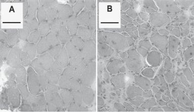
respiratory muscle system influences the function of the lung. Most patients with muscular dystrophy die of respiratory failure. Pulmonary infection often occurs because of food aspiration resulting from the loss of muscular strength responsible for swallowing. A unique feature of Duchenne’s muscular dystrophy is the elevation of the serum c reatine kinase, although the level of the serum creatine kinase is normal at birth. Most patients show a significant increase in serum creatine kinase when muscular weakening occurs. Pathological examinations often demonstrate muscular necrosis, a reduction in the mass of skeletal muscle cells, and an increase in adipocytes and extracellular matrix.
Duchenne’s muscular dystrophy is often associated with cardiac disorders, such as cardiomyopathy, which induces cardiac dilation and a reduction in cardiac contractility. Cardiomyopathy-induced heart failure is the second leading cause of fatality in Duchenne’s muscular dystrophy (note that the first leading cause of death in this disorder is respiratory failure). Mutation or deletion of the dystrophin gene is responsible for the cardiac disorders. Cardiomyopathy is usually induced by truncated or reduced level of dystrophin, which may not cause noticeable skeletal muscle dystrophy. In dystrophindeficient mice, the cardiomyocytes exhibit reduced compliance and enhanced cell contracture, presumably due to increased cell susceptibility to stretch-mediated calcium overload. Prolonged cardiac myocyte contracture induces cell death, a critical mechanism for the development of cardiomyopathy.
Characteristics of muscular dystrophy-related proteins are listed in Table 21.1. Becker’s muscular dystrophy is a disorder similar to Duchenne’s muscular dystrophy
in pathogenesis, pathology, and clinical features. Becker’s muscular dystrophy is induced by mutation of the dystrophin gene. Compared to the Duchenne’s muscular dystrophy, which is induced by complete dystrophin deficiency or mutation of the cysteine-rich domain of the dystrophin gene, Becker’s muscular dystrophy is not associated with complete dystrophin deficiency or mutation of the cysteine-rich domain of the dystrophin gene. Thus, Becker’s muscular dystrophy is not as severe as and progresses slower than the Duchenne’s muscular dystrophy. The Becker’s type of muscular disorder is also referred to as the “benign” form of Duchenne’s muscular dystrophy. Clinical manifestations are usually found after the age of 15. Death due to muscular dystrophy occurs after the age of 40, which is much later than that due to Duchenne’s muscular dystrophy.
Myotonic dystrophy is an autosomal dominant genetic disorder induced by gene mutation in chromosome 19. This disorder is characterized by progressive muscle weakening and the association of disorders in other systems, including cardiac disorders, cataract, intellectual impairment, gastrointestinal disorders, and respiratory failure. Muscle weakening is usually found in the distal extremities at the age of 10–30. Some patients may not show apparent signs or symptoms for a long time. Compared to the Duchenne and Becker muscular dystrophies, the level of serum creatine kinase may remain normal. Pathological examinations often show muscular atrophy or a reduction in the muscle mass. In the heart, conduction system is often involved with a high incidence of conduction block. In the lung, weakening of the respiratory muscles may result in ventilation disorder, hypoxia, cor pulmonale, and respiratory failure.
Facioscapulohumeral muscular dystrophy is a disorder characterized by progressive weakening of the facial, shoulder girdle, and arm muscles. This disorder is induced by autosomal dominant gene mutation and is found in both males and females at any age with a high incidence from age 30 to 40. Some patients may not show any signs of disorder.
882 SKELETAL MUSCLE REGENERATIVE ENGINEERING
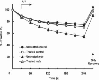
Figure 21.3. Maximum force output during and 300 s after a repeated stimulation fatigue protocol for diaphragm muscle preparations from control and mdx mice (an animal model for Duchenne muscular dystrophy) with and without insulin-like growth factor. Note the comparatively greater reduction in force throughout time in the diaphragm muscles of untreated dystrophic mice compared with untreated control mice (panel A, P < 0.05), and the enhanced resistance of muscles from treated dystrophic mice compared with the untreated dystrophic muscles (panel B, P < 0.05) present from 30 s after commencement of stimulation, to the cessation of stimulation. (Reprinted with permission from Gregorevic P et al: Improved contractile function of the mdx dystrophic mouse diaphragm muscle after insulin-like growth factor-I administration, Am J Pathol 161:2263–72, copyright 2002.)
Molecular Treatment of Muscular Dystrophy [21.4]. The various types of muscular dystrophy as described above are inherited disorders induced by gene deletion or mutation. To date, there are few effective approaches which can be used to treat these disorders. Recent studies have demonstrated that molecular regenerative therapies may be potentially used to treat muscular dystrophy and cardiomyopathy. Numerous genes that are deleted or mutated in muscular dystrophy, contributing to the development of the disorder. The repair and replacement of these genes are potential approaches for the treatment of muscular dystrophy. There are two basic molecular approaches: transfer genes that encode proteins directly responsible for the disorder, and transfer “booster” genes that encode proteins responsible for the survival of muscle cells and the prevention of cell apoptosis. In this section, Duchenne’s muscular dystrophy is used as an examples to demonstrate the principles of molecular therapy for muscular dystrophy.
Transfer of Wildtype Dystrophin Gene [21.5]. For Duchenne’s muscular dystrophy, the primary cause is the deletion or mutation of the dystrophin gene. Thus, direct transfer of a functional wildtype dystrophin gene into the muscular cells is a potential approach for the treatment of the disorder. This gene has been tested in a transgenic animal model, the dystrophin-deficient mdx mouse, which resembles the Duchenne’s muscular dystrophy in
DISORDERS OF THE SKELETAL MUSCLE SYSTEM |
883 |
humans. A number of approaches have been developed and used for the delivery of the dystrophin gene, including virus-, liposome-, and electroporation-mediated gene deliveries. Among these approaches, the virus-mediated gene delivery is the most effective approach. The dystrophin gene can be integrated into modified viral vectors and used to transfer into target muscular cells. Experimental investigations have demonstrated the effectiveness of such an approach. In particular, the transfer of the full-length dystrophin gene with a muscle-specific gene promoter (muscle creatine kinase promoter) has been shown to effectively prevent the progression of muscular dystrophy in the mdx mouse model of muscular dystrophy. The use of the muscle-specific gene promoter can increase the efficiency of gene transfer. However, there are potential problems. These include limited efficiency of gene transfer, immune responses provoked by the expression of the transferred gene and corresponding protein products, temporary gene expression, and poor cell survival. These problems remain to be resolved.
Given the problems with the transfer of the wildtype dystrophin gene, several alternative strategies have been developed and used for the molecular treatment of muscular dystrophy. These include the construction and delivery of truncated dystrophin gene, mutant gene correction by small fragment homologous replacement, correction of mutant genes by chimeraplasty, removal of mutant gene fragments by exon skipping, and compensation for the lost function of mutant dystrophin. These approaches are discussed as follows.
Delivery of Truncated Dystrophin Genes or Microdystrophin Gene Constructs [21.6]. The dystrophin gene is composed of a large number of base pairs, which complicate the preparation and manipulation of the dystrophin gene. Genetic and functional analyses has demonstrated that not all gene sequences are necessary for the production of functional dystrophin. Indeed, selected regions of the dystrophin gene can be deleted and the remaining regions can be recombined to generate a minidystrophin gene. A minimal requirement is that the reconstructed gene must contain critical domains responsible for regulating the structural stability and function of the sarcolemma of muscular cells. The reconstructed minidystrophin gene can be highly functional. When delivered into the mouse mdx muscular dystrophy model, pathological alterations of muscular dystrophy can be significantly prevented and the contractility of the skeletal muscles can be improved (Fig. 21.4). Furthermore, such an approach can be used to reduce muscular cell degeneration in the mdx mouse model of muscular dystrophy. However, it is still debating whether the delivery of the truncated dystrophin gene is more advantageous than that of the full-length dystrophin gene.
Mutant Gene Correction by Small Fragment Homologous Replacement (SFHR) [21.7]. Small fragment homologous replacement is a technique used for constructing and inserting PCR-generated DNA amplicons into the host genome to correct mutant genes. PCR can generate large DNA fragments up to several hundred base pairs. Designed corrective DNA fragments can be constructed and transferred into target cells. When integrated into the genome, these fragments can replace and correct mutant genes, resulting in the generation of functional genes. Such a technique has been applied to cells from the mdx mouse model of muscular dystrophy to correct mutant dystrophin gene. While the mechanisms of gene correction remain poorly understood, preliminary investigations have
884 SKELETAL MUSCLE REGENERATIVE ENGINEERING
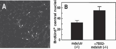
Figure 21.4. Increased integrin promotes the proliferation of satellite cells mdx/utr−/− mice (a animal model of muscular dystrophy) and a7BX2-mdx/utr−/− mice (a muscular dystrophy model with enhanced expression of the α7b1 integrin). Mice were injected with BrdU to label replicating cells. Muscle specimens were collected and analyzed by immunohistochemistry for BrdU incorporation into DNA. Nuclei are stained with DAPI. BrdUrd-labeled central nuclei (arrows in panel A) in 50 random fields were scored for each animal. Mean numbers (±SEM) are given for 11 animals for each genotype. (B) Increased integrin expression enhanced the proliferation of satellite cells and the regenerative capacity of dystrophic muscle. (Reprinted with permission from Burkin DJ et al: Transgenic expression of alpha7beta1 integrin maintains muscle integrity, increases regenerative capacity, promotes hypertrophy, and reduces cardiomyopathy in dystrophic mice, Am J Pathol 166:253–63, copyright 2005.)
provided promising results for this technique. As shown in a study with cultured muscular cells, the delivery of a PCR amplicon into muscular cells with dystrophin deficiency results in the correction of the dystrophin gene in about 20% cells. However, a higher efficiency may be needed to achieve therapeutic effects.
Correction of Mutant Genes by Chimeraplasty [21.8]. Chimeraplasty is a technique used for correcting mutant genes with chimeric RNA–DNA oligonucleotides, also known as chimeraplasts. Short chimeric genetic structures can be constructed by hybridizing complementary 2′-O-methyl ribonucleotide analogues to desired DNA fragments. Such a chimeric complex protects the DNA fragments from exonucleolytic digestion. When the chimeraplasts are delivered to the cell nucleus, the DNA and RNA fragments can anneal to the target site during gene transcription. The chimeraplasts can repair or replace basepair mismatches, if any, resulting in the correction of gene mutation, although the exact mechanisms remain poorly understood. Experimental investigations with in vitro models have demonstrated a correction efficiency about 30%. The efficiency may be further improved when the mechanisms of chimeraplasty are fully understood. Chimeraplasty has been applied to the mdx mouse model of muscular dystrophy. The delivery of chimeraplasts for the dystrophin gene into the skeletal muscle cells results in the correction of mutant dystrophin gene in about 10% cells. Such a manipulation induces an increase in the expression of functional dystrophin gene and a reduction in the symptoms of muscular dystrophy.
Removal of Mutant Gene Fragments by Exon Skipping [21.9]. Exon skipping is a technique used to target selected mutant gene fragments and block the transcription of the
DISORDERS OF THE SKELETAL MUSCLE SYSTEM |
885 |
targeted fragments by introducing specific antisense 2′-O-methyl ribonucleotide analogs to cell nuclei. The antisense ribonucleotides can bind to and block specific homologous DNA exons or sequences in the genome during transcription. Such a process induces a transcription-skip over the ribonucleotide-blocked exons. In other words, the blocked exons can no longer be transcribed. When a ribonucleotide sequence is designed and delivered to target a specific mutant gene fragment, the mutant fragment cannot be expressed and the function of the generated protein may be improved.
For the treatment of experimental muscular dystrophy in the mdx mouse model, a 2′-O-methyl ribonucleotide analogue sequence can be designed to target the exon 23 (at the junction with intron 22), which contains a mutant fragment responsible for the development of muscular dystrophy. The delivery of this ribonucleotide analogue into the mdx mouse model stops the transcription of the exon 23. Such a manipulation results in the generation of a dystrophin form similar to that found in Becker’s muscular dystrophy, which is significantly less severe than Duchenne’s muscular dystrophy. Although the approach does not provide a complete cure of the muscular dystrophy, the pathological changes are reduced and the contractility of the muscle system is improved.
Compensation for Lost Function of Dystrophin [21.10]. There exist proteins that potentially compensate for the function of dystrophin. One of such compensating factors is utrophin, also known as dystrophin-like protein and dystrophin related protein 1. Utrophin is a protein of 3422 amino acid residues and about 395 kDa in molecular weight. This protein is similar to dystrophin in structure and function. As dystrophin, utrophin is expressed in skeletal muscle cells and is localized to the sarcolemma and acetylcholine receptors at the neuromuscular synapses and myotendinous junctions, where it regulates the function of the postsynaptic membrane and, especially, the activity of the acetylcholine receptors. Utrophin is also expressed in the heart, brain, lung, kidney, liver, intestine, and testis. Utrophin can interact with dystrophin at the C-terminus. The suppression or loss of the utrophin activity exacerbates pathological changes of muscular dystrophy in the mdx mouse model of muscular dystrophy. The upregulation of the utrophin gene has been shown to reduce pathological alterations and compensates for functional abnormalities due to dystrophin deficiency in the mdx mouse model of muscular dystrophy. Growth factors, interleukin-6, l-arginine, and nitric oxide can enhance the expression of the utrophin gene promoter.
Transfer of Dystrophin “Booster” Genes [21.11]. In addition to the dystrophin gene, a number of “booster” genes have been discovered and used for the molecular treatment of muscular dystrophy. These genes encode proteins that mediate the survival and enhance the function of the of striated muscular cells. Common “booster” genes include integrin α7β1, ADAM12, calpastatin, nitric oxide synthase, insulin-like growth factor (IGF)I, myostatin, and mini-agrin.
INTEGRIN α7β1. Cell adhesion to extracellular matrix is critical to cell survival. The impairment of muscle cell adhesion to extracellular matrix may induce cell apoptosis, contributing to muscular dystrophy. In skeletal muscle cells, there are two major types of cell–matrix interaction-mediating molecules, including the dystrophin-associated