

Атлас анатомии крупных животных
.pdfChapter 4 THE A B D O M E N / Gastro-intestinalsystem 4-11
4-11 Enterectomy; end-to-end anastomosis
Intussusception [165] is the most frequent indication for small intestinal enterectomy in cattle, although other disorders which interfere with the viability of the intestinal wall may also necessitate resection. In cattle end- to-end anastomosis is usually performed.
Surgery. A right flank laparotomy is carried out on the standing animal under local analgesia (see 4-4). The affected bowel is carefully exteriorized and the extent of the gut to be resected determined. After application of topical analgesia a V-shaped area of the adjacent mesentery, including the vessels, is ligated.
The diseased loop is then isolated from the remainder of the gut by intestinal clamps and resected with its mesentery. The viable ends are flushed with physiologic saline and apposed, with the lumina held toward the surgeon [166]. The adjacent halves of the two wound edges are sutured with a simple continuous suture perforating all layers of the intestinal wall. The
suture begins at the mesenteric border with the knot lying in the lumen of the gut. On reaching the antimesenteric border the lumina are apposed and the continuous suture continues as a Schmieden suture, with which the two other halves are sutured until the mesenteric border has been reached [167]. Using a continuous Lembert suture, the first suture line is completely oversewn [168]. Absorbable suture material is used for both suture lines. The clamps are removed and the anastomosis checked. The defect in the mesentery is closed by tying the long end of Mesenteric ligatures lying opposite to each other. After replacing the intestine into the abdomen the laparotomy wound is closed (see 4-4).
Systemic antibiotics are administered and dietary measures are routinely taken.
Chapter 4 THE A B D O M E N / Castro-intestinal system 4-12 |
47 |
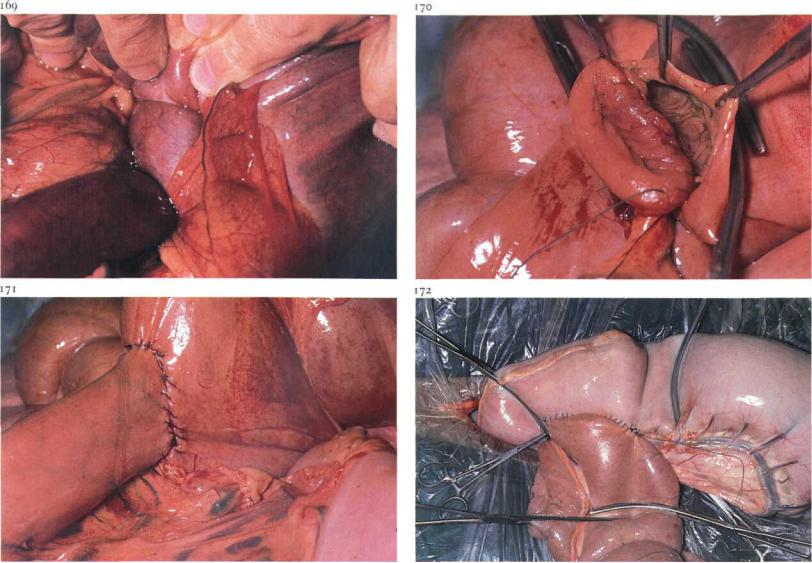
4-12 Jejunocaecostomy; end-to-side anastomosis
Obstruction of the ileum in horses is caused by ileocaecal intussusception [169], involvement of the ileum in internal or external hernias, or ileal obstipation. Because the ileocaecal area is inaccessible, resection of the entire ileum and sparing the ileocaecal valve is impossible. A new junction between the distal jejunum and caecum must therefore be created, and is achieved by either end-to-side or side-to-side anastomosis.
Surgery. Treatment of ileocaecal intussusception by reduction alone rarely succeeds. Usually the intussusceptum is left in place and Jejunocaecostomy is carried out. To perform an end-to-side anastomosis the jejunum is transected as distally as possible, after which the distal stump (ileum) is closed, as described in 4-10. The caecum is then exteriorized. A fold of the wall about 20 cm distal to the ileocaecal valve is clamped off. Half the circumference of the open end of the jejunum is sutured to this fold, using a continuous Lembert suture placed 6-7 mm from the transected edge. The
caecal wall is then incised 6-7 mm from this suture line; the length of the incision corresponds with the diameter of the jejunal end. The caecal and jejunal edges are joined together using a simple continuous suture, which is continued as a Schmieden suture [170], as in 4-10. The Lembert suture is continued to oversewn the Schmieden suture line [171]. The free border of the mesentery of the distal jejunum is sutured to the caecum to obliterate the space between jejunum and caecum.
In ileum obstipation and ileum muscular hypertrophy, a jejunocaecal bypass is performed, leaving the normal ileal pathway open [172]. The bypass is created as a side-to-side anastomosis (see 4-10).
Chapter 4 THE ABDOMEN / Gastro-intestinalsystem 4-13
4-13 Correction of rectum prolapse
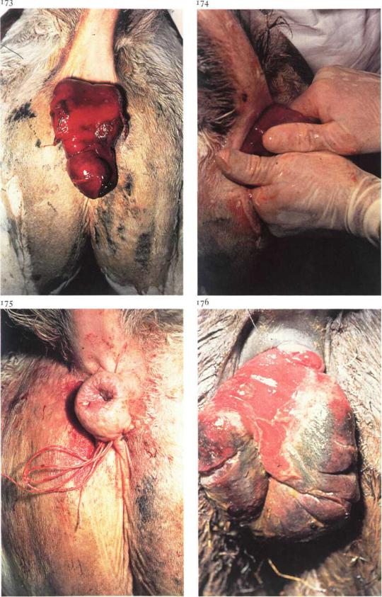
Rectum prolapse involves either the rectal mucosa alone (incomplete prolapse) or the full thickness rectal wall (complete prolapse). Furthermore, the rectum prolapse may contain an intussusception of the rectum, colon or even small intestine.
Treatment depends on the degree of damage to the mucosal layers. Usually, manual reposition and suture retention of the prolapse can be carried out. However, amputation of the prolapse is indicated when reposition is impossible (because of severe swelling or adhesions) or when perforating injuries or necrosis of the mucosal layers are present [176]. In cases of extensive prolapse, the prognosis is guarded due to the possibility of accompanying mesocolon vascular damage; in this condition laparotomy should be considered for definitive diagnosis and possible treatment. Surgery. Correction of rectum prolapse is performed under caudal epidural analgesia (posterior block) or general anaesthesia.
(1) Reposition and retention of the prolapse. Reposition [173, foal] is achieved by careful massage of the prolapse using a vaseline-based ointment [174]. To aid retention of the replaced rectum, a purse-string suture is placed through skin and deep fascia around the anus [175]. Before tying the suture, liquid paraffin is administered rectally with a soft rubber tube to facilitate defaecation. The purse-string suture is then tied in a bow, so that it can be easily loosened and relied. The suture is tied such that two fingers can easily be passed through the anus, thereby allowing some defaecation.
(2) Amputation of the prolapse.
Before resection ([176], horse), a probe is passed between the prolapse and the anal ring to ensure that intussusception is not present. Next, a suitably sized firm rubber tube is passed into the rectum [177]. As slight traction is applied to the prolapse, two long needles are thrust (one vertically, the other horizontally) through the prolapse and the tube. The needles must be close to the anal ring [178], so that healthy tissue is penetrated. The first quadrant of the prolapse is then resected just distal to the needles, and the outer and inner layers of the resected end of the rectum are sutured together, using simple interrupted sutures [178]. The second quadrant of the prolapse is then resected in a similar way [179]. After amputation and suturing of all

Chapter 4 THE A B D O M E N / Gastro-intestinal system 4-13
quadrants have been completed, the ends of the threads are cut off and the two needles and tube are removed. In most cases the stump retracts spontaneously. In those cases in which the stump remains slightly prolapsed, retention of the rectum is achieved by use of a purse-string suture as described above.
Dietary measures and/or liquid paraffin administered either by stomach tube and/or rectally may be used to facilitate defaecation.
It may be necessary to loosen the purse-string suture twice daily to release faeces. The purse-string suture may be removed within 48-72 hours. Plate 180 shows the situation on the tenth postoperative day.

Chapter 4 THE A B D O M E N / Gastro-intestinalsystem 4-14 |
.So |
4-14 Treatment of atresia ani (et recti)
Absence of the anal opening is observed often in the piglet and occasionally in other species. Affected piglets may be up to several weeks old without showing serious illness except progressive abdominal distension [181]. In female piglets even distension may not be evident because of the presence of a recto-vaginal fistula through which some evacuation occurs. In some cases there may be a swelling at the site of the anus [182].
Surgery. After local infiltration analgesia or epidural analgesia a circular piece ofskin is excised over the anal site. Ideally the rectum should be sutured to the skin before the rectum is opened but often faeces are discharged immediately following the skin excision [183]. After sufficient evacuation of the bowel the rectal wall is apposed to the skin by interrupted sutures [184].
If the distal portion of the rectum is also atretic, deeper dissection is required to locate the blind end of the rectum. Gentle traction, assisted by pressure on the abdominal wall, should be used to draw the rectum caudad to the anus, where it is sutured to the skin.
If too large a portion of the rectum is absent, creation of a preternatural anus via a laparotomy under general anaesthesia may be considered. Postoperatively a cicatricial stricture of the anal opening may develop.
Chapter 5 The urogenital system

Chapter^ THE U R O G E N I T A L SYSTEM / The male urogenital system 5-1
186
5-1 Castration: open technique in the pig
Piglets are usually castrated using the open technique, in which the vaginal cavity is left open after emasculation. It is clear that the open technique of castration is contraindicated in cases of inguinal or scrotal hernia. Piglets are generally castrated at the age of 3-6 weeks.
Surgery. Castration is performed under local analgesia of the scrotal skin and both testicles. An assistant holds the animal upside-down by the hindlimbs, with the piglet's back towards the surgeon. The testicle is pushed up into the scrotum with thumb and forefinger [185]. An incision is made into testicular tissue through scrotal skin, tunica dartos and vaginal tunic [186]. The exposed testicle is grasped with the fingers and the scrotal ligament is transected with a scalpel; the incision starts at the site of the mesorchium [187]. To prevent spermatic cord haemorrhage, ligation (with a catgut ligature) or crushing (with an emasculator, which crushes and cuts at the same time) of the spermatic cord is recommended. The emasculator is held as proximal as possible on the spermatic cord [188] for 10-15 seconds. The emasculator is then removed and the severed end of the cord retracts into the inguinal canal.

Chapter^ THE U R O G E N I T A L SYSTEM / The male urogenitalsystem 5-2 |
53 |
5-2 Castration: closed technique in the goat
Castration of goats is in general indicated to reduce the odour originating from the horn glands. Goats should be castrated after the age of six months, since urinary obstruction due to urethral calculi occur more often in young castrated goats.
Goats are usually castrated by the closed technique, in which the vaginal cavity is not opened. This is in contrast to the open castration technique, whereby the vaginal cavity is left open after emasculation (see 5-1).
Surgery. An assistant restrains the goat in dorsal recumbency in his lap: the goat's head is held under one arm and the limbs grasped firmly together. The scrotal skin and both spermatic cords are locally infiltrated with suitable analgesic solution. The tip of the scrotum is tightly stretched distally and is amputated [189]. This allows both testicles to emerge into the incision. The testicle is grasped with a tenaculum forceps and the tunica dartos is stripped from the vaginal tunic with a gauze sponge [190]. The spermatic cord, covered by vaginal tunic, is then crushed by an emasculator or, as in this case, ligated with a ligature of absorbable suture material [191]. The spermatic cord is transected i cm distal to the ligature [192] and the stump checked for bleeding. The opposite testicle is removed in a similar manner.
Tetanus prophylaxis must be provided.

Chapters THE U R O G E N I T A L SYSTEM/ The maleurogenitalsystem 5-3 |
54 |
194
5-3 Castration: half-closed technique in the horse
In the horse several methods of castration may be used: the open technique (see 5-1), closed technique (see 5-2), half-closed technique, which is discussed here, and the primary closure method (see 5-4).
The half-closed technique involves crushing and ligation of the spermatic cord enclosed in the vaginal tunic, with the testicle itself lying outside the opened vaginal tunic. Using this approach, the most serious postoperative complications such as intestinal eventration and spermatic cord haemorrhage are averted.
Surgery. Half-closed castration can be carried out in the recumbent or standing stallion.
(i) Half-closed castration in the recumbent stallion.
For a right-handed surgeon, the horse is positioned in left lateral recumbency. The right leg is pulled tightly against the chest and the foot tied at the level of the shoulder joint. Castration is performed under local infiltra-
tion analgesia or general anaesthesia. The left testicle is held in the scrotum with the left hand, so that the scrotal skin is tightly stretched. A 7-10 cm incision is made i cm lateral and parallel to the median raphe through skin and tunica dartos. A small incision is then made in the vaginal tunic and its edges are grasped with haemostatic forceps [193]. The incision is enlarged sufficiently to allow testicle and epididymis to emerge from the vaginal cavity [194], whereupon the exposed testicle is grasped with a tenaculum forceps. The tunica dartos is stripped from the vaginal tunic with a gauze sponge or blunt scissors [195]. The spermatic cord, covered by vaginal tunic, is then crushed with an emasculator [196]. A ligature of absorbable suture material is applied at the crush site [197]. The spermatic cord is transected 1-2 cm distal to the ligature and the stump checked for bleeding. The opposite testicle is removed in a similar manner.
(2) Half-closed castration in the standing stallion.
The technique is in principle similar to the half-closed castration in the recumbent stallion. Castration is performed under physical restraint and

Chapters THE U R O G E N I T A L SYSTEM / The male urogenitalsystem 5-3 |
55 |
local infiltration analgesia, together with chemical restraint if necessary. The scrotum is grasped firmly with one hand just proximal to the testicle, so that the scrotal skin is tightly stretched. A 7-10 cm incision is made, beginning cranialh, about i cm lateral and parallel to the median raphe through skin, tunica dartos and vaginal tunic into the testicular tissue [198]. The exposed testicle is grasped with a tenaculum forceps and the edges of the incised vaginal tunic are grasped with haemostatic forceps. The tunica dartos is stripped from the vaginal tunic with a gauze sponge and an emasculator is applied to the covered spermatic cord [199]. A ligature of absorbable suture material is applied at the crush site and the spermatic cord is transected 2 cm distal to the ligature [200]. The opposite testicle is removed in a similar manner.
Tetanus prophylaxis is provided. Postoperative management consists of sufficient exercise to avoid scrotal and preputial oedema.