

Атлас анатомии крупных животных
.pdfChapter i THE HEAD / Nose 1-12 |
H> |
057
i-i 2 Treatment of nasolacrimal orifice atresia
Atresia of the nasal opening of the tear duct may be the cause of chronic lacrimation in foals. In some cases the distal part of the duct is also absent. Surgery. Surgery is performed under general anaesthesia. A catheter is introduced into the lacrimal papilla of either the upper or lower eyelid [057]. The catheter is then advanced carefully down the tear duct, until the tip is palpable beneath the nasal mucous membrane [058]. The nasal mucosa and the mucosa of the blind end of the duct are incised over the tip of the catheter [059], after which the catheter is pushed through the opening created [060]. If accessible, the mucous membrane of the duct is sutured to the nasal mucous membrane with simple interrupted sutures of fine absorbable material. The catheter is then sutured to the skin in the nasal and eyelid regions, and left in place for at least two weeks. If suturing of the mucosal layers is impossible the catheter must be left in place for a longer period (3-4 weeks), after which time the wound edges have healed and the opening remains patent.
Postoperatively antibiotic ophthalmic ointments are administered into the conjunctival sac, and corticosteroids may be added for several days to reduce oedema and fibrosis around the created orifice.
Chapter 2 The neck

Chapter 2 THE NECK / Muscles 2-1 |
18 |
062
2-1 Myectomy (Forssell) and accessory nerve neurectomy
In crib-biting the horse grips a fixed object (e.g. manger) with the (upper)incisor teeth, arches the neck and attempts to swallow air; horses which succeed in swallowing air are called windsuckers. Some horses are 'free' windsuckers, these display the vice without cribbing. Initially nonsurgical methods (cribbing strap, aversion therapy) may be used, but are often unsuccessful, and the owner requests surgical treatment. This consists of partial resection of the paired ventral neck muscles: sternohyoideus, omohyoideus, sternothyroideus and sternocephalicus. Instead of myectomy of the latter, neurectomy of the ventral branch of the accessory nerve may be performed.
Surgery. The horse is positioned in dorsal recumbency under general anaesthesia. Excessive extension of the neck should be avoided because of possible stretching of the recurrent nerve, the head should thus be resting at an angle of about 30°.
A midline skin incision of 30-40 cm is made from the hyoid bone caudally. The whole operation may be accompanied by considerable haemorrhage, and careful haemostasis is obligatory. The skin and subcutis are dissected and reflected laterally [061]. The omohyoideus muscle is carefully separated from the jugular vein. The omohyoideus and sternohyoideus are transected near their insertions [062] and reflected back to the caudal edge of the wound, whereafter the entire muscle section is removed [063]. This exposes the cranial part of sternothyroideus [064] which is easily dissected from the trachea and excised [065]. Next the sternocephalicus is freed by blunt dissection after incising its sheath longitudinally. The muscle is transected at the caudal edge of the incision [066], reflected cranially and severed through its tendon [067].
Instead of sternocephalicus myectomy, denervation of the muscle may be performed. The purpose of neurectomy of the ventral branch of the accessory nerve is to diminish the post-operative deformity of the region. The nerve is located on the dorso-medial aspect and runs parallel with the

Chapter 2 T H E N E C K / Muscles 2-1
066
muscle. The neurectomy site is proximal to the entry of the nerve into the muscle; at least 3 cm are removed [068]. The procedure may be carried out before or after myectomy of the other muscles. The skin is closed with interrupted mattress sutures. At both ends of the wound a drain is placed.
Proper functioning of the drains must be checked twice daily, and they must not be removed before 3 days.
Postoperatively the horse is confined for about three weeks, and care is taken that the stall contains no objects that may be grasped with the incisors or on which the wound may be rubbed.

Chapter 2 THE N E C K / Larynx and trachea 2-2
20
070
2-2 Laryngotomy - cricoarytenoidopexy and ventriculectomy
Inspiratory dyspnoea due to laryngeal hemiplegia (roaring) is a common clinical sign in horses requiring surgical treatment to enlarge the reduced laryngeal lumen [o6gA]. Many procedures to alleviate laryngeal hemiplegia have been utilized. Of the various techniques the combination of cricoarytenoidopexy with unilateral or bilateral ventriculectomy has given the best results. Instead of lycra, a double ligature of heavy-sized chromic catgut is preferred for the cricoarytenoidopexy.
Surgery. The horse is positioned in right lateral recumbency in general anaesthesia with the head and neck extended.
A 10 cm skin incision is made parallel and ventral to the linguofacial vein from the cranial border of the larynx to the second tracheal ring [070]. Subcutaneous fascia is incised with a scalpel. The dorsolateral aspect of the larynx is approached by blunt dissection. The muscular process of the arytenoid cartilage is penetrated from medial to lateral with a pointed
Deschamp's needle [071]. The double chromic catgut is threaded through the eye of the needle and pulled through the muscular process. The medial part of the ligature is brought under the crycoarytenoid muscle using the Deschamp's needle. The needle is then passed, from medial to lateral, through the caudal border of the cricoid cartilage, about 2 cm lateral to the median ridge. The needle passes through the cartilage, but not through mucous membrane into the laryngeal lumen. The needle emerges approximately i cm cranial to the caudal border of the cricoid [072]. The medial part of the thread is threaded into the needle and pulled through the cricoid cartilage [073]. The two ends of the ligature are tied [074] with sufficient tension to fully retract the arytenoid cartilage; this can be checked by laryngoscopy [0693].
A vacuum drain is placed in the wound cavity. The subcutaneous and deep fascial tissues are closed with a simple continuous suture and the skin with interrupted sutures, using synthetic absorbable material.
The patient is then positioned in dorsal recumbency; the nose is supported

Chapter 2 THE NECK / Larynx and trachea 2-2
074
to prevent extreme extension of the neck. The laryngeal cavity is opened (see 2-3); the crycoid cartilage is not incised.
The mucous membrane of the left laryngeal saccule is removed. The rim of the laryngeal saccule is incised on its caudal border [075] and the index finger is brought submucosally to free and then evert the mucous membrane. The everted mucous membrane is resected with scissors as close to the base as possible without damaging the adjacent cartilage [076]. To prevent foreign body aspiration during recovery and recuperation the skin is closed with a few non-absorbable interrupted sutures. If postoperative dyspnoea occurs a tracheotomy tube is inserted through the laryngotomy wound, or tracheotomy (see 2-5) is performed.
Antibiotics are administered. The vacuum drain is removed after two to three days. The laryngotomy wound is cleansed daily and heals satisfactorily by second intention. The horse is confined to a box for 4 weeks. After two months at pasture the horse may be returned to training.

Chapter 2 THE NECK / Larynx and trachea 2-3
°77
2-3 Laryngotomy - extirpation of subepiglottal cyst
Inspiratory and/or expiratory noise at work and dyspnoea due to epiglottal lesions (subepiglottal cyst, abscess, and epiglottic entrapment) are occasionally observed in horses. To enable surgery in the area of the soft palate and epiglottis, a laryngotomy must be performed. The surgical technique for removal of a subepiglottal cyst is described here. Diagnosis is made by endoscopic examination [077].
Surgery. The patient is positioned in dorsal recumbency under general anaesthesia. A 10 cm midline incision is made over the larynx through the skin and through the midline junction of the sternohyoid muscles. The crycothyroid membrane is incised in the midline. In this condition, incision through ventral midline of the crycoid cartilage is necessary to gain space. A self-retaining wound retractor exposes the laryngeal cavity [078]. After the endotracheal tube has been removed exposure of the subepigottal cyst is possible. The apex of the epiglottis is drawn caudad from the naso-
pharynx into the laryngeal lumen, thereby presenting the cyst on the ventral surface of the epiglottis in the surgical field. Traction on the epiglottis is accomplished by grasping the aryepiglottic fold, on the lateral wall of the pharynx between the base of the epiglottis and the arytenoid, with a sponge forceps. A Lakey traction forceps applied to the apex of the epiglottis retains it in position.
The mucosa surrounding the cyst is carefully grasped with Allis tissue forceps [079], incised and the cyst is then dissected bluntly from the surrounding tissue [080]. Excess mucosa may be excised. The wound is left open. Only the skin is closed with a few non-absorbable interrupted sutures. The laryngotomy wound heals by second intention. The patient is box rested for 4 weeks.

Chapter 2 THE NECK / Larynx and trachea 2-4
2-4 Laryngotomy - in bovine necrotic laryngitis
Necrotic laryngitis associated with calf diphtheria is the most important cause of laryngeal dyspnoea in young cattle. The results of medical treatment are often disappointing. The aim of surgical treatment is to excise necrotic cartilage and/or granuloma and produce a permanent widening of the glottis.
Surgery. Prior to laryngotomy, a tracheotomy is performed (see 2-5). The patient is placed in dorsal recumbency under general anaesthesia, or under sedation in combination with local infiltration analgesia. If laryngeal intubation is impossible an endotracheal tube must be inserted via the tracheotomy wound. The inflated cuff prevents the passage of blood into the lungs during surgery.
The skin and subcutanuous tissue are incised in the midline and the sternohyoid muscles are separated. A midline incision is made through the crycothyroid ligament, thyroid cartilage and the first 2 tracheal cartilages
[081]. (The thyroid cartilage is cut with a strong pair of scissors). Topical analgesia of laryngeal mucosa is necessary if general anaesthesia has not been used. The laryngeal cavity is exposed by means of Volkmann retractors. In most cases the.necrotic lesions and granulomas are clearly visible. The depth of (a) possible fistulous tract(s) is determined with a probe [082]. Necrotic debris, including cartilage sequestra, are removed by curettage [083]. After cleaning the laryngeal cavity, the skin is closed with nonabsorbable material in an interrupted pattern [084].
Systemic antibiotics are administered. The tracheotomy tube is removed as laryngeal swelling reduces and has less effect on respiration.
Chapter 2 THE NECK / Larynx and trachea 2-5
2-5 Tracheotomy
Tracheotomy is usually an emergency procedure and is indicated to relieve dyspnoea caused by acute nasal, laryngeal or proximal tracheal obstructions. Tracheotomy is also indicated prior to some operations on the nose, paranasal sinuses or larynx.
Surgery. Tracheotomy is usually performed on the standing animal under local infiltration analgesia, but may also be carried out on the recumbent patient. The head and neck of the animal are extended and an approximately 7 cm ventral midline skin incision is made in the cranial third of the neck at the level of the 4th-6th tracheal ring. After incising the thin cutaneous muscle in the midline, the longitudinal junction of the sternohyoid muscles is divided and the trachea exposed. The muscles and skin are spread with a wound retractor.
If temporary tracheotomy is indicated, a tracheal annular ligament is pierced with a scalpel and a tracheal tube (ovoid in cross-section) is inserted.
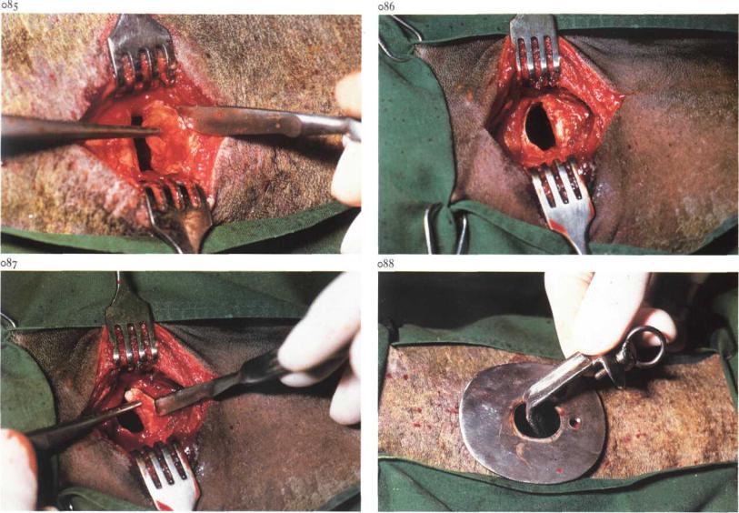
If the tracheotomy tube is to remain for a longer period, partial resection of a tracheal ring is performed, (i) A tracheal window is produced by partial resection of one tracheal ring [085]. The disc to be removed is grasped with forceps. (2) To prevent the tracheal ring from collapsing, resection of a semi-disc of the tracheal rings proximal and distal to an incision through the annular ligament is recommended [086,087].
After tracheotomy a self-retaining tube is inserted [088] and the skin edges are sutured around the tube in a simple interrupted pattern. Since there is considerable mucous secretion for the first few postoperative days, the tube must be cleaned frequently. Later on, when the discharge has reduced, air passage through the tube is checked daily, but the intervals of cleaning may be prolonged. After the tube is withdrawn, the tracheotomy wound heals by second intention.
Chapter j The thorax