

Атлас анатомии крупных животных
.pdfChapter 7 THE MUSCULOSKELETAL SYSTEM / Metacarpus and metatarsus 7-17 |
126 |
474 |
|
7-17 Fracture treatment by the walking cast technique
The walking cast technique is used in treating fractures of the distal radius and tibia, metacarpal/metatarsal bones and phalanges. This method is useful in treating comminuted fractures which can not be treated by other osteosynthesis techniques. It is also used where it is necessary to protect the digit from weightbearing (e.g. protection of osteosynthesis). The concept of the method is that two or three Steinmann pins, inserted proximal to the fracture site and connected to a U-shaped steel frame, allow the body weight of the patient to rest on the frame and thus relieve the fracture site. In distal radius and tibia fractures the pins are placed in the proximal radius and tibia, in cannon fractures in the distal radius or tibia, and in phalangeal fractures the pins are placed in distal metacarpus/metatarsus. One of four sizes of Steinmann pins can be used, depending on the weight ofthe patient: standard factory Vitallium® pins with a diameter of 3.96 mm or 4.76 mm and stainless steel pins of 6 or 8 mm diameter. The pins are cut
pre-operatively such that both ends will protrude approximately 2.5 cm outside the steel frame, the size of which depends on the weight of the patient. The cast and frame should reach to the elbow, stifle, carpal, or tarsal joint to prevent secondary fractures at the site ofthe proximal pin. The technique is demonstrated on a distal cannon bone fracture in a young
bull [473].
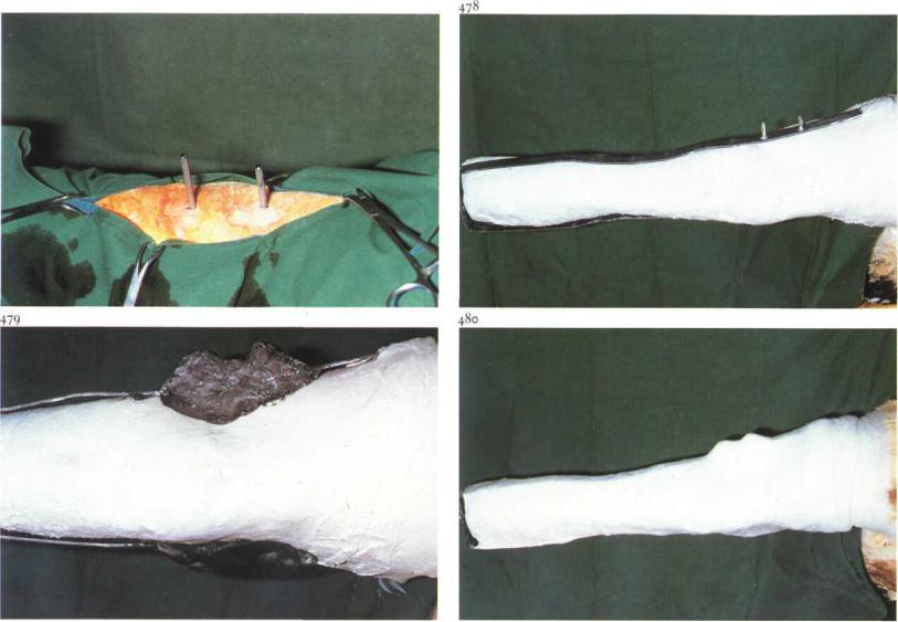
Surgery. Surgery is performed with the patient in lateral recumbency under general anaesthesia, or (for hindlimb fractures in cattle) under caudal epidural (anterior block) analgesia. A piece ofsteel wire is fixed to the hoofwall, and traction can be applied to the limb if necessary to facilitate reduction [474]. The reduction is checked radiographically, as are the sites of pin placement.
After stab incisions, the holes are drilled through the distal part of the tibia [475]. The diameter of the drill is the same as the diameter of the pins. After the pins have been placed [476,477], the frame is fitted and the sites where holes must be drilled in the frame are marked. The holes in the
Chapter 7 THE M U S C U L O S K E L E T A L SYSTEM / Metacarpus and metatarsus 7-17 |
127 |
477
frame must be about 3 mm larger than the pin diameter to prevent torsion between pins and frame. If necessary, the skin wounds are closed with synthetic absorbable suture material, and covered with sterile tampons.
A sterile layer of cotton wool and gauze bandage are used as padding. A plaster cast approximately 3 mm thick is applied to the leg from hoof to stifle. The frame is moulded to the shape of the casted leg and placed on the pins [478]. The space between the pins and frame is filled with Technovit® to fasten the frame to the pins. The protruding ends of the pins are covered with Technovit® to protect the animal as well as the pins [479]. A second layer of plaster incorporates the frame and fixes it to the first layer. The distal part of the frame is not covered by the cast and the hoof is not fixed to the frame [480]. After hardening of the cast, the steel traction wire is cut. Systemic antibiotics are administered.
The patient is confined to stall rest during the healing period. The walking cast is not removed until X-ray control reveals that sufficient callus has been formed. There may be osteolysis around the pins, accompanied by
slight purulent secretion. The skin and subcutaneous wounds close some days after the pins have been removed. After removal of the cast the healed leg can be put in a supporting bandage.
Chapter 7 THE MUSCULOSKELETAL SYSTEM / Metacarpus and metatarsus 7-18 |
128 |
7-18 Sequestrectomy
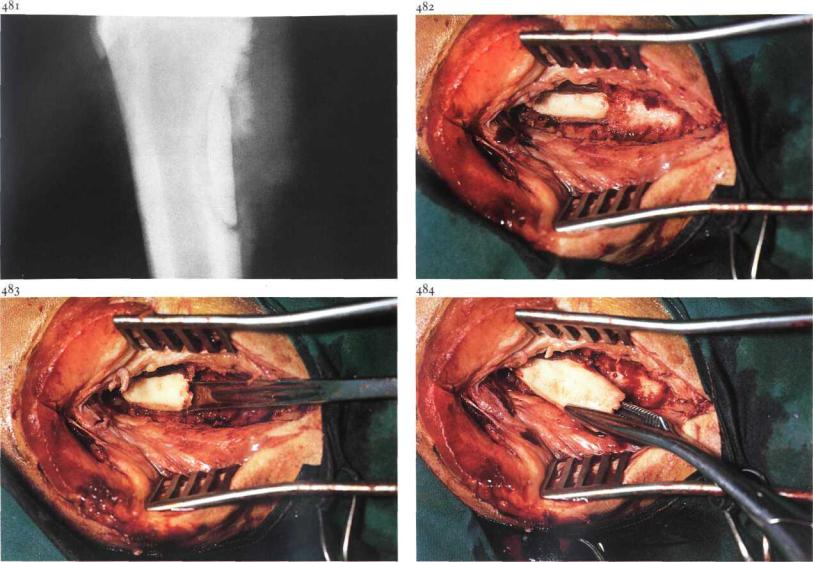
Bone infection in large animals is a sequel of penetrating wounds, haematogenous infection (usually metaphysitis), or local extension of infection from neighbouring tissues. The resulting devitalised bone, separated from the parent bone and surrounded by granulation tissue, is called a sequestrum [481], which may become enclosed by new bone (involucrum), penetrated by channels (cloacae). The escape ofexudate results in a draining tract to the skin surface. In most cases removal of the sequestrum is obligatory.
Surgery. The method of restraint and analgesia depends on the site and size of the sequestrum, presence or absence of an involucrum, and tractability of the patient. Esmarch's ligature may be useful. If a draining tract is present, it is removed in toto. The skin incision is lengthened and the subcutaneous tissues are incised. Periosteal new bone, if present, is removed with a bone chisel, allowing visualisation of the sequestrum [482], which is often
discoloured and located in a bed of pus and/or granulation tissue. The sequestrum is loosened [483] and removed with a suitable forceps [484]. Very large sequestra may be fragmented with a bone chisel and hammer to facilitate removal.
After removal of all necrotic bone and debris, which should be checked bytaking radiographs, the wound is partly closed with simple interrupted skin sutures of non-absorbable material, leaving the distal commissure open to allow drainage. An antiseptic bandage is firmly applied. Systemic antibiotics may be indicated. The bandage is changed on the second postoperative day and thereafter as needed.
Chapter 7 THE M U S C L L O S K E L E T A L SYSTEM / Fetlock and phalanges 7-19 |
129 |
7-19 Ostectomy of apical fracture of proximal sesamoid bone
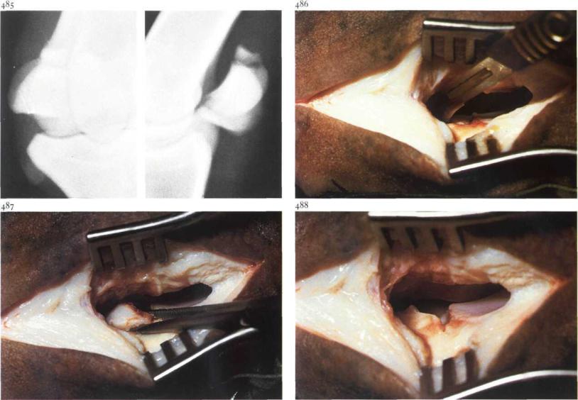
Fractures of the proximal sesamoids are most common in standardbreds and thoroughbreds and are classified as apical, midline or basilar fractures. Treatment consists of either lag screw fixation, plaster casting in slight flexion or removal ofthe fracture fragment. Apical fractures [485] involving one third or less of the bone are amenable to treatment by surgical resection of the fragment.
Surgery. The patient is placed in lateral recumbency, with the affected sesamoid bone uppermost, under general anaesthesia. An Esmarch bandage may facilitate surgery.
An incision approximately 5 cm long is made, between the caudal border of the third metacarpal (metatarsal) bone and the cranial border of the suspensory ligament from just below the button of the splintbone to the proximal border of the collateral sesamoidean ligament. The joint capsule is incised and a Weitlander retractor is positioned to facilitate exposure of the proximal sesamoid bone.
The fragment should be dissected free from its attachment to the suspensory ligament and intersesamoidean ligament using a hooked scalpel blade [486]. When the soft tissue attachments are severed, the fragment is removed with Ochsner forceps [487,488]. Flushing of the joint cavity may be indicated.
The fibrous capsule is closed with simple interrupted sutures of synthetic absorbable material. The subcutaneous tissue is closed in a simple continuous pattern using synthetic absorbable material, and the skin with simple interrupted sutures. A sterile dressing is placed over the incision, and a firm bandage is applied.
Aftercare consists of at least 3 weeks' bandaging and box rest. Training may begin after 6 weeks in some cases.
Chapter 7 THE MUSCULOSKELETAL S Y S T E M / Fetlock and phalanges 7-20
7-20 Treatment of first phalanx fractures
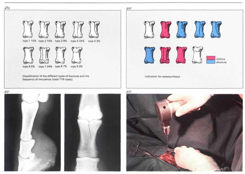
First phalanx fissures and fractures in horses are usually the result of injuries sustained during locomotion and may be classified into nine types [489]. There are two types of fissure (type i and type 2), which result from the effects of torsion of the sagittal ridge of the loaded metacarpus and metatarsus ('screwdriver effect'). The fracture types 3, 4 and 5 occur in the sagittal plane and may also result from the screwdriver effect. Type 6 is a fissure or fracture in the frontal plane; type 7 is a comminuted fracture, type 8 an avulsion fracture. Type 9 is a transverse fracture, and most often occurs in young foals, due to trauma caused by the mare. There are two possibilities for treatment of first phalanx fissures and fractures.
(1)Conservative treatment (non-surgical). Types i and 9 are treated by externalfixation.
(2)Surgical treatment (osteosynthesis). Relative indication for osteosynthesis means that the prognosis will not be much different in using either
osteosynthesis or conservative treatment [490], but osteosynthesis reduces the time of healing. Absolute indication for osteosynthesis means that the prognosis is significantly less favourable ifthe lag screw technique is not employed. Types 2 and 7 may be treated surgically (relative indication); lag screw fixation is used in type 2 fissures, whereas for type 7 the walking cast technique may be employed (see 7-17).
Types 3, 4, 5 and 6 have an absolute indication for lag screwing. In type 8 lag screwing is used only if the fragment is large enough; if the fragment is small, it is surgically removed.
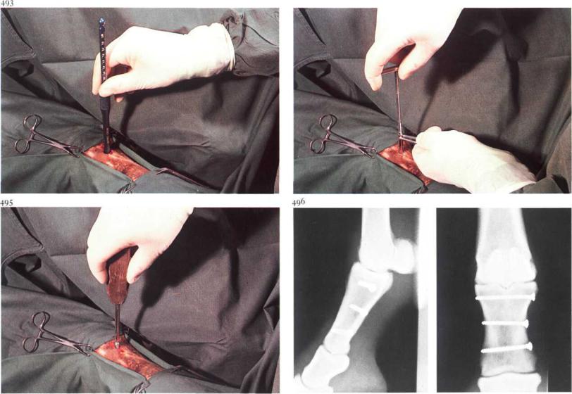
Surgery. The lag screw technique, in this case for type 4 fracture [491], is carried out under general anaesthesia with the patient in lateral recumbency. At the site of insertion of the screw into the smaller fragment, an approximately 1.5 cm incision is made through the skin into the periosteum, which is then retracted. With the soft tissues protected by a drill guide, a 4.5 mm diameter hole is drilled through the smaller fragment [492]; this is referred to as the gliding hole. In the larger fracture fragment,
Chapter 7 THE M U S C U L O S K E L E T A L SYSTEM / Fetlock and phalanges 7-20
494
the thread hole is drilled, using a 3.2 mm drill guide placed in the gliding hole. The length of the cortex screw is determined with the depth gauge [493]. In the larger fragment the thread is cut using a tap [494]. After countersinking, the screw is screwed in [495]. The length should be such that i-i threads of the screw protrude on the cortex of the larger fragment. Radiographic monitoring during surgery should be used for exact positioning of the screws. The osteosynthesis is protected by an external fixation for about six weeks. The best chances for full functional recovery occur after contact healing with minimal or no callus formation [496].
Healing is radiographically visible at approximately eight weeks. Patients with comminuted fractures (type 7) remain lame and can be used only for breeding purposes.
Chapter 7 THE M U S C U L O S K E L E T A L SYSTEM / Fetlock and phalanges 7-21 |
132 |
497
7-21 Arthrotomy of fetlock joint in chip fracture
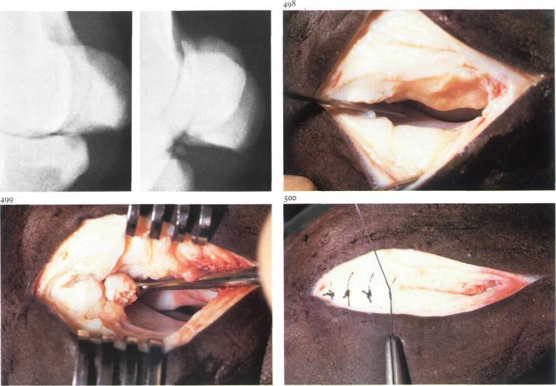
Arthrotomy of the fetlock joint is indicated in e.g. anterior or posterior chip fractures of the first phalanx, apex fracture, and basilar chip fracture of the proximal sesamoid bone. Arthrotomy to remove a posterior chip fracture of the first phalanx is presented here. The presence of such a fragment is often the cause of lameness. Radiographic examination shows a dis-placed bony fragment at the level of the base of the proximal sesamoid bone [497]. Surgery. The patient is placed in lateral recumbency with the affected side uppermost, under general anaesthesia. An Esmarch bandage may facilitate surgery. The joint is opened with the same technique used in the removal of the apical chip fracture of the proximal sesamoid bone (see 7-19) but in this operation the incision must be extended distally through the collateral sesamoidean ligament to provide exposure of the posterior distal joint cavity [498]. Wound retractors are positioned to facilitate exposure of the fragment. The fragment should be dissected free from the joint capsule
using a hooked scalpel blade, after which the fragment is removed using Ochsner forceps or a Brun curette [499]. After removal of the fragment the joint cavity is flushed with sterile physiologic saline.
The fibrous capsule and the collateral sesamoidean ligament is closed with simple interrupted sutures using synthetic absorbable material [500]. The subcutaneous tissue is closed in a simple continuous pattern using synthetic absorbable material and the skin with simple interrupted sutures. A sterile dressing is placed over the incision, and the leg is firmly bandaged. Postoperattive management is similar to that for removal of apical chip fracture (see 7-19).
Chapter 7 THE M U S C U L O S K E L E T A L SYSTEM / Fetlock andphalanges 7-22 |
'33 |
7-22 Treatment of third phalanx fracture in the horse
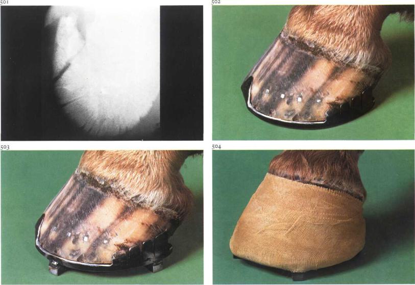
Trauma is the predominant cause of fractures of the third phalanx. Wing [501], sagittal, and extensor process fractures are most common. Large and small extensor process fracture fragments may be treated by lag screw fixation and ostectomy respectively. The failure of bony union of wing and sagittal fractures is caused by continuous movement of the fracture fragments by expansion of the hoof. Therapy aims to immobilize the fracture fragments by preventing expansion of the hoof. Stability can be also achieved by lag screw fixation of the fracture fragments. Use of a lag screw may be considered in case of sagittal (intra-articular) fractures in older horses. Precise implantation of the screw demands a special guide apparatus (see 7-23) and radiographic monitoring during surgery.
However, most fractures heal successfully with conservative treatment using a full bar shoe with quarter and heel clips [502]. Further reduction of hoof expansion can by achieved by applying a rigid cast over the clip shoe.
Four calks are used to prevent wear and tear to the cast [503,504]. Care is taken to leave the coronary band free of the cast to avoid pressure sores.
Chapter 7 THE MUSCULOSKELETAL SYSTEM / Fetlock and phalanges 7-23
7-23 Lag screw fixation of navicular bone fracture
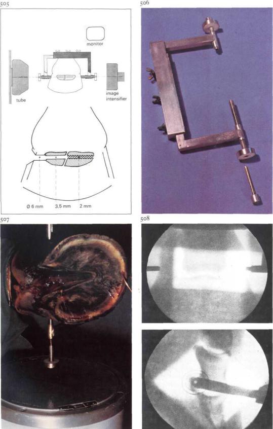
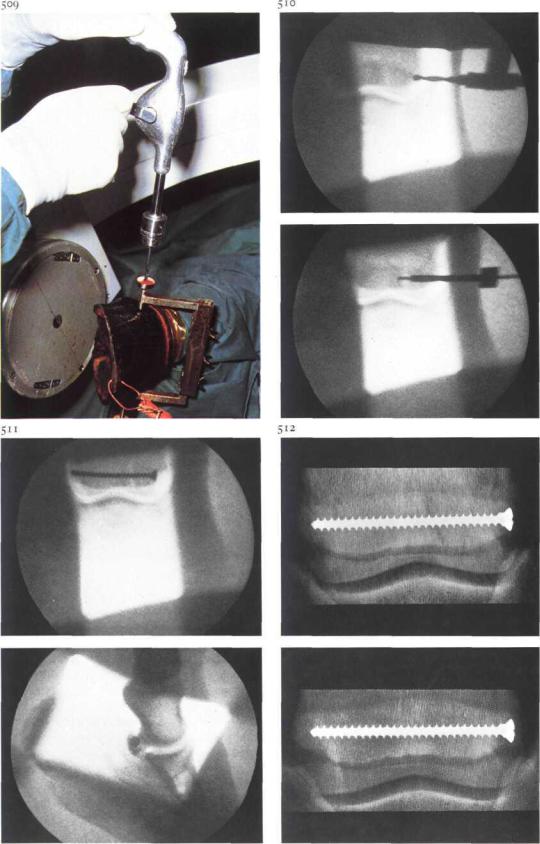
Navicular bone fractures in the horse occur infrequently. These fractures are presumably the result of sudden extreme unequal pressure from the second and third phalanges and deep flexor tendon upon the navicular bone. Osteoporosis as a result of severe navicular disease or local osteolysis may be predisposing factors. Most fractures are sagittal, located in the latero-central or medio-central area of the navicular bone, and are minimally displaced. Healing occurs mainly by fibrous callus of endosteal origin, resulting in permanent lameness. The failure of bony union of navicular fractures is caused by continuous instability of the fracture fragments. Stability can be achieved by lag screw fixation of the fracture fragments. Implantation of the screw precisely along the transverse axis of the navicular bone demands radiographic monitoring during surgery as well as a specially developed apparatus [505,506] to ensure perfectly accurate insertion of the drill. With the help of two 3.5 mm threaded drill guides, the guide system can be fixed to the hoof. The screw on the drill guide is made of nylon, which is not radiopaque. A stainless steel ring around the nylon screw facilitates centering. Surgery. The horse is placed in lateral recumbency under general anaesthesia. By means of a latero-medial fluoroscopic view, the ends of the navicular bone are located and marked with hypodermic needles in the lateral and medial area of the hoof wall. The threaded drill guides of the guide apparatus are placed over the needles. The needles are removed and the position of the drill guides is adjusted, using latero-medial, dorsopalmar and caudal proximo-distal fluoroscopic views. Exact alignment of both drill guides along the transverse axis of the navicular bone is required [507,508].
Through the 3.5 mm drill guide, a hole is drilled through the hoofwall, sensitive laminae, lateral cartilage or wing of ?3, and through the navicular bone as far as the fracture line [509]. The progress of the drilling is monitored radiographically [SIOA]. With the 2 mm drill guide in the 3.5 mm guide, a hole is drilled into the other fragment of the navicular bone [5108]. The 3.5 mm drill is reinserted, and the hole in the tissue peripheral to the navicular bone is widened using a 6 mm flexible (intramedullary) reamer. The 3.5 mm hole in the navicular bone is countersunk.
Chapter 7 THE M U S C U L O S K E I . E T A L SYSTEM / Fetlock andphalanges 7-23 |
135 |
The lag fragment is tapped, and a cortical screw of suitable length (usually about 50 mm) is used to exert compression between the two fragments
Systemic antibiotics are administered. Postoperatively, bandages covering the coronary band and hoofwall are applied and the animal is box rested until the fracture is healed.
Bony union of the fracture fragments is visible on radiographs taken approximately 6 weeks after surgery [5128].