

Атлас анатомии крупных животных
.pdfChapter 3 THE T H O R A X 3-1
2(1
OQO
3-1 Diaphragmatic herniorrhaphy
In large animal surgery thoracotomy is seldom used. Its main indication is diaphragmatic hernia, although the technique is occasionally used in cases of traumatic injury with or without foreign body penetration into the thorax, approach to the thoracic oesophagus, and experimental surgery.
Surgery. The patient is placed in lateral recumbency under general anaesthesia with positive pressure ventilation.
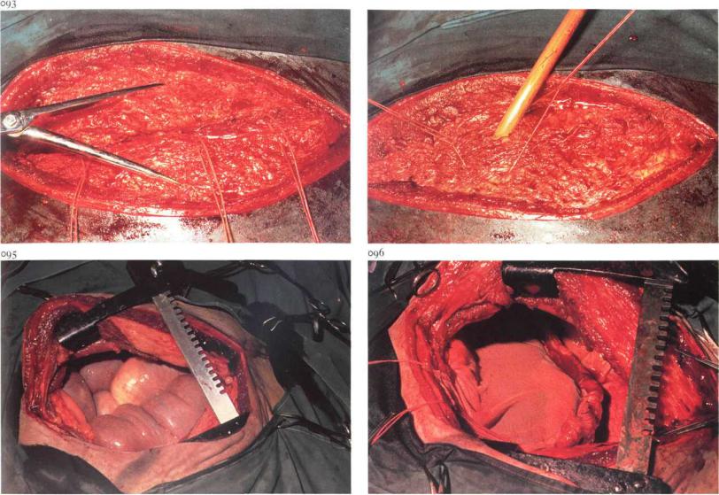
The thorax is approached through the left or right 8th, Qth or loth intercostal space, depending on the localisation of the hernia. Skin, subcutis, fascia, intercostal muscles and pleura are incised in the middle of the intercostal space. In young animals access to the thorax is facilitated by one or two rib retractors [089]. In older animals, partial resection of one or two ribs may be necessary. Plate 089 shows a part of colon in the thorax of a horse, between the lung and diaphragm. After partial reposition of the intestine the diaphragmatic hernial ring is visible [090].
A second surgeon, working through a laparotomy incision, may be needed to aid with reposition of the abdominal organs (intestines and liver) and in placing the sutures in the hernial ring. The hernia is closed by tying the pre-placed interrupted sutures of non-absorbable material [091,092]. When the diaphragmatic defect is extensive or can not be closed with sutures, closure is achieved using a synthetic mesh [095,096] (see also 4-1). Reposition of the ribs is done with the help of reposition forceps. The ribs are held in position by several interrupted sutures [093]. Prior to tying the last two sutures of the intercostal muscles, air must be removed from the thorax by either evacuation or by inflating the lungs [094]. Closure of the wound is completed by suturing of the fascia, subcutis and the skin with interrupted sutures of absorbable material. Systemic antibiotics are indicated. Intrathoracic drains are not routinely used, and postoperative complications have not arisen. Residual air in the thoracic cavity is resorbed within a few days.
Chapter j THE T H O R A X 3-1
094

Chapter j THE T H O R A X 3-2 |
28 |
097
3-2 Treatment of fistulous withers
Infection of tissues in the region of the withers may be the result of traumatic injury, pressure necrosis of skin and underlying tissues due to illfitting saddles and/or prolonged riding [097], infection of the supraspinous bursa (e.g. Brucellosis) and invasion of the nuchal ligament with filariae (Onchocerca sp).
Pockets and compartments of exudate are formed between the tissues of the withers as inflammation extends. The tissues involved (ligament, bursa, spines of first thoracic vertebrae) and the depth and direction of the tracts may be identified with a probe [098, recumbent horse]; (contrast)radiography is an additional diagnostic aid. Surgical therapy consists of drainage of pockets and fistulous tracts.
Surgery. Short interventions and superficial incisions may be carried out on the standing animal under sedation and physical restraint. Drainage of deeper tissues demands recumbency and general anaesthesia. The general
principle of drainage should be followed: opening of pockets and counterincision^) at the lowest point to achieve drainage [099]. Transverse incisions over the withers must be avoided. Necrotic tissue, which may include the tips of spines, is removed, but excision should not be too radical. Drainage openings are kept open by gauze drains or rubber tubing [100] to allow daily irrigation with a mild disinfectant until exudation changes from a purulent to mucous character and finally ceases. Surrounding skin should be protected with vaseline ointment.
Anthelmintic drugs may be indicated, and systemic antibiotics are administered in cases of acute inflammation.
Treatment of fistulous withers is usually time-consuming: several surgical sessions may be necessary.
Chapter 4 The abdomen

Chapter 4 |
THE A B D O M E N / Abdominal wall 4-1 |
101 |
102 |
4-1 Umbilical herniorrhaphy
Umbilical hernias may occur in all domestic animals, especially pigs, cattle [101] and horses, and may be reducible or non-reducible. A hernia is reducible if the hernial contents can be returned into the abdomen. Hernias are non-reducible because of either adhesions between hernial contents and internal hernial sac (hernia accreta), or incarceration of viscera by the hernial ring (incarcerated hernia). If spontaneous recovery of the umbilical hernia does not occur, or in cases of incarcerated hernia, surgical correction is indicated.
Surgery. Herniorrhaphy is performed with the patient in dorsal recumbency under general anaesthesia (pig, horse) or epidural analgesia (anterior block) in combination with a field block cranial to the umbilicus (cattle). An incision, usually elliptical, is made through the external hernial (cutaneous) sac and dissection of the internal hernial sac is continued down to the hernial ring [102].
(i) Amputation of the internal hernial sac.
Amputation is indicated in cases of hernia accreta or incarcerated hernia. The internal hernial sac is carefully incised without damaging the hernial contents and amputated along the edge of the hernial ring using dissection scissors. The adhesions between hernial sac and hernial contents are separated and the hernial contents returned to the abdomen. If it is expected that the internal hernial sac can not be incised without damaging the hernial contents (usually in cases of incarcerated hernia), the abdomen is opened in the linea alba just cranial to the hernial ring, which is then enlarged using a blunt-pointed scalpel. If incarcerated viscera appear to be devitalized, the internal hernial sac is not separated from the hernial contents, but is removed together with the resected viscera at the time of enterectomy. The hernial ring is closed using horizontal mattress sutures, which perforate both abdominal wall and peritoneum [103,104]. When all sutures have been inserted, steady traction is applied on all sutures to close the hernial ring, whereafter the sutures are tied. Non-absorbable suture
Chapter 4 THE ABDOMEN / Abdominal wall 4-1 |
31 |
material or stainless steel is used. In horses with small hernias, use of synthetic absorbable suture material is preferable.
(2) Replacement of the internal hernial sac.
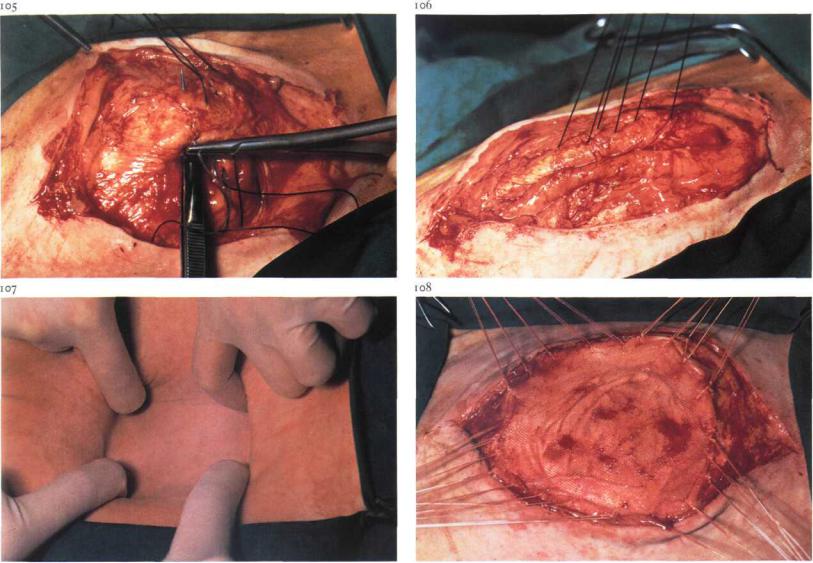
In cases of reducible hernia, the internal hernial sac is usually replaced into the abdomen. The hernial ring is then closed using horizontal mattress sutures. The needle is introduced into the hernial ring 1-2 cm from its edge and runs deeply through the ring without perforating peritoneum. The index finger or the handle of a thumb forceps can be used as a guide [105]. When all sutures have been inserted, they are tightened [106] and tied.
(3) Closure of the hernial ring using alloplastic material.
When the hernial ring is too large for closure with horizontal mattress sutures [107], repair may be successful when alloplastic meshes are used. If possible, the internal hernial sac should be left intact. The mesh is cut 2 cm larger than the hernial ring and the edges are sutured to the hernial ring with simple interrupted sutures (non-absorbable material), without perforating the peritoneum [108].
In all three techniques, subcutaneous tissues are sutured in a continuous pattern, in which the ridge of the closed hernial ring or the central part of the mesh is included to obliterate dead space. The skin is closed using simple interrupted sutures. In female cattle and horses, a belly bandage is recommended for support and to prevent excessive oedema. Tension on the wound edges of large hernias may be reduced by restricting dietary intake preand postoperatively.
Chapter 4 THE A B D O M E N / Abdominal wall 4-2
no
4-2 Resection of urachal fistula
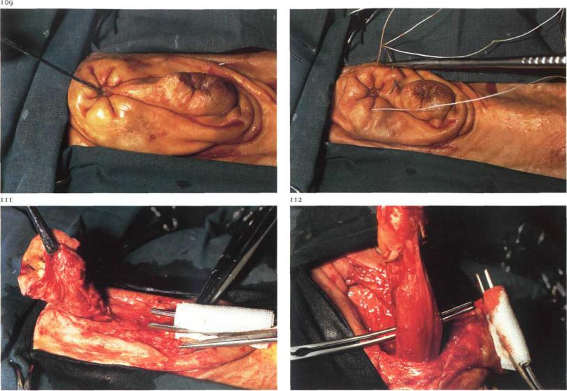
Infection of the umbilical cord in calves may cause inflammatory processes involving the umbilical vessels, urachus, bladder or liver. Chronic cases may result in urachal abscessation, and surgical treatment is indicated. Abscessation of the urachus frequently results in urachal fistula, in which case purulent exudate is visible at the umbilicus. In this bull calf [109], the fistula opening is visible cranial to the preputial orifice. The direction and depth of the fistula can be determined with a probe [109]. Urachal fistulas course caudo-dorsally towards the bladder, and are frequently accompanied by umbilical hernia.
Surgery. Resection of urachal fistula is performed under caudal epidural analgesia (anterior block) in combination with a field block cranial to the umbilicus. The calf is restrained in dorsal recumbency with the legs tied in an extended position. To prevent contamination of the operative area by the urachus, a purse-string suture is placed around the fistula opening
[no]. An intestinal clamp is placed over the preputial orifice to avoid possible contamination by urine. An elliptical skin incision is made around the umbilicus and is extended paraprepudally. To facilitate dessection of the affected umbilical cord, the cranial part of the prepuce is freed from the underlying tissues. Traction is applied to the periumbilical skin, using a tenaculum forceps. The umbilical cord is dissected towards the abdominal body wall [i 11]. The abdominal cavity is entered by incising in the midline cranial to the umbilical cord, and after digital exploration, the body wall directly adjacent to the umbilical cord is excised.
Urachal fistulas often extend to the serosa of the bladder, in which case partial cystectomy is indicated. In order to gain access to the bladder it may be necessary to extend the laparotomy wound caudally. The umbilical cord is dissected from peritoneum and/or greater omentum towards the bladder [112]. The distinction between the affected urachus and the bladder is clearly visible [i 12]. Intestinal clamps are placed on the apex of the bladder and the urachus [113], and the apex is transected between the two

Chapter 4 THE A B D O M E N / Abdominal wall 4-2 |
33 |
|
114
clamps. Plate 114 shows the incised bladder, the mucosa of which appears to be normal. The bladder is closed with a Schmieden intestinal suture, and oversewn with a Lembert seromuscular suture in a continuous or simple interrupted pattern [115] using absorbable material. Finally, the abdominal wall is sutured as described for umbilical herniorraphy (see 4-1). Because surgery has taken place in a possibly heavily contaminated area, systemic antibiotics should be administered.
Plate 116 shows the severely enlarged urachus, which has been incised longitudinally. The umbilicus is visible on the left and the resected apex of the bladder on the right. The lumen of the urachus is necrotic and contains purulent exudate.

Chapter 4 THE A B D O M E N / Abdominal wall 4-3 |
34 |
4-3 Ventral midline laparotomy
Laparotomy in the linea alba is the method of choice in abdominal surgery in horses. Abdominal exploration and exposure of the intestines are easily performed through a ventral incision. The paramedian and flank approach should be considered only for specific indications e.g. bilateral abdominal cryptorchidism (see 5-7), caesarian section. The incision through the linea alba may be umbilical (through the navel), pre-umbilical and postumbilical, depending on the location of the abdominal disorder. The advantage of the median incision is the ease of extension craniad or caudad, if necessitated by the abdominal situation.
Surgery. The animal is placed in dorsal recumbency under general anaesthesia. The legs must be tied with the forelimbs extended and the hindlimbs slightly abducted and flexed.
The skin and subcutis is incised over the desired length, followed by a small incision precisely through the linea alba [117]. After palpation of the
dorsal aspect of the linea alba with the index finger this incision is extended, using a blunt-pointed scalpel [i 18]. Blunt dissection of the retroperitoneal adipose tissue reveals the round ligament (lig. teres hepatis). The ligament is incised [119] and then split with the fingers or blunt scissors [120]. The split round ligament [121] later functions as a reinforcement of the peritoneum and prevents tearing out of the sutures. The wound edges and viscera to be exteriorised are protected by introducing the ring of a sterile plastic drape into the abdomen and spreading the drape over the ventral abdominal wall [122].
The wound is closed by suturing the distinct layers, preferably with synthetic absorbable material. The peritoneum is closed in a simple continuous pattern [123]; the linea alba with simple interrupted sutures [124] or in a simple continuous pattern with double-stranded material of maximum strength; a simple continuous suture closes the subcutis and apposes this layer to the linea alba; finally the skin is sutured with simple interrupted or horizontal mattress sutures. Systemic antibiotics are usuallyindicated.

Chapter 4 THE A B D O M E N / Abdominal wall 4-3 |
35 |
1 2 1