

Атлас анатомии крупных животных
.pdfChapter 4 THE A B D O M E N / Abdominal wall 4-4
4-4 Flank laparotomy
Laparotomy in cattle is most often carried out through a flank incision. The choice between the left or right flank, the selection of the specific site and the type of incision depend upon the procedure to be performed and, in some cases, on the preference of the surgeon.
Surgery. Flank laparotomy is carried out in the standing animal. Local analgesia is achieved by either infiltration, inverted L field block, or paravertebral nerve block.
(1) Left flank.
The standard method for left flank laparotomy is the 'through-and-through' incision. A vertical skin incision is made ventral to the lumbar transverse processes [125]. The external [126] and internal oblique muscle are transected in the same direction. Bleeding vessels may be clamped with haemostats or ligated. The transversus muscle is then carefully incised vertically [127]. The transversalis fascia and peritoneum are elevated and lifted with thumb forceps, and incised with a scalpel; care must be taken not to incise underlying viscera. The incision is enlarged dorsally and ventrally with scissors [128]. Each incision in the separate layers of the abdominal wall is shorter than the preceding one.
The wound is closed in three or four layers. The peritoneum and transversalis fascia are closed together with the transversus muscle using a simple continuous suture. The oblique muscles are closed together using simple interrupted sutures. Either absorbable or non-absorbable suture material may be used for these sutures. If the laparotomy is carried out in the lower part of
the flank the subcutis, which is more prominent there, is sutured in a simple continuous pattern with absorbable suture material. Finally the skin is closed, using simple interrupted sutures with non-absorbable suture material.
(2) Right flank.
Rightflanklaparotomy is usually executed byperforming either a 'true grid' or a 'modified grid' incision. In both methods a 15-20 cm skin incision is made vertically. In case of a 'true grid' incision the external oblique muscle is split in the direction of its fibres (caudo-ventrally), while in case of a 'modified grid' incision this muscle is incised vertically. Both the internal oblique muscle and the transversus muscle are split in the direction of their fibres (i.e. cranio-ventrally and vertically respectively). The transversalis fascia
Chapter 4 THE A B D O M E N / Abdominal wall 4-4 |
37 |
|
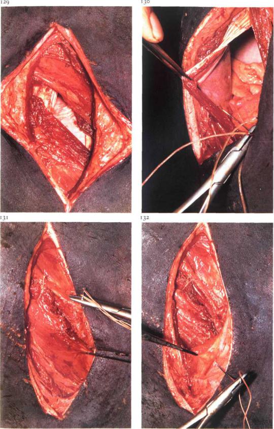
and peritoneum are incised vertically as described for left flank laparotomv. Plate 129 shows a 'modified grid' incision.
The right flank laparotomv wound is always closed in four separate layers. Peritoneum, transversalis fascia and transversus muscle are sutured together in a simple continuous pattern [130], The internal oblique muscle is sutured with two or three simple interrupted sutures [131]. The external oblique muscle is also closed with simple interrupted sutures, the number of which depends upon the direction of muscle incision : in a 'true grid' incision two or three sutures are sufficient, while in a 'modified grid' incision more sutures must be placed [132]. The skin is closed with simple interrupted sutures.

Chapter 4 THE A B D O M E N / Abdominal wall 4-5
4-5 Paramedian laparotomy
The main indications for paramedian laparotomy are abomasoand omentopexy in bovine left or right abomasal displacement, cystic surgery and abdominal cryptorchidectomy in horses (see 5-7). Paramedian parapreputial laparotomy in the horse is discussed here.
Surgery. The paramedian laparotomy is performed under general anaesthesia with the horse in dorsal recumbency. A 15 cm skin incision is made caudally from the level of the preputial orifice, 7-10 cm lateral and parallel to the midline. The abdominal tunic (tunica flava) and the closely adherent ventral sheath of the rectus abdominis muscle are incised in the same direction. The underlying rectus muscle is split in the direction of its fibres by blunt dissection [133]. Subsequently, the internal (dorsal) rectus sheath is incised at right angles (i.e. along its fibres) to the skin incision, after which the peritoneum is bluntly split in the same direction [134].
The wound is closed in four separate layers using synthetic absorbable
suture material. Peritoneum and internal rectus sheath are sutured together in a simple continuous pattern [135]. The rectus muscle and external rectus sheath are sutured together using simple interrupted sutures [136].
Finally, subcutis and skin are closed with continuous and simple interrupted sutures respectively.
Chapter 4 THE ABDOMEN / Gastro-intestinal system 4-6 |
39 |
4-6 Rumenotomy
Rumenotomy is indicated for the removal of penetrating foreign bodies (traumatic reticuloperitonitis), neoplasms (papillomas at the site of the cardia) or abnormal ruminal ingesta (plastic, rope, toxic plants).
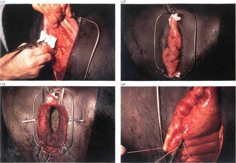
Surgery. Laparotomy is performed in the upper left flank (see 4-4). The abdomen is explored for the presence and extent of peritonitis and other possible disorders. To prevent peritoneal contamination by ruminal contents, Weingart's apparatus is used. The fixation frame is inserted into the dorsal commissure of the skin wound. A fold of the wall of the dorsal ruminal sac is drawn outside the skin incision [137] and fixed with two rumen forceps, first in the lower and then in the upper eye of the frame [138]. The ruminal wall is now incised, beginning ventrally; as the incision is lengthened Weingart hooks are placed through the full thickness of the rumen wound edges and attached onto the frame [139]. Ruminal contents are now visible, and abnormal ingesta can be removed. In cases of reticuloperitonitis all
penetrating and loose foreign bodies must be removed from both the reticulum and the rumen.
Ingesta is removed from the edges of the rumen wound by flushing. The two lowest hooks are removed and a Schmieden suture or a continuous seromuscular suture (Lembert or Gushing) is begun. As suturing progresses dorsally the other hooks are removed. The first suture line is oversewn with a continuous seromuscular suture [140]. Either absorbable or non-ab- sorbable material may be used. The rumen fold is replaced in the abdomen, after which the Weingart frame is removed and the laparotomy wound closed (see 4-4). Administration of antibiotics is indicated. Dietary measures are taken routinely.
Chapter 4 THE ABDOMEN / Gastro-intestinal system 4-7 |
4° |
4-7 Correction of left displaced abomasum
Procedures used for correction of left displaced abomasum include omentopexy through the left or right flank, and omentopexy or abomasopexy by right paramedian laparotomy. In addition to these methods techniques of percutaneous abomasopexy have been developed.
Surgery. The Utrecht method (left flank laparotomy and omentopexy) and percutaneous abomasopexy using a bar suture are discussed here,
(i) Utrecht method.
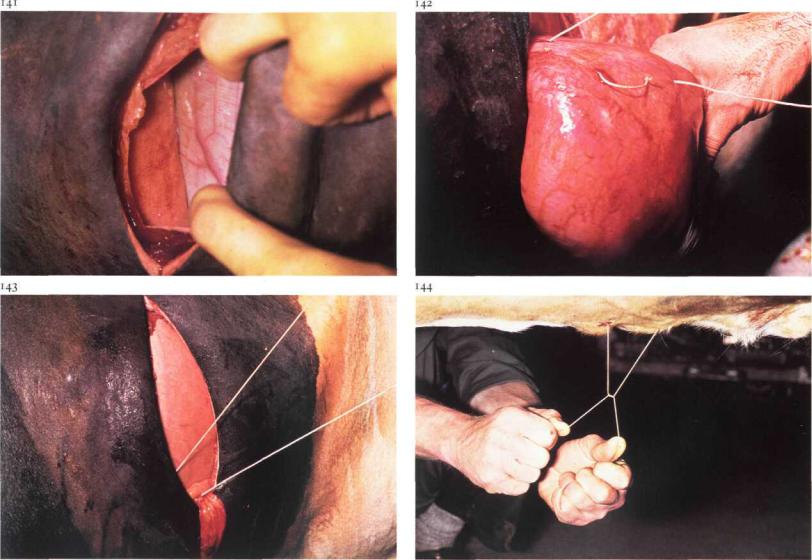
In the standing animal left flank laparotomy (see 4-4) is carried out one handbreadth behind the last rib and about 20 cm ventral to the lumbar transverse processes. In most cases the abomasum appears directly in the laparotomy wound [141]. With the right hand and forearm some pressure is applied on the greater curvature in the direction of the xiphoid to expel gas and achieve partial replacement. Only if distension is excessive, is gas aspirated with a needle affixed to a long tube; the puncture opening is closed with a seromuscular purse-string suture.
At the level of the i ith-i2th rib, a fold of the greater omentum is grasped and pulled into the laparotomy wound. A two metre long heavy thread of non-absorbable suture material with a needle at both ends is then passed a few times through this fold over a length of about 10 cm close to its attachment to the abomasum [142]. Care must be taken to avoid blood vessels.
The abomasum is pushed ventral to the ruminal antrum into its normal position. Both ends of the thread still protrude from the laparotomy wound
[143].The needles, shielded by the hand, are taken separately along the left abdominal wall to the ventral midline and inserted through the abdominal wall about 10 and 15 cm cranial to the umbilicus respectively. The mattress suture is drawn tight and tied on the outside of the body by an assistant
[144].Care must be taken that no other structures are interposed in the omentopexy area. The laparotomy wound is closed as described in 4-4. Postoperative dietary measures are routinely taken. The fixation suture and skin sutures are removed on the tenth postoperative day.
Chapter 4 THE ABDOMEN / Gastro-intestinal system 4-7
(2) Percutaneous abomasopexy using a bar suture.
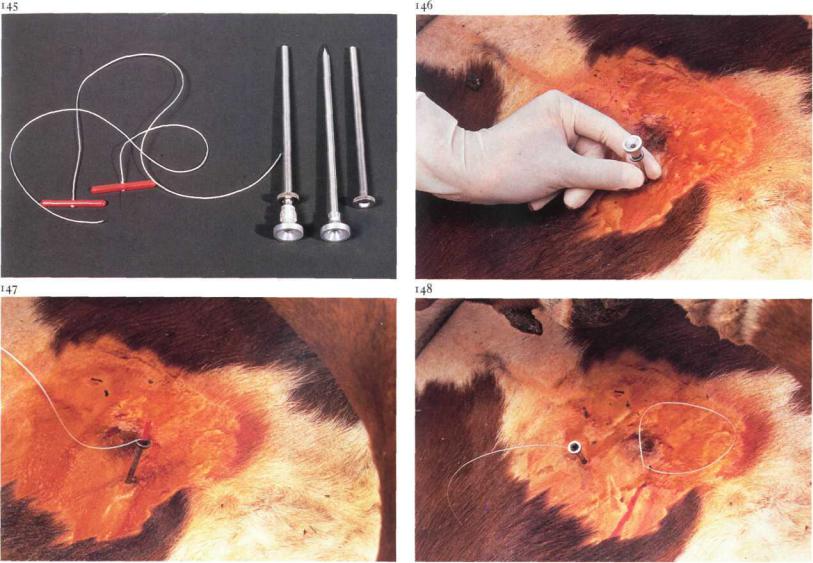
The cow is cast, restrained in right lateral recumbency, and then carefully rolled into dorsal recumbency. Percussion and auscultation of the body wall between the umbilicus and xyphoid for detection of a 'ping' reveals the location of the abomasum and the site of trocarization. In the region of the loudest 'ping', usually on the right side ofthe ventral midline, a 4 mm diameter trocar-cannula [145] is inserted through the abdominal wall into the abomasum, avoiding the subcutaneous abdominal vein [146]. The trocar is then removed with the cannula left in place. Perforation of the abomasum is suggested by the distinctive odour of escaping abomasal gas, and is confirmed by determination of a pH of 2-3 in aspirated content. A bar suture [145] is placed in the cannula [147] and pushed into the abomasal lumen with the trocar. The cannula and trocar are then removed, leaving the suture in place. In a similar manner, a second suture is placed about 7 cm from the first [148]. Prior to removal of the second cannula, abomasal gas is allowed to escape. The sutures are tied loosely. The cow is rolled into left lateral recumbency and allowed to stand.
Postoperative care consists of dietary measures. After about 10 days the sutures are cut close to the skin allowing the sutures to withdraw into the lumen of the abomasum.
Chapter 4 THE A B D O M E N / Gastro-mtestinal system 4-8
4-8 Correction of right displaced abomasum
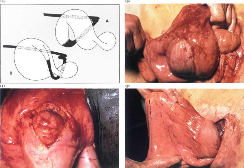
Right abomasal displacement always starts with a 'flexio', i.e. a displacement about a horizontal axis running cranio-caudally; the greater curvature of the abomasal corpus moves dorsally along the right abdominal wall [i49A]. In many cases the flexio is followed by a 'rotation', i.e. the abomasum turns about an axis perpendicular to its greater curvature and running through a point halfway between the omaso-abomasal junction and the sigmoid curve of the duodenum; the pyloric part moves cranially along the right abdominal wall [1493].
Surgery. After right flank laparotomy one handbreadth behind the last rib (see 4-4) the abdomen is first explored to determine the type of displacement and to detect possible complications. If necessary the abomasum is punctured before reposition is started (see also 4-7). A flexio is corrected bypushing with the whole lower arm the greater curvature in a cranioventral direction along the right abdominal wall. If a flexio with rotation is present,
the abomasal corpus is first pushed in a cranial and ventral direction, after which the pyloric part is grasped and pulled caudally across the abdominal floor. In case of severe stasis of abomasal contents a pyloromyotomy may be performed. The pylorus is grasped and brought extra-abdominally [150].
An approximately 8 cm long incision is made through the serosa and muscularis of the pyloric region until the mucosa bulges along the entire length of the incision [151]. The pylorus is replaced into the abdomen. In order to prevent recurrence of the displacement a fold of the greater omentum about 20 cm caudal to the pylorus [152 - dotted line] is incorporated into the first layer of the abdominal wall closure, i.e. the simple continuous suture of the peritoneum and transversus muscle. The other muscular layers and the skin are sutured as described in 4-4.
Further supportive treatment, such as administration of fluids and electrolytes, is based upon the patient's general condition and biochemical findings.
Chapter 4 THE A B D O M E N / Gastro-intestinalsystem 4-9
4-9 Caecotomy in cattle
Caecotomy is indicated in cattle suffering from dilation and torsion of the caecum and ansa proximalis of the colon. In some cases only a dilation is present, but usually a torsion also occurs; this may be clockwise or counterclockwise seen from the animal's right side.
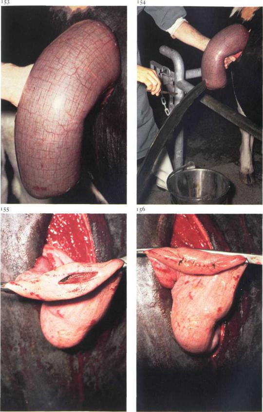
Surgery. Laparotomy is carried out on the standing animal in the caudal part of the right flank (see 4-4). The abdomen is explored to detect whether torsion is present or not; if present its direction is determined. The free part of the caecum is brought extra-abdominally and held in a ventral position [153], after which the caecal apex is incised to release ingesta{i54]. The intra-abdominal part of the caecum and the ansa proximalis of the colon are also emptied as well as possible, by massaging the ingesta in the appropriate direction. An intestinal clamp is then placed proximal to the incision [155], which is closed by either a Schmieden suture oversewn with a continuous Lembert suture, or with two continuous Lembert sutures [156]. The surface of the caecal apex is flushed with physiologic saline. The caecum is replaced in the abdomen, and the reposition of the torsion is completed. The laparotomy wound is then closed (see 4-4). Antibiotic administration is optional. Dietary measures are routinely taken.
Chapter 4 THE A B D O M E N / Gastro-intestinalsystem 4-10 |
44 |
4-10 Enterectomy; side-to-side anastomosis
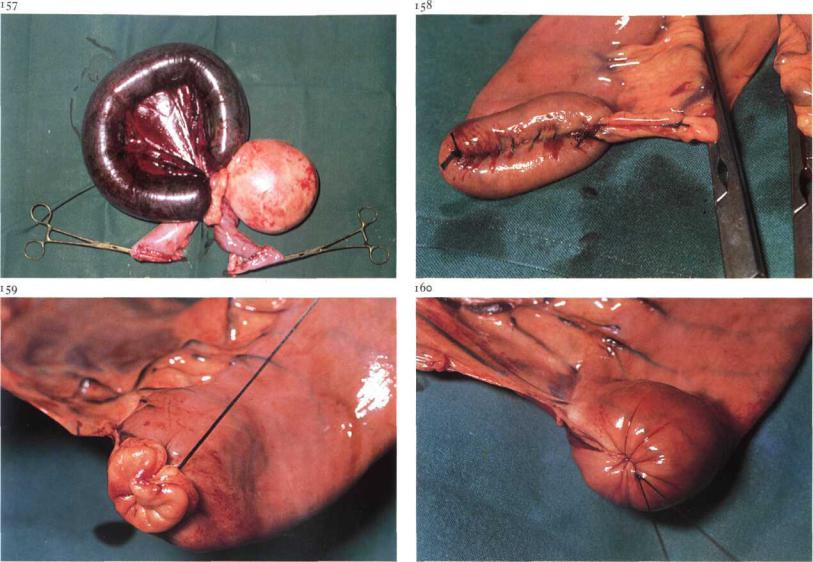
Enterectomy is indicated in cases of irreversibly compromised viability of the intestinal wall, which may be caused by incarceration, torsion, intussusception, strangulation by fibrous bands or pedunculated tumours, etc. Plate 117 shows strangulation ofa jejunal loop by a pedunculated lipoma. In horses, side-to-side anastomosis of the jejunum is often preferred, to avoid postoperative stenosis.
Surgery. The operation is carried out with the animal in dorsal recumbency under general anaesthesia. A laparotomy is performed in the ventral midline (see 4-3). After correction of the primary disorder and emptying of the jejunum, the intestinal wall is examined for viability, in which special attention is paid to the state of circulation and motility. If enterectomy is required the affected gut is brought sufficiently extra-abdominally to allow resection through healthy tissue and to prevent peritoneal contamination from intestinal contents. After the extent of resection is determined, the
vessels of the adjacent mesentery are ligated; the mesentery to be removed is usually wedge-formed. The blood supply of the proposed sites of resection must not be compromised. The affected intestine is isolated by carefully placing two pairs of intestinal clamps over adjacent bowel. The mesentery is transected distal to the ligatures, and the diseased gut is resected between the two clamps, both proximally and distally. After flushing with physiologic saline the remaining ends are closed using a Schmieden suture oversewn with a continuous Lembert suture, both with absorbable suture material [158]. An alternative method of resection is as follows: the intestinal loop to be resected is isolated from the remainder of the gut by ligatures instead of intestinal clamps. The gut is then transected between the ligatures [159]. The ligatures of the remaining ends are invaginated after which closure of the ends is performed with a seromuscular purse-string suture [160].
The ends of the gut are now laid side by side with their stumps either in the same direction (anisoperistaltic = functional end-to-end anastomosis) or in
Chapter 4 THE A B D O M E N / Gastro-intestinal system 4-10 |
45 |
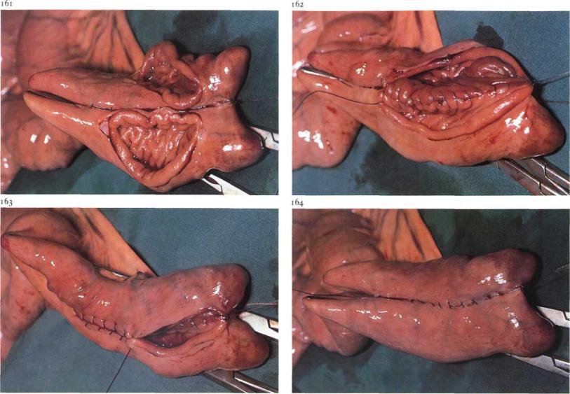
opposing directions (isoperistaltic). Intestinal content is milked back and isolated with intestinal clamps. The two apposed loops are sutured together with a continuous Lembert suture. They are then incised about i cm from and parallel to the Lembert suture. The incisions must be of equal length and shorter than the Lembert suture [161]. A simple continuous suture, including all layers of the intestinal wall, is started at one end of the incision, thus suturing together the wound edges on the luminal side of the Lembert suture [162]. On reaching the other end of the incision the continuous suture continues as a Schmieden suture [163], which is continued up to the origin of the simple continuous suture. The previously placed Lembert suture, with which the simple continuous suture is already oversewn, is now continued to oversew the Schmieden suture [164]. Absorbable suture material is used for both suture lines. After removal of the clamps the intestine is flushed again and the anastomosis is checked for patency and leakage. The mesentery is closed with interrupted sutures. The intestine is replaced into the abdomen and the laparotomy wound closed (see 4-3). Systemic antibiotics are indicated.