

Атлас анатомии крупных животных
.pdfChapter^ THE UROGENITAL SYSTEM / Thefemale iirtigenitul system 5-21
5-21 Caesarean section in cattle
Different techniques have been developed for caesarian section in cattle: flank, paramedian and midline approach in the recumbent animal, and right or left flank laparotomy in the standing position. Each method has certain advantages and disadvantages. Choice depends on the type of dystocia, breed and condition of dam and foetus, and in some cases on the preference of the surgeon. In many cases of dystocia, however, foetotomy is the method of choice. In dairy cattle, standing left flank caesarian section is most commonly carried out and is described here.
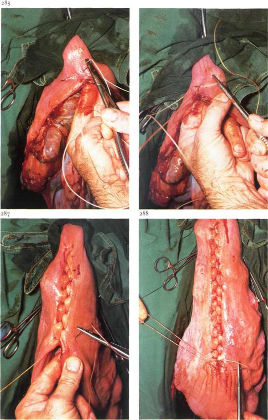
Surgery. In addition to paravertebral or infiltration anaesthesia, a caudal epidural analgesia (posterior block) is given to reduce abdominal straining. A vertical through-and-through incision of adequate length is made (see 4-4). If the apex of the gravid horn (left or right) is positioned in the right side of the abdomen, the uterus must be rotated, by bringing the right hand under the uterus, and grasping the dorsomedial surface of the tip of the horn. With the hand so placed, the uterus is rotated about 90 by pulling the apex under the uterus, which is elevated and pushed to the opposite side by the arm and elbow. The apex of the gravid horn is elevated and exteriorized: the right hand locates the foetal digits in the horn tip, follows the metatarsal bone and grasps the calcaneus. The left hand then grasps the tip of the horn from the medial side [281] and presents the tip of the horn into the wound. The calcaneus is brought into the ventral commissure of the wound with the right hand-[282]. The incision through the uterine wall along the greater curvature runs from 5 cm below the digits to the calcaneup, and is large enough to allow extraction [283]. (In cases of posterior presentation, the head is supported with the right hand, the uterus is exteriorized bytraction on the metacarpus with the left hand, and an incision in the greater curvature is made over the metacarpus.) Sterile chains are applied to the limbs and the foetus is slowly extracted [284], Thereafter the uterus is kept in position by means of sponge forceps: escaping placental fluids must not enter the abdominal cavity. The placental edges are excised. The uterus is closed with a modified Gushing suture (Utrecht method), using plain catgut. The suture begins about 2 cm from the upper end of the wound [285]. Oblique bites are used [286] so that the knot is
Chapters THE U R O G E M T A L S Y S T E M / Thefemale urogenital system 5-21
286
77
buried by the inverted tissue. The next bite begins about 2 cm from the incision edge and is directed at an angle of 45° to it [287]. The bite on the other side is placed similarly but is inserted some distance back from the emergence of the previous bite. It is important that each suture be pulled tightly following its insertion. This inversion suture prevents any leakage and leaves minimal exposure of suture material. Before closure is complete intrauterine tetracycline is administered. The final knot is buried in the same way as at the beginning of the suture line [288].
The uterus is replaced in the abdominal cavity, and oxytocin is given intramuscularly. The laparotomy wound is closed (see 4-4). Systemic antibiotics are administered. The placenta is normally expelled after 4 hours. Retention of the placenta is usually not treated: after about 10 days it is expelled as a necrotic mass. In some cases repeated intra-uterine application of tetracycline is necessary.
Chapters THE U R O G E N I T A L SYSTEM / Thefemale urogenital system 5-22
290
5-22Vestibuloplasty
Pneumovagina is the involuntary aspiration of air, sometimes contaminated with faeces, into the mare's vagina, and results from faulty conformation and closure of the vulva and vestibulum. It is seen in mares ofall ages, and may cause infection of the genital tract, and occasionally pneumometra.
The aim of vestibuloplasty is to reduce the vestibular introitus (vulvar cleft) by 30-50%, and to lower the vestibular roof. Infection should be treated pre-operatively.
Surgery. The mare is restrained in stocks, and the tail wrapped and tied to the side. The vestibular mucosa is flushed with a mild antiseptic.
Analgesia is achieved by infiltration (using a fine needle) of the dorsal vestibular mucosa and the mucocutaneous junction of the upper vulvar cleft. Sedation may be indicated.
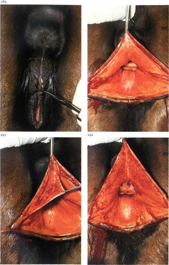
Firstly, the site of the new dorsal commissure is marked on the lips with scissors [289]. If a scar of an old firstor second degree laceration is present, this site is at a point halfway between the dorsal and ventral vulval commissures.
In a non-ruptured vulva, the upper third of the vulvar cleft is closed, and the new dorsal commissure will be at the level of the floor of the bony pelvis.
The vestibulum is exposed by retraction with a forceps in the dorsal commissure and two stay sutures in the vulval lips, placed just ventral to the scissor marks. Incision starts in the vestibular roof 3 to 6 cm caudal to the vestibulevaginal junction and runs caudally to the mark on the vulval lip. An identical incision is made on the other side. Finally the dorsal commissure and muco-cutaneous junction of the labia are incised [290]. A double-triangular section of mucosa is then dissected submucosally from the dorsum and dorsolateral aspects of the vestibulum [291,292]. The left and right horizontal wound edges are carefully apposed and sutured with absorbable material in a simple interrupted pattern, beginning cranially [293]. The knots are tied in the vestibular cavity. After 3-4 knots, simple interrupted sutures are placed dorsal to this suture line to appose the dissected surfaces [294]. Suturing is completed by alternate suturing of the mucosa and deeper layers [295]. The skin is closed with simple interrupted sutures of absorbable material [296].
Tetanus prevention is obligatory. Complete

Chapters THE U R O G E N I T A L SYSTEM / Thefemale urogenital system 5-22
293
79
functional healing usually takes place in 4-8 weeks; during that time natural covering is prohibited.
Episiotomy is necessary at parturition, to prevent irregular tearing of the vulva (see 5-24).
Chapter5 THE U R O G E N i T A L SYSTEM / Tkefemale urogenitalsystem 5-23 |
So |
297
5-23 Perineal reconstruction after third degree laceration
Rupture of the perineal septum and body, vestibular walls, and rectum may occur during parturition at all ages, and is most common in the primiparous mare. The foal's forefoot or nose catches the vestibular roof caudal to the vaginovestibular junction. If unattended, the abdominal straining pushes the foot or nose through the vestibular roof and rectal floor. If the foal is born in this position, the perineum ruptures and a third degree laceration occurs. Immediate reconstruction is not recommended, because of contamination and oedema. Haemostasis, tetanus prevention, debridement, and systemic antibiotics are of importance directly following
i n j u r y .
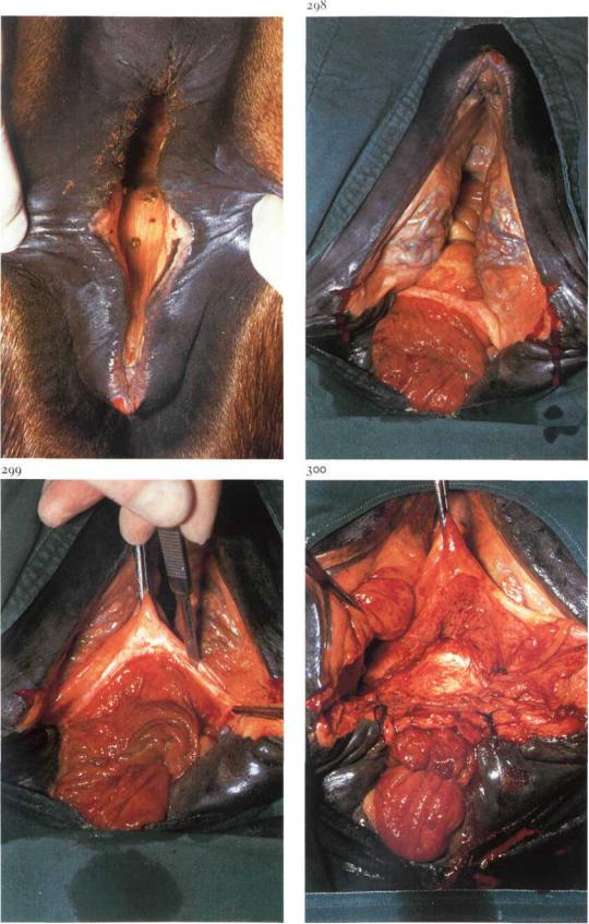
Surgical reconstruction is carried out after weaning of the foal, or healing by second intention (in case of a dead foal). Plate 297 shows the cloaca in the standing mare. Preoperative fasting for 9-10 days is necessary to ensure uneventful wound healing.
Surgery. Positioning in dorsal recumbency under general anaesthesia is preferred because surgery in the standing mare is difficult due to the depth of the injury and the need for sufficient exposure. The site of the new dorsal commissure is marked on the vulva, and retraction is effected by two stay sutures [298]. The operation consists of 3 phases.
(1) Incision of the rectovestibular septum and sidewalls is begun cranially, in the middle of the shelf remaining between vestibule and rectum. The dissection is extended caudallv and horizontally along the junction ofrectal and vaginal mucosa to the points marked on the vulva [299]: the dissection is extended sufficiently to create flaps of tissue which can be apposed and sutured without tension.
The next incisions run in the mucocutaneous junction of both vulval lips, from the mark to the anal sphincter, from where an incision runs cranially in the vestibular mucosa on both sides, at an angle of 30 degrees to the horizontal incision. The triangles of vestibular mucosa are resected.
(2) The most cranial edge of one lateral flap is apposed to the adjacent half of (the dorsal i.e. rectal part of) the 'frontal' flap, using absorbable synthetic material in a simple continuous pattern. The rectal mucosa is not included. The same

Chapter 5 THE U R O G E N I T A L SYSTEM / Thefemale itrogenital system 5-23 |
81 |
procedure is followed on the opposite side [300], and the continuous suture continues caudad, apposing the more caudal parts of both side flaps.
The thus created Y-formed suture line is oversewn with a second continuous suture. The remaining part of the dissected surfaces of both lateral walls (vaginal side) are then apposed with layers of continuous sutures. The mucosa of the vestibular roof is then apposed and closed with everting interrupted sutures, using non-absorb- able material [301].
(3) The perineum (perineal body) is reconstructed by suturing skin and subcutaneous tissue in a simple interrupted pattern, using non-absorb- able material [302]. The anal sphincter is reconstructed with a large supporting mattress suture, which passes through muscle tissue and is tied over two surgical swabs [303].
Systemic antibiotics are administered and tetanus prophylaxis is obligatory. Fasting is continued for 4 days postoperatively, after which the mare is allowed small portions of hay, daily increasing to full rations by the tenth postoperative day. On the 6th postoperative day the supporting suture in the anus is removed. Perineal skin sutures and vestibular mucosa sutures are removed on the loth [304] and 2Oth day respectively. If bacteriological examination of uterine mucus is negative 6 weeks postoperatively, natural covering is allowed. Just prior to the next parturition the vulvar cleft must be opened (see 5-24).
Chapters THE U R O G E N I T A L SYSTEM / Thefemale urogemtal system 5-24,5-25 |
82 |
5-24 Episiotomy
Surgical widening of the vulvar cleft at parturition is sometimes indicated in primiparous mares and is very often necessary after vulval surgery (see 5-22,5-23).
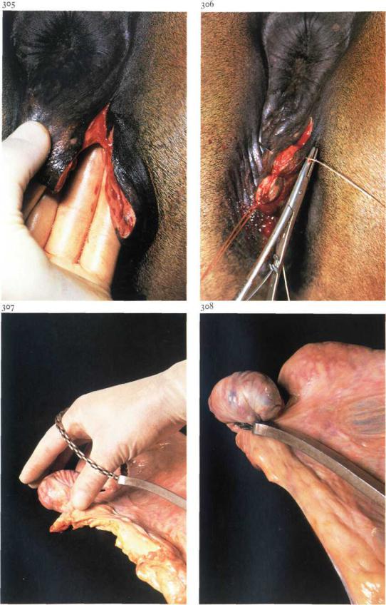
Surgery. During parturition one vulval lip is incised in a dorso-lateral direction beginning 3 cm below the dorsal commissure ([305] plate shows non-foaling mare). The incision through skin and vestibular mucosa is sufficiently long and deep to allow delivery. The wound must be sutured within 4 hours after parturition. The first suture is placed in the mucocutaneous junction. The wound edges are stretched by applying slight traction in a ventro-lateral direction. The other sutures are then placed in a simple interrupted pattern, penetrating all layers, including the vestibular mucosa [306].
Tetanus prophylaxis is provided.
5-25 Ovariectomy
Bilateral ovariectomy may be indicated in various types of nyphomania. A vaginal approach (colpotomy) is described here.
Surgery. The operation may be performed on the standing mare, but dorsal recumbency and general anaesthesia is preferred. Bladder and rectum are emptied and the vagina is prepared. The vaginal roof is stretched and thus lifted from the underlying rectum by displacing the cervix cranially with an instrument introduced through
the vagina. A 5 cm incision is made through the dorso-lateral vaginal wall, 3 cm caudal to the cervix. After careful manual enlargement of the perforation, the ecraseur, with the loop of the chain covered by hand, is inserted and the ovary is brought into the loop [307], which is tightened around the mesovarium. The chain is tightened step by step, at decreasing intervals [308]. The vaginal wound heals by second intention. Systemic antibiotics are administered and tetanus prophylaxis is provided. During the first days the mare is kept standing to avoid the risk of eventration.
Chapter 6 The common integument

ChapterG THE COMMON INTEGUMENT / Skin 6-1 |
84 |
309 |
310 |
6-1 Skin grafting
Autologous skin grafting is often a very useful procedure in the treatment of accidental wounds in horses, especially larger granulomatous wounds of the lower legs. Various types of grafting techniques have been developed: most involve split thickness grafting (Thiersch) and pinch grafting (Braun's implantation technique).
(i) Split skin grafting.
The appropriate time for grafting is when the wound is in an advanced state of second intention healing, i.e. the surface should be covered with healthy granulation tissue [309]. The procedure requires recumbency and general anaesthesia. After preparation for sterile surgery, a superficial layer of granulation tissue is removed [310]. Bleeding is prevented by a tourniquet. A donor site (abdominal wall, caudal surface of the thigh) is prepared for surgery and a split thickness graft of about 0.5 mm thickness is harvested with a dermatome [311], after which the transplant is trimmed to
shape and sutured to the wound edges. In larger wounds several grafts may be necessary to cover the surface. Small parallel incisions in the transplant allow some increase in area of the graft, exact fitting of the donor skin to the receptor site, and drainage of fluids [312]. The operation site is covered with sterile paraffin gauze and an elastic bandage, which is changed every 2-3 days for the first two weeks postoperatively. Excessive movement should be prevented by confinement in a small box. Systemic antibiotics are advisable.
Even when transplantation is not followed by complete adhering of the donor skin, the healing process, i.e. epithelialisation and contraction, is quickened considerably. The procedure usually leads to an acceptable cosmetic appearance without excessive scar formation [313].
(2) Pinch grafting.
When bandaging is impossible and/or larger areas e.g. withers and back [314] must be covered, seed grafting may be preferred. The pinch grafts are harvested from a surgically prepared site using a biopsy punch or sim-

Chapter 6 THE COMMON I N T E G U M E N T / Skin 6-1
313
315 |
316 |
ply by excising a piece of skin elevated with forceps, so that a split skin seed of 4-6 mmis obtained. These grafts are implanted in healthy granulation tissue, in stab incisions made at about 30° to the surface and to a depth of 4-5 mm. If movement can be prevented sufficiently, most seeds will take and epithelial islands become visible after a few weeks [315]. This technique usually controls granulation tissue, accelarates epithelialisation and may lead to complete skin cover of extensive wounds [316].