

Атлас анатомии крупных животных
.pdfChapter i THE H E A D / Guttural pouch 1-3
1-3 Drainage and fenestration
Guttural pouch drainage is indicated in chronic empyema, chondroids and tympany, the latter of which is observed only in foals [017].
Surgery. Surgery is usually performed with the animal in lateral recumbency under general anaesthesia. The guttural pouch is commonly opened through Viborg's triangle, which is bounded by the linguofacial vein, the tendon of the sternocephalic muscle, and the vertical ramus of the mandible. A 3-5 cm incision is made dorsal and parallel to the vein through the skin and subcutaneous fascia. The connective tissue at the ventral border of the parotid is bluntly dissected, until the guttural pouch submucosa has been reached. A fold is carefully elevated as far as possible, and opened with scalpel or scissors [018]. If possible the wall of the guttural pouch is sutured to the edges of the skin wound. Abnormal contents are removed by flushing with a mild disinfectant. A rubber tube is inserted into the drainage opening, and fixed to the skin with sutures [019]. Postoperative flush-
ing is carried out until exudation has ceased; during this period the tube is left in place.
In unilateral tympany the tympanic pouch is opened through Viborg's triangle, and a fenestration is made in the septum between the right and left guttural pouch, using long tissue forceps and scissors [020]. In bilateral tympany one pouch is opened, fenestration performed, and the mucous membrane flap (valve) at the outlet of the Eustachian tube of the opened pouch dissected. In both cases the opened guttural pouch is drained and flushed until exudation has ceased.
Chapter i THE HEAD / Face 1-4
021
O22
1-4 Trephination of the frontal sinus in cattle
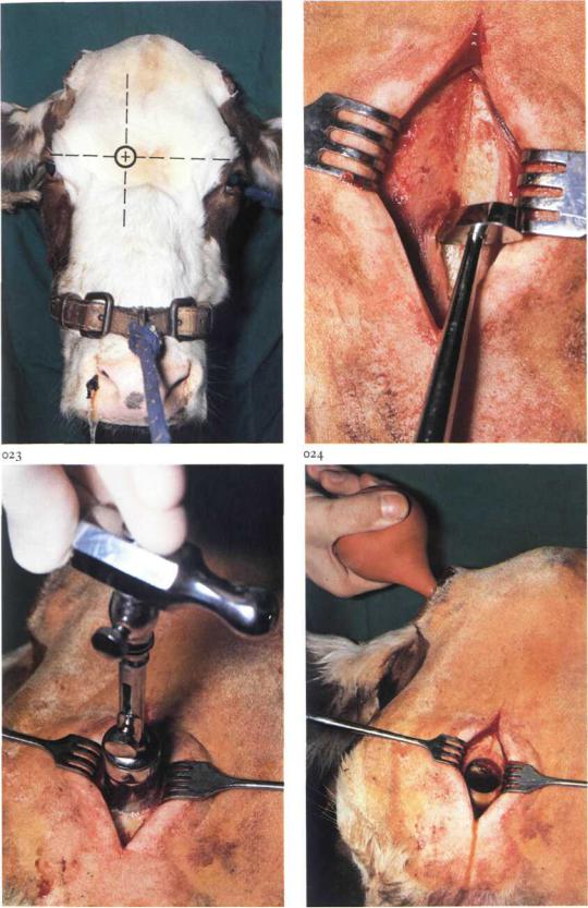
Trephination of the frontal sinus is indicated in chronic empyema, which in adult cattle is caused usually by infection of the sinus following dehorning or horn fracture. Initially the sinusitis is often confined to the caudal part of the sinus, but in long-standing cases the entire sinus may be involved. In the latter case drainage of the sinus is obtained by trephining 2 cm from the midline on a line passing through the centre of the orbits [021]. If the original opening to the sinus at the site of the dehorning wound is narrowed or closed by granulation tissue, it is enlarged or re-opened under cornual nerve block to facilitate adequate flushing of the sinus.
Surgery. Trephination is carried out on the standing animal under local analgesia. An approximately 5 cm long vertical incision is made through skin, subcutis and periosteum. The periosteum is dissected from the bone with a periosteal elevator [022] and drawn aside, together with the skin, with wound retractors. The point of the trephine is inserted into the bone. Trephination is performed by rotating the trephine [023]. After a circular groove has been cut into the bone, the point of the trephine is retracted, and trephination is continued through the full thickness of the bone. The disc is removed with a bone screw inserted into the hole made previously by the point of the trephine. Sometimes the disc must be levered out because it remains fixed to a bony sinus septum.
To remove exudate and necrotic tissue the sinus is flushed thoroughly with a disinfectant solution [024].
To prevent premature closure of the openings they are packed with gauze bandage plugs. Postoperative flushing is repeated daily, until the sinus has healed, as evidenced by absence of purulent discharge.
Chapter i THE HEAD / Face 1-5
1-5 Trephination of the maxillary sinuses and repulsion of teeth in the horse
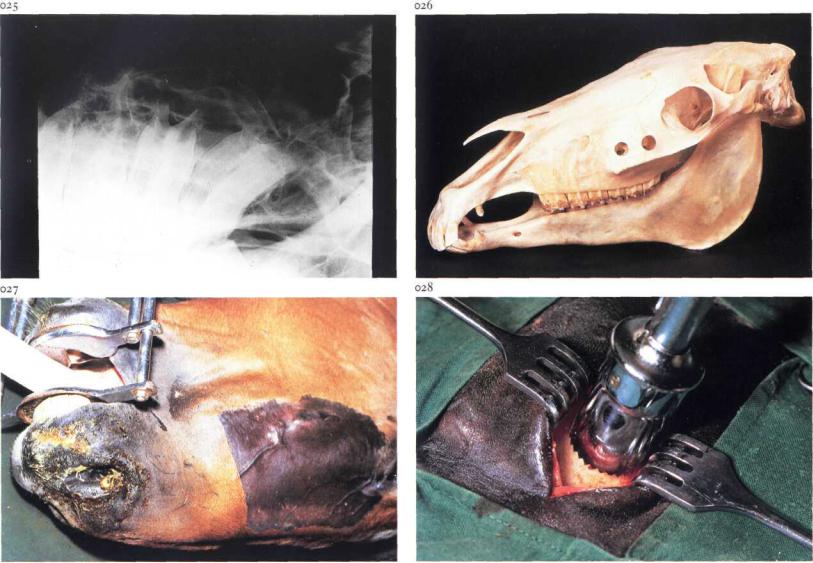
Trephination of the maxillary sinuses is indicated in cases of empyema, cysts or neoplasms, and for repulsion of upper molar teeth. Plate 025 represents a radiograph showing chronic alveolitis of the first upper molar. The rostral maxillary sinus is trephined about 2-3 cm dorsal to the rostral end of the facial crest; the caudal maxillary sinus is trephined 2-5 cm rostral to the medial canthus and 2-3 cm dorsal to the facial crest [026]. Care must be taken to avoid damage to the nasolacrimal duct.
Surgery. The operation may be carried out either on the standing animal under local infiltration analgesia, or on the recumbent animal [027] under general anaesthesia. In case of tooth repulsion general anaesthesia is required.
At the selected site an approximately 4 cm long incision is made parallel to the facial crest through the skin and subcutaneous tissue. Depending on
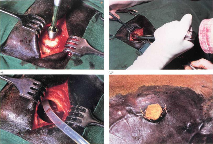
the site of surgery it may be necessary to retract the levator labii maxillaris muscle in order to expose the periosteum. The periosteum is then incised with a scalpel and separated from the bone with a periosteal elevator. The wound edges of the skin and periosteum are drawn aside with wound retractors [028]. Trephination is performed as described in 1-4. The disc is removed with a bone screw [029]. In empyema caused by alveolitis, the sinus is flushed and the affected tooth is carefully located. A punch is then introduced into the sinus and placed upon the roots of the tooth to be repelled. To prevent damage to adjacent teeth and the maxillary bone, the punch must be placed accurately; it may thus be necessary to enlarge the trephination hole with rongeurs. The tooth is repelled from its alveolus with firm, but careful, blows [030]. The course of repulsion is constantly checked by the surgeon's hand in the oral cavity. After removal the tooth is examined to determine if it is complete. Any tooth or bony fragments must be removed. Intra-operative radiography is recommended to ensure that no fragments remain. The sinus and alveolus are copiously flushed with a
Chapter i THE HEAD / Face |
1-5 |
029 |
030 |
disinfectant solution [031]. The alveolus and trephination hole are then packed with povidone iodine soaked gauze bandage plugs [032]. Postoperatively the sinus and alveolus are repeatedly flushed after removal of both plugs. The plug placed in the alveolus after flushing must be somewhat smaller than the previous one, in order to enable granulation tissue to gradually fill the alveolus; the plugs in the trephination hole are of constant size. Only when the alveolus is closed off by granulation tissue and exudation in the sinus has ceased is the trephination hole allowed to close.
Chapter i T H E H E A D / Mandible i -6 |
10 |
034
1-6 Treatment of premaxilla and mandibular body fractures
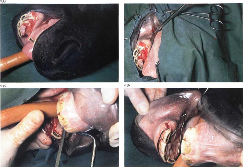
Fractures of the maxilla and mandible have been observed in all large animals but occur most frequently in horses and cattle. Self-inflicted trauma and external violence are the most common causes. In horses, fractures involving the incisor teeth and a variable sized fragment of premaxilla or mandible occur frequently [033]. The deciduous teeth in young animals are frequently involved. Because these teeth have short roots the injury is often minor and of little consequence. Clinically, dislocation of the incisor teeth is obvious. The wound may be packed with feed if the animal has attempted to eat. Teeth may be loose, broken or missing.
Surgery. The operation should be performed in lateral recumbency under general anaesthesia. Debris and granulation tissue, if any, are removed and the wound is carefully cleansed and disinfected. The fragment should be fixed to the premaxilla or mandible by wiring the incisor teeth, but compression in a caudal direction may also be necessary. A canine tooth is used
for the caudal fixation, but if absent (as in this case) a cortical screw is placed in the interalveolar space [034]. To prevent the wire from slipping off the teeth, grooves are made with a hack saw or file in the neck of both third incisors [035] and in the canine teeth. By tightening the stainless steel cerclage wire around the unfractured teeth the fragment is stabilised laterally, and additional stabilisation and compression is achieved by applying and tightening the wire around the canine tooth or the screw [036]. Postoperatively, there is usually no problem associated with prehension or mastication. Healing of the fracture takes place in 4-8 weeks depending on the age of the patient. After healing the wire must be removed.
Chapter i THE HEAD / Mandible 1-7
037
1-7 Treatment of mandibular interdental space fracture
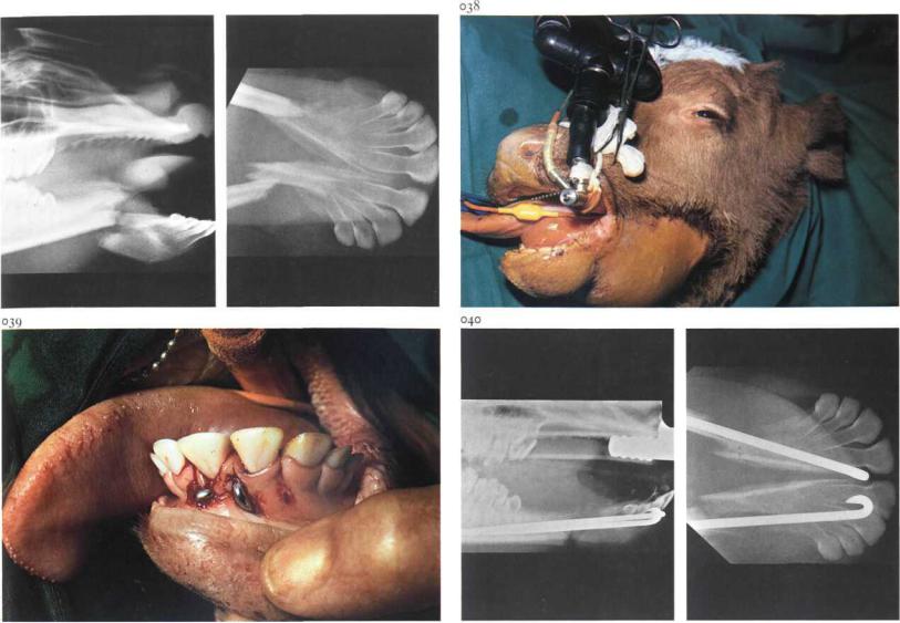
Fracture in the interdental space is the most common fracture involving the horizontal rami of the mandible. The fractures may be unilateral or bilateral [037] and usually compound into the mouth. Unilateral interdental space fractures without severe dislocation heal spontaneously. Bilateral fractures of the interdental spaces cause dislocation of the rostral part [038], and require osteosynthesis.
Surgery. The operation should be performed with the patient in lateral recumbency under general anaesthesia. In this case intramedullary nailing was chosen because the rostral fracture fragment was too short for plating or transfixation.
Implantation of the nails precisely in the mandibular rami without damaging the roots of the teeth demands radiographic monitoring during surgery to ensure accurate insertion of the drill. After reposition of the fracture a hole is drilled in each mandibular ramus beginning medioventral to the
two first incisor teeth or between the first and second incisor teeth. The two Rush nails, previously cut to the required length and contoured correctly, are inserted with a hammer and impactor [039]. It is important that the nails do not damage the roots of the premolars [040]. Postoperative feeding must be modified, but in sucklings nursing can be permitted. In the event of compound fracture bone sequestration often occurs, in which case purulent material usually escapes from draining tracts inside and/or outside the mouth. Sequestra must be removed, after which discharge ceases. Healing of the fracture takes place in 4-8 weeks depending on the age of the patient; implants may then be removed.
Chapter i THE HEAD / Mouth 1-8 |
12 |
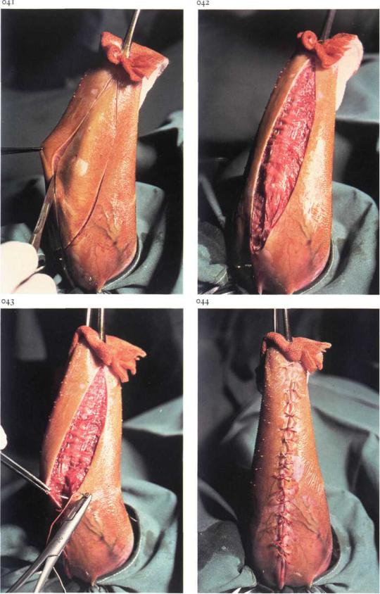
1-8 Lingual mucosa resection in cattle
Because of changes in housing facilities in the last ten years the problem of cattle which suck themselves or each other has increased. Numerous mechanical devices have been invented to prevent sucking. Because the herd may be-
come restless due to the pain these devices inflict, and feeding and drinking may be impaired, surgical treatment is preferable.
Surgery. The patient is sedated and restrained in lateral recumbency. Traction on a modified sponge forceps applied to the tip of the tongue facilitates exposure of the ventral lingual surface. A tourniquet is placed around the base of the tongue as close to the frenulum as possible, and the operative area is submucosally infiltrated with local analgesic. The elliptical incision begins a few centimetres caudal to the tip of the tongue, ends just cranial to the attachment of the frenulum, and is about 5 cm wide at its widest part in adult cattle [041,042]: it is important that sufficient mucosa is excised so that a convex shape of the dorsum of the tongue is produced after suturing is complete. The wound edges are apposed with single interrupted sutures of synthetic absorbable material. The sutures must include not only the mucosa but also some muscle to prevent tearing of the tissue by the sutures [043,044].
For 12-24 hours postoperatively the patients receive only water. Nearly all animals will eat immediately thereafter, and show no major problems in prehension and mastication of food.
Chapter i THE HEAD / Eye i -9
045
1-9 Suturing of eyelid lacerations
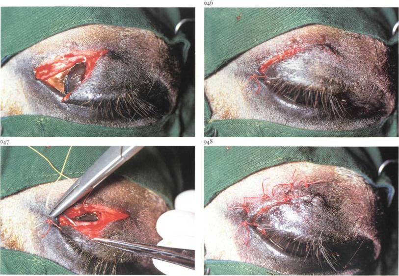
Eyelid lacerations in horses are often full-thickness, i.e. involving skin, orbicularis oculi muscle and conjunctiva. Usually the upper eyelid is torn [045]. In cases of recent laceration re-apposition and careful suturing must be attempted.
Surgery. Treatment may be carried out either on the standing animal under local analgesia (infiltration or frontal nerve block), or on the recumbent animal under local analgesia or general anaesthesia. The wound and conjunctival sac are flushed with physiologic saline. Sharp superficial excision of the wound edges is then carried out with a scalpel, to produce fresh, bleeding surfaces. Suturing is begun at the site of the palpebral margin, where a simple interrupted suture is placed [046]. The remainder of the wound is then closed. Usually the conjunctiva is not sutured: in any case perforation should be avoided to prevent damage to the cornea. The orbicularis oculi muscle and skin are sutured together, either with interrupted vertical mat-
tress sutures or with deep simple interrupted sutures alternating with superficial simple interrupted sutures. The deep bites of the mattress suture involve the skin together with the orbicularis oculi muscle [047], and the superficial bites only the skin. Either absorbable or non-absorbable suture material may be used.
The sutures are left in place for at least one week [048]. During this period ophthalmic antibiotic ointment may be administered twice daily.
Chapter I THE H E A D I Eye |
i-io, i-n |
049 |
050 |
i-io Excision of the nictitating membrane
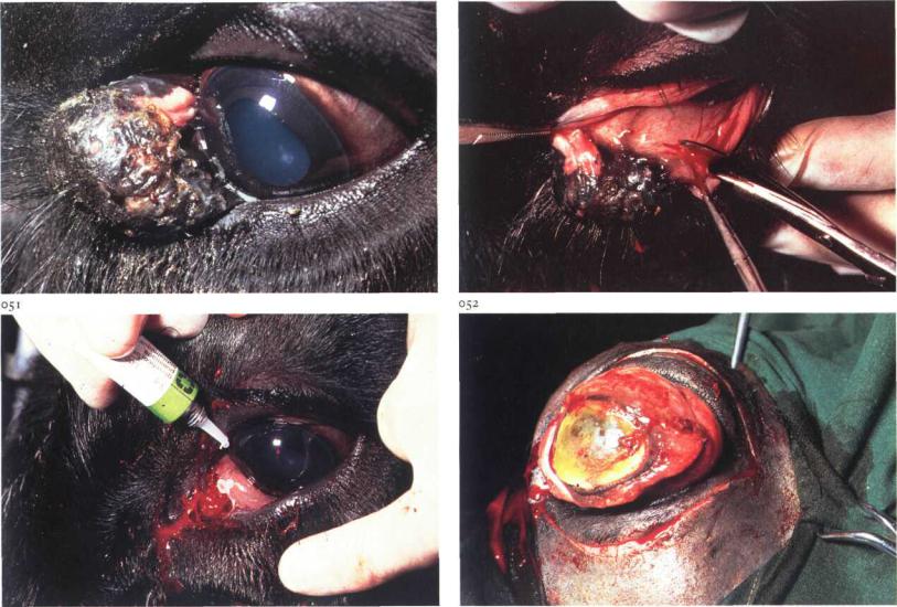
Surgery of the third eyelid is indicated in case of tumorous growth, which in horses and cattle usually involves squamous cell carcinoma. Small neoplasms can be removed leaving an intact nictitating membrane, but larger tumours require total excision [049].
Surgery. The operation is carried out on the standing or recumbent animal under local analgesia. The base of the third eyelid is infiltrated with a local analgesic after instillation of topical analgesic in the conjunctival sac.
The nictitating membrane is held with a forceps and drawn from the conjunctival sac as far as possible. Complete excision deep to the cartilage is performed using a pair of curved blunt-pointed scissors [050]. Haemorrhage is controlled by pressure with a gauze swab soaked in o.oi per cent adrenalin solution.
Ophthalmic antibiotic ointment is administered in the conjunctival sac for several days [051].
Chapter i THE H E A D / Eye i-n
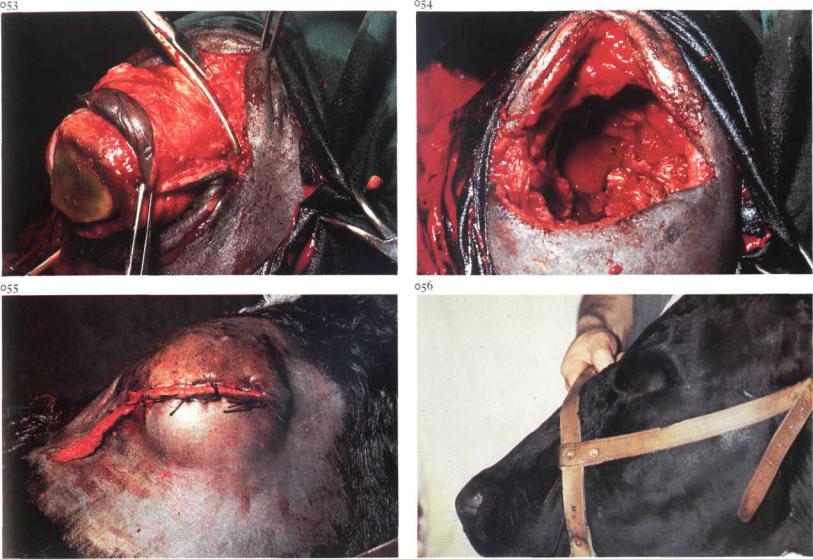
i-i i Enucleation of the eyeball
Enucleation of the eyeball usually includes removal of the globe together with the bulbar and palpebral conjunctiva, the nictitating membrane and the lacrimal gland. The operation may be indicated in cases of eyelid or eyeball neoplasia, gross injuries of the eyeball (e.g. corneal rupture) and panophthalmitis [052].
Surgery. Surgery is carried out with the animal recumbent under either general anaesthesia, or under ophthalmic nerve regional analgesia and infiltration analgesia ofthe lower eyelid and medial canthus. If possible, the upper and lower eyelids are sutured together with a continuous suture. An elliptical incision, 0.5-1 cm from and parallel to the margin of the eyelids, is made through the skin and palpebral muscle [052]. By blunt dissection in the direction of the orbita! ridge the orbit is entered [053]. The retrobulbar tissues and extra-ocular muscles are dissected bluntly and transected as close to the globe as possible. Finally the retractor bulbi muscle and en-
closed ophthalmic vessels and optic nerve are clamped with a curved crushing forceps and transected between forceps and globe. The eyeball is then withdrawn from the orbit [054]. The lacrimal gland is then removed. The forceps is removed; haemorrhage may be controlled either by vessel ligation or packing the orbit with sterile gauze bandages. The eyelids are sutured together with interrupted sutures, leaving a small opening medially for the gauze drain [055].
The gauze packing is removed after 2 to 3 days. Plate 056 shows the induced artificial ankyloblepharon several months postoperatively.