

Атлас анатомии крупных животных
.pdfChapierf) THE COMMON I N T E G U M E N T / Bovine digit 6-12 |
96 |
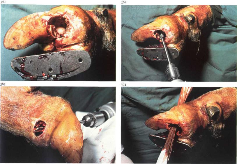
6-12 Resection of the distal interphalangeal joint and navicular bone
Purulent necrotic inflammation ofthe distal interphalangeal (pedal) joint is usually a complication of other diseases such as foreign body penetration, chronic necrotic pododermatitis [357], and interdigital necrobacillosis. When chronic arthritis of the pedal joint is present, or when structures of the joint (cartilage, subchondral bone, joint capsule) are necrotic and antibiotic therapy is unlikely to be successful, amputation of the digit (see 6-13) or resection of the pedal joint is indicated.
Surgery. The approach to the joint, dictated by the primary cause of the arthritis, is in most cases as described here; occasionally (e.g. following coronet wounds or extension of interdigital necrobacillosis) navicular bone resection is unnecessary, and the joint is approached from the dorsolateral or medial aspect respectively. The patient is restrained in lateral recumbency. A tourniquet of rubber tubing is placed around the limb in the midmetatarsal (metacarpal) region and local intravenous analgesia is administered.
The joint is approached through a circular incision (about 3 cm diameter) in the sole of the hoof about i cm distal to the plantar (palmar) coronary border [358]. The remnants of the deep flexor tendon are resected using a scalpel; the partially necrotic navicular bone becomes visible [359]. The navicular bone is removed by sectioning the distal navicular ligament first and then the remaining navicular ligaments [360,361]. Care must be taken not to penetrate the digital synovial sheath. All articular cartilage and subchondral bone of the pedal joint is removed with a 12 mm gouge driven by a low-speed electric drill [362]. With a scalpel a 2 cm diameter circular section of skin is excised just proximal to the coronet on the dorsolateral aspect [363]. It is important that all remaining necrotic tissue and the joint cartilage on the plantar aspect of the distal surface of the second phalanx are removed, using a curette. The cavity is dressed with antiseptic-soaked gauze drains [364]. The sound digit is shod with a block to avoid damage to the operated digit and to facilitate healing. Systemic antibiotics are administered.
Chapter 6 THE COMMON I N T E G U M E N T / Bovinedigit 6-12 |
97 |
If the deep flexor tendon has been transected the toes must be wired together for 6 weeks to prevent upturning of the operated hoof. A firm bandage is applied, and is changed after 2 days and thereafter weekly for 3-4 weeks. When the bandage is renewed, the wound is cleaned and the drain changed. When new granulation tissue has filled the depths of the wound the drain is not replaced. The block is removed after about 6 weeks.
If it appears (following navicular bone resection) that other parts ofthe joint are not necrotic, it may not be necessary to resect the entire joint: intra-articular antibiotics may be effective. The wound isfilledwith antibiotic-soaked gauze swabs and the digit is bandaged with sterile cotton. Systemic antibiotics are administered for 10 days. The wound must be dressed sterilely till the joint capsule is closed by granulation tissue formation.
Chapter 6 THE COMMON I N T E G U M E N T / Bovine digit 6-13
6-13 Amputation of digit
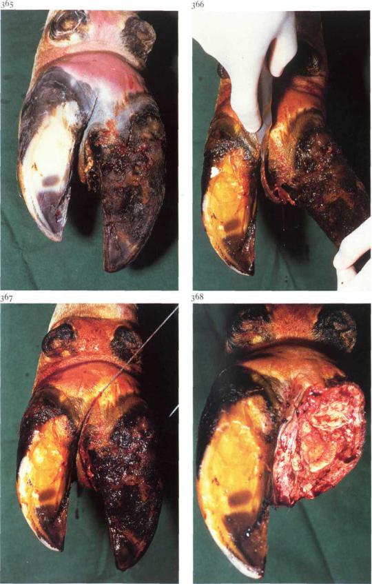
Digital amputation is indicated in cattle suffering from severe disorders of the digit such as purulent necrotic inflammation of the distal or proximal interphalangeal joint, or severe pedal trauma. Plate 365 shows a chronic necrotic pododermatitis, which has resulted in a purulent necrotic inflammation of the distal interphalangeal joint. Advantages of digital amputation in comparison with other surgical methods, are the simplicity of the technique and the relatively rapid return to normal general condition and milk yield.
Surgery. The cow is restrained in a standing position with the limb elevated and secured. A rubber tourniquet is applied in the mid-metatarsal (metacarpal) region and local intravenous analgesia is administered. Using a scalpel, a 2-3 cm deep incision is made along the whole length of the interdigital space through skin, subcutaneous tissue and interdigital ligaments, as close as possible to the affected claw [366]. An embryotomy wire is inserted in the incision and the digit is sawn off, directing the wire towards a point 2-3 cm proximal to the abaxial aspect of the coronet [367]. The transection courses through the middle phalanx, flexor tendons, digital flexor synovial sheath and skin [368].
In cases of purulent necrotic inflammation of the proximal interphalangeal joint, the level of amputation is the distal third of the proximal phalanx. The embryotomy wire is inserted in the interdigital incision, and the transection plane runs from 1-2 cm proximal to the axial aspect of the proximal interphalangeal joint to a point 3-4 cm proximal to the joint space abaxially. The wound is dressed with povidone iodine soaked gauze, and a pressure bandage is applied. The first bandage is renewed after 2 days. The stump is checked for inflammatory processes, such as infection of the digital flexor synovial sheath. After 1-2 weeks no further dressing is required.
Chapterd THE COMMON INTEGUMENT / Bovine digit 6-14
370
6-14 Resection of digital flexor tendons and digital synovial sheath
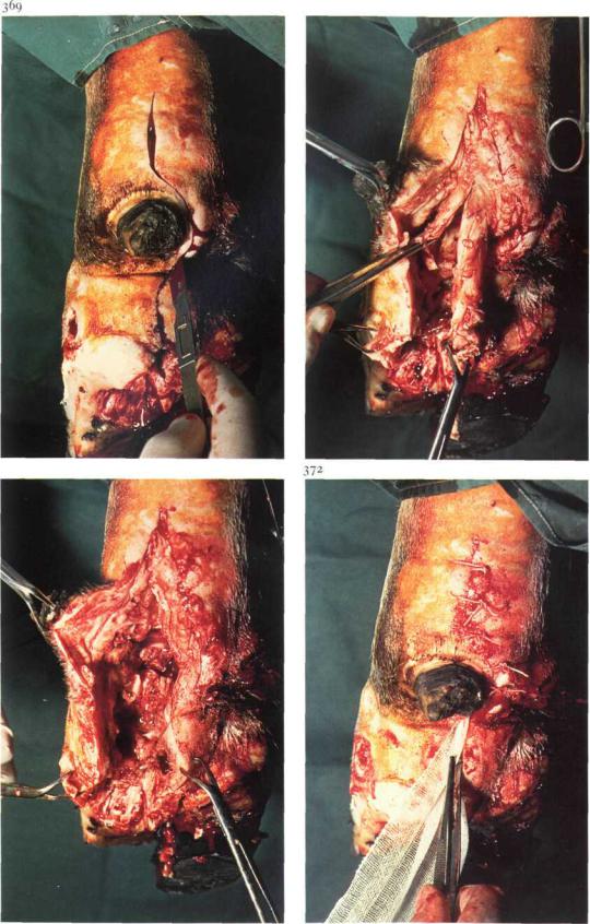
Infection of the digital synovial sheath develops from a proximal extension of a sole lesion (navicular bursitis and pedal arthritis), b penetrating wound into the sheath or c haematogenous infection. If conservative therapy fails - the affected tendon and sheath being necrotic - excision of the necrotic structures is the prime requisite. Surgery. The patient is placed in lateral recumbency. A tourniquet of rubber tubing is placed around the limb in the mid-metatarsal (metacarpal) region and local intravenous analgesia is applied. The skin and subcutaneous tissue over the tendon sheath are incised from the bulb (or sole ulcer) to a point about 5 cm proximal to the accessory digit, i.e. immediately distal to the bifurcation of the deep flexor tendon [369]. The sheath itself is then opened along its plantar (palmar) aspect. The superficial flexor tendon (which envelops the deep flexor at the level of the fetlock) is sectioned longitudinally; the deep tendon is transected adjacent to the bifurcation [370] and subsequently transected distally and removed. The superficial tendon is then likewise
371
transected proximally and at its insertion at the second phalanx [371].
After all necrotic debris and tissue have been removed, the wound is dressed with povidone iodine soaked gauze and the skin is closed with interrupted sutures [372]; distally the wound remains open for drainage. The toes are wired together for about 6 weeks to prevent upturning of the affected digit, and the sound digit is shod with a block to avoid damage to the operated digit and to facilitate healing.
The digit is bandaged firmly. Systemic antibiotic administration is advisable. The bandage and drain are changed on the second postoperative day, and thereafter as needed.
Chapter 6 THE COMMON INTEGUMENT / Mammary gland 6-16 |
101 |
377
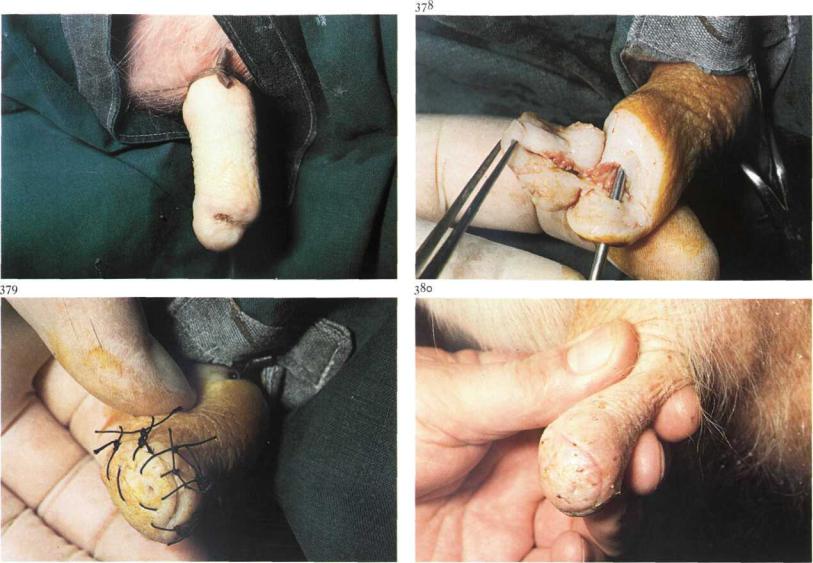
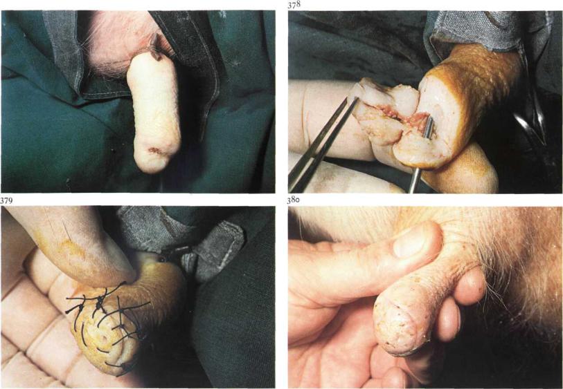
6-16 Repair of traumatic teat fistula
When milk flows continually through a perforating teat wound, failure of wound healing results in a teat fistula [377]. Congenital teat fistula is associated with a supernumerary mammary gland (see 6-17). Treatment of a teat fistula consists of dissection of the fistula and subsequent suturing of the wound. It is preferable to operate after the cow has been dried off.
Surgery. The operation is performed under local ring block analgesia at the base of the teat. A rubber tourniquet is applied to the teat base [377]. A milk canula is placed through the streak canal into the sinus, and an elliptical incision is made around the fistula. It is important that all scar tissue (in skin, muscular layers and mucosa) is removed to ensure primary intention healing [378]. After dissection, the fresh wound surfaces are apposed with several vertical mattress sutures and simple interrupted sutures [379] (see 6-15). Because of anticipated postoperative wound swelling, it is important to tie the sutures loosely to prevent wound edge necrosis. The tourniquet is
removed and long-acting antibiotics are injected into the teat sinus.
In dry cows a self-retaining plastic teat canula is usually not used, although in cases of streak canal damage a teat canula may prevent stenosis during repair.
The teat is bandaged with a slightly elastic bandage (see 6-15). Postoperative management consists of daily checking for mastitis. Plate 380 shows primary intention healing on the tenth postoperative day, after all sutures have been removed.
Chapter 6 THE COMMON INTEGUMENT / Mammary gland 6-16 |
101 |
377
6-16 Repair of traumatic teat fistula
When milk flows continually through a perforating teat wound, failure of wound healing results in a teat fistula [377]. Congenital teat fistula is associated with a supernumerary mammary gland (see 6-17). Treatment of a teat fistula consists of dissection of the fistula and subsequent suturing of the wound. It is preferable to operate after the cow has been dried off.
Surgery. The operation is performed under local ring block analgesia at the base of the teat. A rubber tourniquet is applied to the teat base [377]. A milk canula is placed through the streak canal into the sinus, and an elliptical incision is made around the fistula. It is important that all scar tissue (in skin, muscular layers and mucosa) is removed to ensure primary intention healing [378]. After dissection, the fresh wound surfaces are apposed with several vertical mattress sutures and simple interrupted sutures [379] (see 6-15). Because of anticipated postoperative wound swelling, it is important to tie the sutures loosely to prevent wound edge necrosis. The tourniquet is
removed and long-acting antibiotics are injected into the teat sinus.
In dry cows a self-retaining plastic teat canula is usually not used, although in cases of streak canal damage a teat canula may prevent stenosis during repair.
The teat is bandaged with a slightly elastic bandage (see 6-15). Postoperative management consists of daily checking for mastitis. Plate 380 shows primary intention healing on the tenth postoperative day, after all sutures have been removed.
Chapter6 THE COMMON I N T E G U M E N T / Mammary gland 6-17 |
102 |
6-17 Management of congenital teat fistula
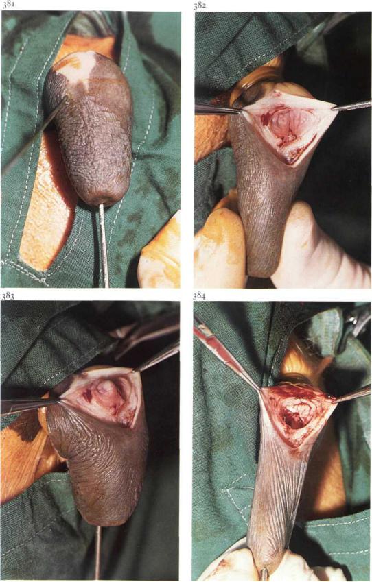
A congenital teat fistula is in general caused by the presence of a supernumerary mammary gland and is usually localized in the proximal part of the teat [381]. Milk flows through the fistula. Surgical correction is possible only when the lactiferous sinus of the supernumerary gland is situated close to the teat sinus.
Surgery. After local ring block analgesia at the base of the teat, a rubber tourniquet is applied. An elliptical incision is made around the fistula into the lactiferous sinus of the supernumerary gland. After dissection of the fistula, a fresh perforating teat wound is produced. In the depth of the wound, the mucosal membrane between the teat sinus and the sinus of the supernumerary gland is visible [382]. With a milk canula, brought through the streak canal into the teat sinus, the mucosal membrane is elevated into the wound [383]. By excising the membrane with scissors or scalpel, a connection is made between the two sinuses [384], and milk can drain from the supernumerary gland to the teat sinus. The wound is sutured using vertical mattress and simple interrupted sutures, loosely tied (see
6-15)-
The tourniquet is removed and antibiotics are injected into the teat sinus. In lactating cows a self-retaining plastic teat canula is placed in the streak canal and the teat is bandaged (see 6-15). Postoperative management consists of prevention of mastitis.
Chapter6 THE COMMON I N T E G U M E N T / Mammary gland 6-18 |
103 |
6-18 Management of teat obstructions
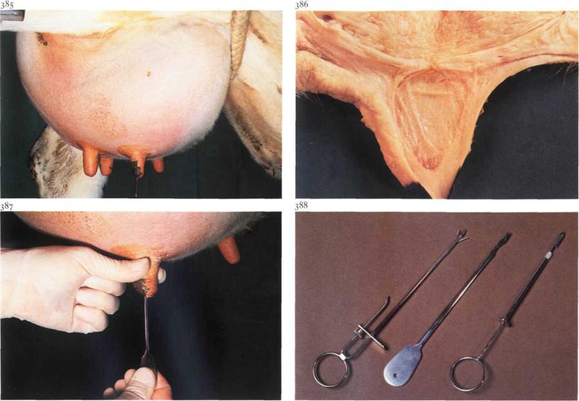
Teat obstructions in the cow may be congenital or acquired. Congenital obstruction at the base of the teat is caused by a completely closed annular fold of mucous membrane. Acquired basal obstructions develop during the dry period and are due to chronic inflammation of the annular fold. Localised obstructions in the teat sinus ('peas') may be due to local neoplasia or to chronic proliferative inflammation. Chronic inflammatory processes of the 'rosette of Furstenberg' or the streak canal may also lead to stenosis. Surgery. In cases of basal teat obstructions the affected quarter is under considerable tension [385] and milk flow is absent. Plate 386 shows a congenital basal teat obstruction at the annular fold. The membrane is perforated with a milk canula. Ifthere is no outflow ofmilk, the prognosis is unfavourable. If milk flow is present, Hug's teat knife [388,centre] is inserted through the membrane, which is then slit radially in three of four directions [387]. To ensure a permanent communication, it is recommended the
quarter is not milked completely for about ten days. In general the prognosis for a productive quarter is guarded, but in cases of thin membranes the prognosis is more favourable.
Teas' are usually localized in the mid-teat region. Large lesions may require open teat surgery, but the smaller ones are removed from the wall (with Hug's teat knife, Hug's tumour extractor [388,right] or a curette) and massaged out.
Cows with apical obstructions (traumatic or congenital) are usually presented as 'hard milkers'. The stenosis may be temporarily relieved by longitudinal radiate incisions of the sphincter or the fibrosed canal with a Danish teat knife [388,left] or by widening the opening with a teat dilator. A selfretaining teat canula prevents postoperative stenosis.
Chapter6 THE COMMON I N T E G U M E N T / Mammary gland 6-19 |
104 |
390
6-19 Amputation of the mammary gland
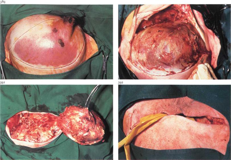
The main indication for amputation of the mammary gland is a severe lifethreatening mastitis, often seen in sheep and goats. Plate 389 shows an acute necrotizing mastitis in a ewe, characterised by a dark blue discoloration of the skin and a severely swollen right mammary gland. Surgery. Amputation of the mammary gland is performed with the patient under epidural analgesia (anterior block) or general anaesthesia and in dorsal or dorso-lateral recumbency. An elliptical skin incision is made around the discoloured area [389], followed by dissection of the glandular tissue along the suspensory ligament medially and the lateral ligament laterally [390]. The gland is grasped with a tenaculum forceps and dissected from the body wall, beginning caudally. The major vessels (external pudendal artery and vein, ventral perineal artery and vein, and subcutaneous abdominal vein) are connected by numerous anastomoses, and double ligation is essential. The vessels are transected between the ligatures and dissection of
the gland is continued craniad [391]. The large external pudendal artery, which enters the udder on the cranio-lateral aspect after passing through the inguinal canal, should be respected. After dissection of the mammary gland is complete, subcutis is sutured in a simple continuous pattern using absorbable suture material. Sterile gauze or latex drains are placed in the wound, one deeply, the other superficially [392], and the skin is closed with simple interrupted sutures. Systemic antibiotics are administered.
105
Chapter 7 The musculoskeletal system