

Атлас анатомии крупных животных
.pdfChapter 6 THE COMMON I N T E G U M E N T / Skin 6-2 |
86 |
6-2 Cryosurgery of sarcoids
Equine sarcoid is the most common tumour of the horse: it has a nonmalignant character but recurrence is often observed after surgical excision. Far better results are obtained by destruction of tissue through freezing (cryonecrosis). Controlled freezing of tissue has the advantage of absence of haemorrhage, reducing the likelihood of dissemination of tumour cells. A disadvantage is the necrosis and subsequent sloughing of tissues. The cryonecrosis results from direct cellular damage (ice crystal formation, dehydration etc.) and anoxia due to destruction of blood vessels. Cryogens most often used include liquid nitrogen and nitrous oxide. Surgery. Cryosurgery may be carried out under sedation and local analgesia, but recumbency under general anaesthesia may sometimes facilitate the procedure. Tissue should be frozen rapidly and this may be achieved by direct spraying of the tumour with liquid nitrogen [317]. The surrounding skin should be protected, e.g. by a styrofoam cup, cut to a fitting shape
[317]. The tumour is frozen to 2010-30° C, the temperature is monitored by thermocouples in and deep to the 'iceball' [317]. The frozen tumour is then removed at the level of the surrounding skin [318], whereafter the base is frozen with a circulating contact probe [319]. A double freeze-thaw cycle is used. The larger tumours may need several sessions of Cryosurgery, after each of which a layer of tissue will become demarcated over a period of weeks. The aftercare is usually minimal: the post-freezing oedema resolves quickly, and separation of the necrotic tissue occurs in 7-14 days [320]. The time needed for complete healing depends on the lesion. Scarring is minimal, but white hair will often grow at the site ofthe lesion.
Chapter 6 THE COMMON I N T E G U M E N T / Skin 6-3
6-3 Extirpation of acquired carpal bursa
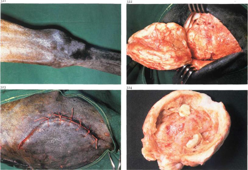
Subcutaneous acquired carpal bursa is localised on the cranial surface of the carpus [321] and is the result of (repeated) trauma. Rest, aspiration of the bursa, and administration ofmedicaments (e.g. corticosteroids) together with a firmly placed carpal bandage often produces unsatisfactory results. Surgical extirpation of the bursa is recommended if further treatment is requested.
Surgery. The patient is placed in right or left lateral recumbency under general anaesthesia, depending on the affected site. An Esmarch's bandage facilitates surgery. The carpus is slightly flexed and a curvilinear incision [323] is made through skin and subcutaneous tissue. On the dorsal surface ofthe bursa blunt dissection from the surrounding tissue is difficult. When the dissection plane is found on the edges of the bursa, separation of the capsule from the dorsal surface of the carpus may be simpler. The irregular borders of the bursa may be the cause of opening the bursa despite the
widest possible excision. To avoid opening of the flexor tendon sheaths or joint cavity care should be taken when dissecting the bursa from the dorsal surface of the carpus [322]. Plate 324 shows the bursa, opened after extirpation.
After complete excision dead space is closed with simple interrupted sutures, subcutaneous tissue with continuous and the skin with interrupted sutures, using absorbable synthetic material [323]. A vacuum drain may be placed before closure of the dead space is complete. Systemic antibiotics are administered.
After surgery the limb is immobilized (plaster cast) in extension for about 3 weeks. The drain is removed 3 days postoperatively. The cast is replaced by firm bandaging for 3 weeks.

Chapter 6 THE COMMON I N T E G U M E N T / Equine hoof 6-4,6-5 |
ss |
6-4 Treatment of pododermatitis
Septic pododermatitis may result from foreign body penetration, cracks in the white line or sole, local bruising (e.g. corns), or chronic laminitis. Trauma causes haemorrhage and/or necrosis of the pododerm, rendering it prone to bacterial infection. The result is severe pain caused by exudate under pressure between pododerm and sole. The foot of the affected leg may be swollen, due to oedema or extension of the septic process. Puncture wounds in the sole or cracks in the white line always result in a black dot or line at the site of initial injury [325]. A hoof tester is helpful in locating the involved area.
Surgery. Analgesia may not be required. The hoof is trimmed. When present the tract must be followed, if not the horn over the most painful spot must be removed. The (black) exudate is usually under pressure and has a putrid odour. Proper drainage may be achieved through a rather small opening of the undermined area, followed by applying a wet disinfecting cotton bandage for two days. Tetanus prophylaxis is provided.
If the patient remains very lame after this period and purulent exudate is still present, all undermined horn must be removed (under analgesia), even if this results in a large defect [326]. The edges of the defect are thinned and the wound should be dressed with disinfectant-soaked gauze and a waterproof pressure bandage.
In this case healing was delayed due to bone sequestration, which made removal of necrotic material necessary [327].
Dressings are changed weekly. As soon as the defect is covered with newly formed horn, the hoof is shod with a sole protecting pad.

Chapter 6 THE COMMON I N T E G U M E N T / Equine hoof 6-5
330
6-5 Extirpation of keratoma
A keratoma is an abnormal proliferative growth of the horn which develops on the inner aspect of the hoofwall [328]. The white line is deflected inward by the keratoma, which appears as a cylindrically shaped growth between the wall and laminar corium. The keratoma may extend a variable distance up the wall from the bearing surface towards the coronet.
When the keratoma increases in size lameness may gradually develop. Pressure of the keratoma results in localized atrophy of the pedal bone, which can be demonstrated by radiography [329]. Contamination through defects in the white line may result in secondary septic inflammation of the damaged sensitive laminae which usually leads to development of a fistulous tract [328]. Therapy consists of surgical extirpation of the keratoma.
Surgery. The operation can be performed on the standing horse under local analgesia or in lateral recumbency under general anaesthesia.
The horny wall on both sides of the keratoma is removed almost to the laminar corium [330]. The keratoma is then dissected from the corium [331,332]. If it is confirmed that the 'tumour' extends to the coronary band, removal may be achieved by grasping the distal border with a large pincer and reflecting it proximad. To prevent hoofwall movement as much as possible, a shoe with a clip on either side of the defect is used. The defect is dressed with a disinfectant and pressure bandage. The bandage is changed weekly.
Bandages are left off as soon as the thin layer of newly formed horn is sufficiently hard, but desiccation should be avoided to prevent cracking. The special shoe is used until the defect in the wall has grown out.

Chapter 6 THE COMMON I N T E G U M E N T / Equine hoof 6-6
334
6-6 Management of penetrating wounds of the sole
Puncture wounds in the foot are quite common in horses and are caused by a variety of foreign bodies, such as nails and fragments of glass.
These foreign bodies may penetrate to the third phalanx, which may result in an infectious pedal osteitis. Wounds in the middle third of the frog are most serious because of the possibility of puncture of the navicular bursa [333], and infectious arthritis of the coffin joint. The depth and direction of the tract can be determined by radiography [334] and exploration with a sterile probe [335].
Surgery. In the early stages, treatment consists of superficial drainage of the lesion, which is accomplished by wide trimming of the horny walls of the tract opening. The horn in the surrounding area should be thinned to prevent a prolapse of the pododermal tissue. The wound is then flushed, and a disinfectant bandage is applied. Tetanus prophylaxis is provided, and antibiotics are given systemically.
If considerable improvement of lameness has not occurred within two days, surgery is indicated under local or general anaesthesia. In cases in which the navicular bursa is affected, drainage of the bursa should be performed. The foot is disinfected and the frog trimmed out. All necrotic tissue should be removed.
Drainage of the bursa is achieved by fenestration of the deep flexor tendon performed at the site of the tract (by excising a 'window' of i X i cm), the flexor surface of the navicular bone is now visible [336]. If the cartilage has been damaged, curettage of the navicular bone is carried out. The bursa is then flushed with sterile physiologic saline, containing antibiotics. The wound is packed with sterile surgical gauze [337].
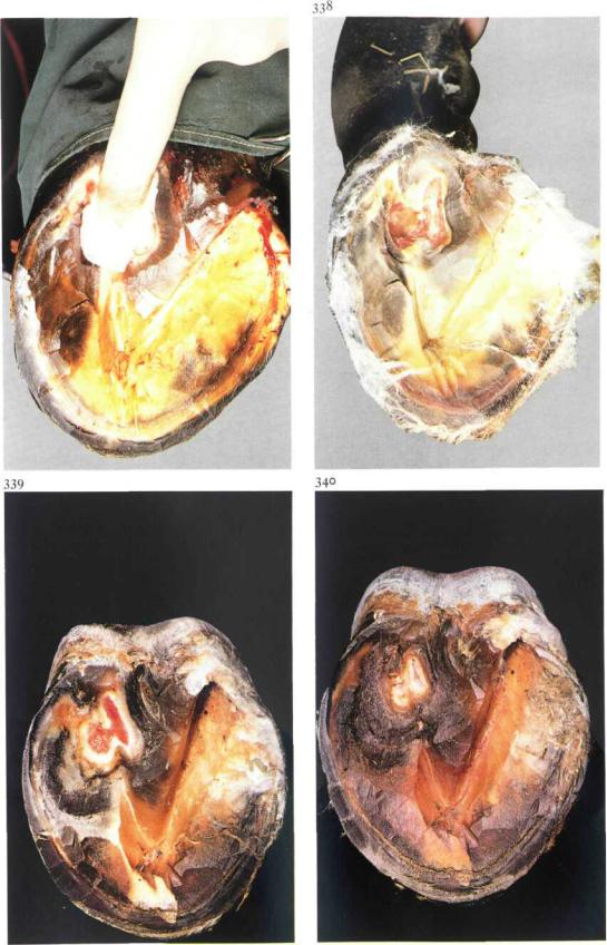
The foot is kept under a sterile bandage and systemic antibiotics are administered as long as the bursa remains open. Five days postoperatively newly formed granulation tissue is visible [338]. After 18 days, a zone of new pododermal tissue has developed [339]. If the wound has granulated in completely, which means that the navicular bursa is closed, the bandage is replaced by a shoe with a removable steel plate. This permits prolonged treatment, if necessary. If the horse remains lame, due to inflammation of the deep flexor tendon, raising the heels of the shoe with calks to spare the tendon is recommended. After
Chapter 6 THE COMMON I N T E G U M E N T / Equine hoof 6-6
337
40 days, epithelialisation is almost complete [340]. At this stage protective shoeing (shoe with a leather pad) is provided for a period of4-6 weeks.
The prognosis of superficial puncture wounds and pedal osteitis is favourable. In cases of inflammation ofthe navicular bursa and infectious arthritis of the coffin joint, the prognosis is guarded and unfavourable respectively.
Chapter 6 THE COMMON I N T E G U M E N T / Equine hoof 6-7
6-7 Treatment of canker
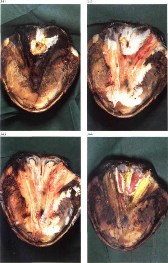
Canker may be considered as a chronic hypertrophic pododermatitis, but also as parakeratosis because of the abnormality of the horn produced. The disease begins usually in the frog, but may extend to all other areas of the hoof. As lameness is not present initially, the lesions maybe well advanced before they are detected. In severe cases the pododerma may show a cauli- flower-like hypertrophy offoul-smelling weak caseous horn [341].
Aetiologically the disease is presumably related to unhygienic stabling and neglected foot care, and must be differentiated from thrush (see 6-8) and from so called chronic progressive pododermatitis. The latter has been seen especially in the fore feet of trotters and is associated with lameness.
Surgery. The treatment of both canker and progressive pododermatitis is based on removal of the diseased tissue. Regional analgesia may be necessary if part of the hypertrophic pododerm must be removed. The procedure begins with removal of loose and abnormal horn, followed by thinning of the surrounding horn [342]. Severely affected pododerm is also removed [343]. Surgical removal of abnormal tissues is preferable to the use of caustic agents. Less hypertrophic pododerm may be left to the beneficial influence of a pressure bandage, which is necessary in all cases. Astringent and drying powders may be added to the bandage. In cases of extensive lesions a plaster cast may be used. Dressings should be changed once a week. Plate 344 shows the epithelialization of the treated area of the frog one week after surgery.
The treatment of canker may be time-consum- ing, but the prognosis is usually not unfavourable, although recurrence is possible. As a matter of course prevention lies in good hoofcare, including hygienic stabling.
Chapter 6 THE COMMON I N T E G U M E N T / Equine hoof 6-8,6-9
345
93
6-8 Treatment of thrush
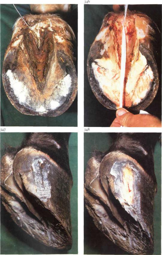
Thrush is a degenerative condition of the frog, usually starting in the central sulcus and resulting from poor hoof care and unhygienic management. The affected areas are characterized by the presence of grayish-black degenerate horn [345] with a distinct odour.
Surgery. The hoof should be trimmed to normal conformation to achieve normal pressure on the frog; all affected and undermined horn should be removed and the surrounding horn trimmed. Thorough cleansing of the sulci [346] is followed by application of disinfectants and astringents. Deep sulci may be packed with cotton swabs soaked in the medication. Treatment should be continued until the frog is covered with normal horn. The prognosis is good, provided hoof care and stalling are improved.
6-9 Treatment of sand crack
Sand crack consists of a fissure (fracture) of variable length commencing at the coronet or at the bearing surface of the wall. The cracks are identified as toe, quarter or heel cracks depending on their location. Lameness may not be present, but it will develop if the crack extends into the sensitive laminae [347].
Surgery. If the crack extends into the coronary band and/or into the sensitive laminae radical excision of the crack is the prime requisite. The operation can be performed under local regional analgesia. The horny wall on both sides of the crack is thinned with rasp and knife and the damaged sensitive laminae is removed. The bearing surface caudal to the crack should be trimmed approximately 0.5 cm shorter than the rest of the foot, so that it does not bear weight until the crack has grown out [348].
The defect is dressed with disinfectant and a pressure bandage is applied until the defect is covered by horn. Shoeing is beneficial to prevent recurrence (a full bar shoe is used in quarter crack).

Chapter 6 |
THE COMMON I N T E G U M E N T / Bovine digit 6-10 |
349 |
350 |
94
6-10 Treatment of pododermatitis
Septic pododermatitis can result from foreign body penetration, or from generalised bruising which may follow irregular weightbearing and / or chronic laminitis.
Bruising causes haemorrhage and/or necrosis of the sensitive laminae; bacterial infection ofthe damaged sensitive laminae can occur primarily (in penetration trauma) or secondarily by contiguous or haematogenous infection. The result is serious lameness caused by pain brought about by exudate under pressure between sensitive laminae and sole [349]. The heel of the affected side may be swollen, because of extension of the septic process.
Surgery. Analgesia may not be required. When present, the tract must be followed along its full length [350]. All separated sole horn should be removed. If the affected area is small, horn trimming, dressing the defect with a disinfectant, and confining the animal on soft bedding or dry pasture for some days may be sufficient to allow healing. Often however the exudate has extensively under-run the horn. Because of the presence of necrotic pododerma this horn must be removed, even if a large defect results [351]. Necrotic material in deeper structures should be removed similarly, as in this case of bone sequestration of the pedal bone [352]. The defect should be dressed with disinfectant and packed with gauze, cotton wool, and a waterproof pressure bandage.
In cases ofextensive inflammation and/or severe lameness, the sound digit is shod with a block to avoid damage to the affected digit and to facilitate healing. The dressing should be renewed after one week. Severe cases of bone infection may require prolonged treatment.
Chapter 6 THE COMMON I N T E G U M E N T / Bovine digit 6-11 |
95 |
354
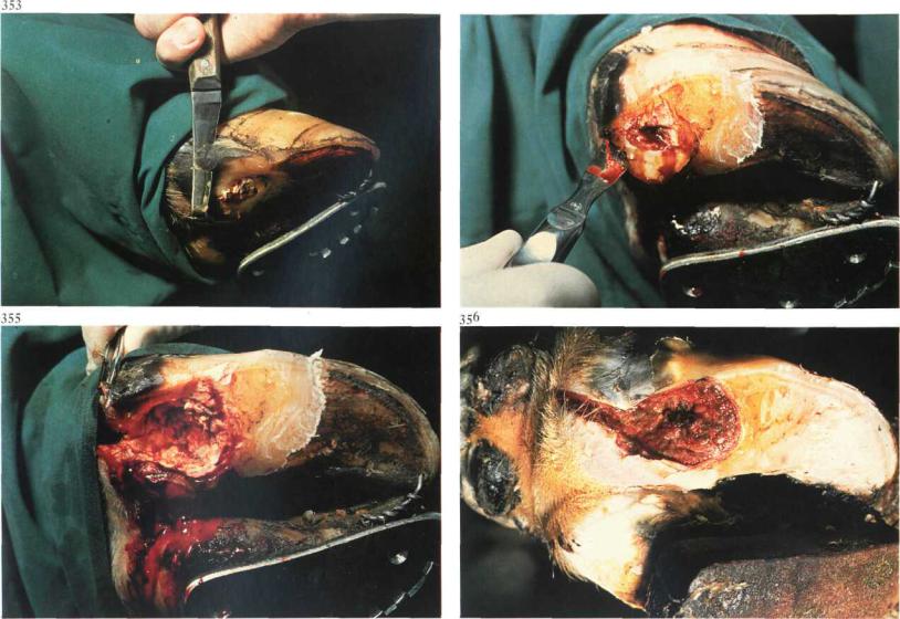
6-11 Treatment of heel abscess
Abscessation of the heel results from necrotic pododermatitis, interdigital necrobacillosis, or puncture wounds of the heel, and causes prominent localised swelling of the whole heel extending over the coronet to the haired skin. The heel itself may be firm or fluctuating. The abscess is often deepseated and associated with a purulent necrotic process originating in the sole, as in this case.
Surgery. The patient is restrained in standing or recumbent position. A tourniquet of rubber tubing is placed around the limb in the midmetatarsal (metacarpal) region and local intravenous analgesia is administered. The horn of the heel is thinned [353]. An opening is made around the necrotic ulcer and an incision is made into the abscess [354]. Necrotic tissues and debris are removed from the cavity with a curette [355]. The sound digit is shod with a block to avoid damage to the operated digit and to facilitate healing [356].
If the deep flexor tendon has been destroyed by necrosis, the toes are wired together for about 6 weeks to prevent upturning of the operated digit.
The abscess space is irrigated, packed with sterile gauze swabs soaked in povidone iodine, and bandaged for a few weeks. The shoe is removed after healing is complete.
Complications such as extension of necrosis towards the pedal joint (causing septic arthritis) and/or towards the digital sheath (infectious tenosynovitis) may occur.