

Атлас анатомии крупных животных
.pdfChapter-; THE U R O 3 E N I T A L SYSTEM / The male urogemtal system |
5-4 |
201 |
202 |
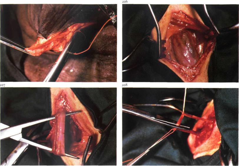
5-4 Castration: primary closure method in the horse
Castration in which primary closure of the scrotal wound is attempted should be carried out only under strictly aseptic conditions. The technique for the stallion is discussed here.
Surgery. Castration is performed under general anaesthesia with the horse in dorso-lateral recumbency. The uppermost leg is secured in flexion and abduction.
The testicle is grasped firmly in one hand, so that the scrotal skin is tightly stretched. A 7-10 cm incision is made through skin and tunica dartos in the caudal part of the scrotum, parallel to the median raphe. A small incision is then made through the vaginal tunic and its edges grasped with tissue forceps [201]. The incision is then enlarged sufficiently to allow testicle and epididymis to emerge from the vaginal cavity, whereupon the exposed testicle is grasped with a tenaculum forceps. The scrotal ligament is severed with dissection scissors [202], and the mesorchium is carefully torn from
the vaginal tunic and the spermatic cord is ligated with absorbable suture material as proximally as possible. A peritoneum forceps is applied to the spermatic cord about 3 cm distal to the ligature [203] and the spermatic cord is transected between forceps and ligature. The stump and mesorchium are checked for bleeding. The wound is closed in three separate layers using synthetic absorbable suture material: the vaginal tunic [204] and tunica dartos [205] are sutured in a continuous pattern, and the skin with interrupted mattress sutures.
Aftercare consists of routine postcastration management.
Chapter^ THE U R O G E N I T A L SYSTEM / The male ungenital system 5-4,5-5 |
57 |
205
5-5 Vasectomy
Vasectomy is indicated for production of heat detector (teaser) bulls or rams.
Surgery. The operation can be performed under local analgesia, with the animal either cast or standing.
The spermatic cord is located. A 2-3 cm skin incision is made medially to reveal the vaginal tunic [206]. The ductus deferens is palpated as a firm, non-pulsating cord-like structure. After careful incision of the tunic, the ductus deferens is easily separated and exteriorized [207]. A segment of about 2 cm is removed and the incised ends are ligated [208].
It is not essential to suture the vaginal tunic, but the skin is closed with a few interrupted sutures.
Chapters THE U R O G E N I T A L SYSTEM / The male urogenital system |
5-6 |
209 |
2IO |
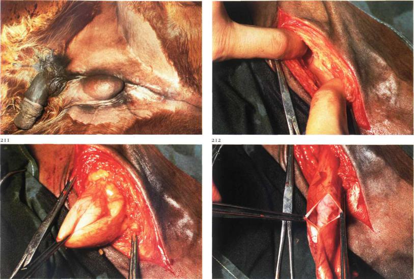
5-6 Inguinal cryptorchidectomy in the horse
Cryptorchidism may be classified into two main types: a testicle and epididymis retained between the internal inguinal ring and the scrotum ('high flanker'); b testicle and epididymis retained within the abdominal cavity (abdominal cryptorchid). In a partial abdominal cryptorchid, the epididymis descends into the inguinal canal with the testicle remaining in the abdominal cavity.
Inguinal cryptorchidectomy is discussed here.
Surgery. Surgery is performed with the patient under general anaesthesia and in dorso-lateral recumbency with the leg on the cryptorchid side uppermost and secured in flexion and abduction [209]. A 10 cm incision is made over the external inguinal ring through skin and scrotal fascia. Care should be taken to avoid trauma to the large vessels in this region. Blunt dissection of the connective tissue is continued towards the inguinal canal [210]. The testicle and epididymis, covered by vaginal tunic, may be en-
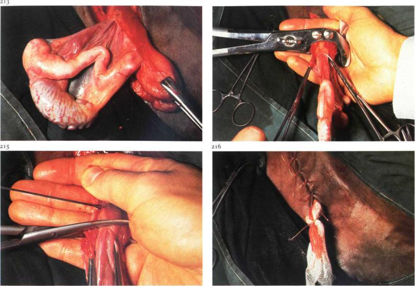
countered adjacent to the external inguinal ring or can be located in the inguinal canal. The vaginal tunic is grasped with forceps [211] and incised longitudinally [212]. The cryptorchid testicle (which is usually much smaller than the descended testis) and epididymis are then drawn outside the vaginal cavity [213]. Half-closed castration is then performed (see 5-3): the spermatic cord, covered by the vaginal tunic, is crushed [214], ligated and transected 2 cm distal to the ligature [215]. The stump is checked for haemorrhage and the long ends of'the ligature cut. A sterile gauze or latex drain is placed in the wound and the skin is closed with simple interrupted sutures [216].
Ifthe vaginal sac can be drawn sufficiently beyond the external inguinal ring, the closed technique or primary closure method of castration can be used instead of the half-closed method. Tetanus prophylaxis is provided. The drain is removed after 48 hours. Aftercare consists of routine postcastration management.
Chapter5 THE U R O G E N I T A L . SYSTEM / The male urogenitalsystem 5-6
214
Chapters THE U R O G E N I T A L SYSTEM / The male urogemtalsystem 5-7
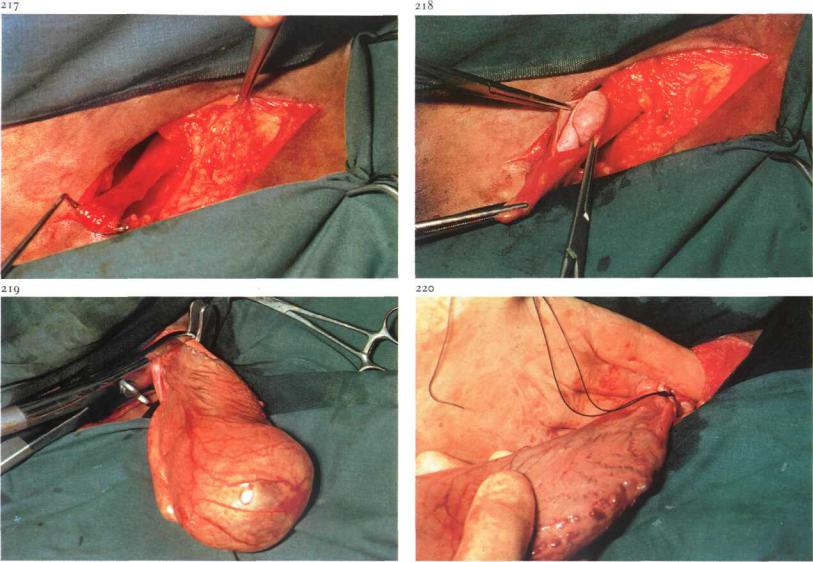
5-7 Abdominal cryptorchidectomy in the horse
The inguinal approach is most commonly used for abdominal cryptorchidectomy in the equine, while paramedian laparotomy is indicated for bilateral abdominal cryptorchid or grossly enlarged testicles.
Surgery.
(i) Inguinal approach. The position, preparation and incision is as for inguinal cryptorchidectomy (see 5-6). Frequently, in an abdominal cryptorchid the extra-abdominal part of the gubernaculum can be located in the inguinal canal. Careful blunt dissection may allow visualization of this structure. By exerting moderate traction on the gubernaculum [217], the vaginal process can be drawn through the external inguinal ring. If the extra-abdominal part ofthe gubernaculum can not be recognized, dissection is continued and the vaginal process is searched for at the level of the internal ring. The vaginal process is then grasped with sponge forceps and withdrawn as far distally as possible. The vaginal process is then incised,
exposing the epididymis [218], which is grasped by forceps. Gentle traction is applied until the testicle is delivered. Cystic degeneration had caused enlargement and retention of this testicle [219].
When neither the extra-abdominal part of the gubernaculum nor the vaginal process can be recognized in the inguinal canal, the peritoneum is perforated at the level of the internal inguinal ring with the finger. One or two fingers (or even the entire hand) in the abdominal cavity search for the gubernaculum, epididymis, testicle or ductus deferens. After one of these structures has been recognized, it is brought into the inguinal canal, grasped with forceps and withdrawn further until both epididymis and testicle are exteriorized. The exposed spermatic cord is crushed [219], ligated [220] and transected 2 cm distal to the ligature. Suture closure of the external inguinal ring with simple interrupted or continuous sutures is recommended. A drain is placed in the wound and the skin is closed with simple interrupted sutures. Tetanus prophylaxis is provided. The drain is removed after 48 hours. Aftercare consists of routine post-castration management.
Chapter^ THE U R O G E N I T A L SYSTEM / The male urogenitalsystem 5-7,5-8 |
61 |
221 |
|
(2) Paramedian approach. A paramedian parapreputial laparotomy [221] is performed under general anaesthesia with the horse in dorsal recumbency (see 4-5). The surgeon's hand is introduced into the abdominal cavity and a search is made for the testicle. Most retained testicles are located close to the internal inguinal ring. The testicle is brought through the incision [222], and the exposed spermatic cord is crushed with an emasculator, ligated with absorbable suture material and transected 2 cm distal to the ligature. The laparotomy wound is closed in four separate layers (see 4-5).
5-8 Abdominal cryptorchidectomy: flank approach in the pig
Because cryptorchidism in the pig is usually abdominal, a lateral flank laparotomy is recommended in this species.
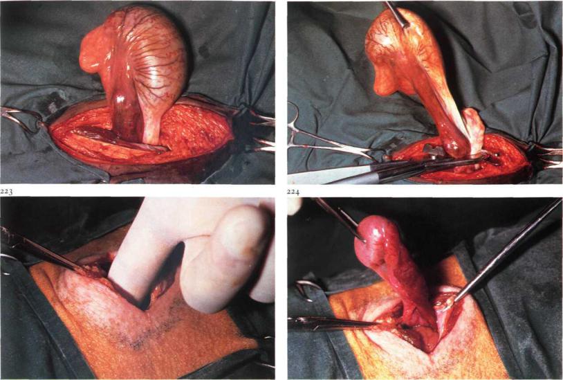
Surgery. The operation is done under general anaesthesia with the pig in lateral recumbency and the cryptorchid side uppermost. Flank laparotomy is performed, using a through-and-through incision (see 4-4). The forefinger is introduced into the abdominal cavity [223] and a search is made for the testicle, which usually can be found between the caudal pole of the kidney and the pelvic inlet. When the testicle has been located, it is delivered into the flank incision [224] and the spermatic cord is either ligated and transected, or severed with an emasculator. Peritoneum and all muscle layers are sutured together, using absorbable material in a simple interrupted pattern. The skin is then closed with interrupted sutures.

Chapters THE UROGENITAL SYSTEM / The male urogenital system 5-9 |
62 |
226
5-9 Inguinal herniorrhaphy in the pig
Inguinal or scrotal hernias frequently occur in pigs and are often discovered at the time of castration. Inguinal hernias also occur in female pigs. In general, inguinal and scrotal hernias in pigs are reducible, and the visible swelling in the groin can be increased by applying slight pressure to the abdominal wall, which allows viscera to pass into the scrotum via the inguinal canal, unior bilaterally [225].
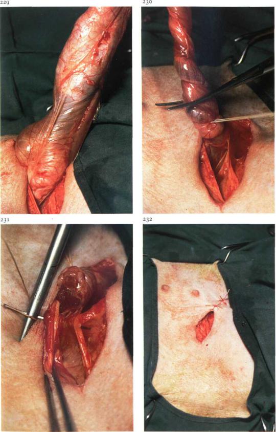
Surgery. Inguinal herniorrhaphy in pigs is performed under general anaesthesia. The pig is tied in dorsal recumbency. A 4 to 6 cm incision is made over the external inguinal ring through skin, subcutaneous tissue and scrotal fascia. The incision through the scrotal fascia must be done carefully, so that the vaginal tunic is not inadvertently incised: it is thus preferable to split the scrotal fascia with dissection scissors [226]. The spermatic cord, covered by the vaginal tunic, is loosened from the surrounding tissues by blunt dissection with the forefinger [227]. Steady traction is exerted on the tunic, testicle and spermatic cord to distract the scrotal fascia from the scrotal skin [228]. Small intestine can now be visualised through the vaginal tunic [229]. The hernial contents (intestine) are replaced into the abdomen by twisting the vaginal sac [230]. The vaginal sac and spermatic cord are ligated as close as possible to the external inguinal ring and the cord, covered by vaginal tunic, is transected approximately i cm distal to the ligature [230]. To prevent inguinal herniation, the size of the external inguinal ring is reduced. This is achieved by suturing the external inguinal ring in a simple continuous pattern using both ends of the spermatic cord ligature [231]. Finally, the skin incision is partly closed with simple interrupted sutures: the most ventral point is left open to allow drainage of exudate [232].
Chapters THE U R O G E N I T A L SYSTEM / The male urogenital system 5-9-
Chapter^ THE U R O G E N I T A L SYSTEM / The male urogemtalsystem 5-10
5-10 Treatment of incarcerated inguinal hernia in the horse.
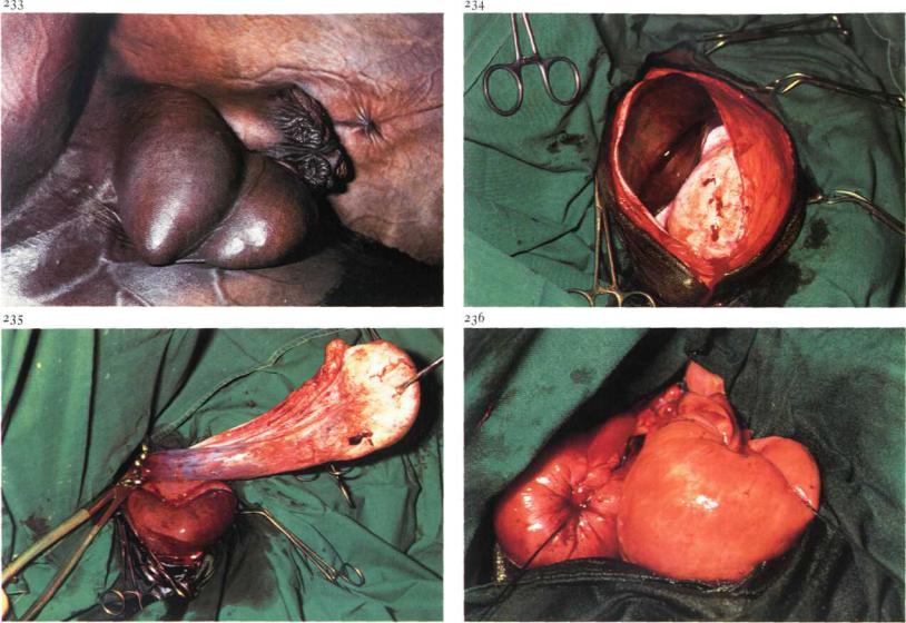
Incidence of incarcerated inguinal hernia is greatest in the equine [233]. Incarceration of herniated intestine (often ileum) is caused by the hour- glass-like constriction of the vaginal tunic.
Surgery. Herniorrhaphy is carried out in the horse under general anaesthesia in dorso-lateral recumbency. The hindleg on the affected side is uppermost and secured in flexion and abduction. A 15-20 cm skin incision is made from the external inguinal ring to the distal aspect of the scrotum. After carefully incising the vaginal tunic, the testicle and incarcerated intestine are exposed [234]. Testicle and epididymis are then drawn outside the vaginal cavity. The spermatic cord is crushed, ligated and transected
2 cm distal to the ligature [235]. If the bowel appears to be vital, replacement takes place through the vaginal cavity into the abdomen. If replacement is difficult, the hourglass-like constriction of the vaginal tunic is carefully incised in a cranial direction with a blunt-pointed scalpel. If the
intestinal loop is necrotic [234], enterectomy (see 4-10) is performed, the remaining ends are closed [236] and replaced into the abdomen. If rupture of the devitalized loop is unlikely, the hernial contents are reduced before enterectomy. Resection and anastomosis is performed after ventral midline laparotomy (see 4-3).
The vaginal tunic is bluntly dissected, crushed, ligated and transected as proximally as possible. If the hourglass-like constriction of the vaginal tunic has been incised, suture closure of the external inguinal ring is recommended. A drain is placed in the wound and the skin is closed using simple interrupted sutures. Systemic antibiotics are administered and tetanus prophylaxisisprovided.
Chapter5 THE U R O G E N I T A L SYSTEM / The maleurogenitalsystem 5-11
237
5-11 Inguinal herniorraphy in foals
In male foals two types of inguinal hernia may be observed.
(1)Most frequently a true hernia is seen, i.e. a protrusion of intestine and/or omentum through the vaginal ring into the inguinal region. The contents are enclosed by the vaginal tunic. In persons this is called an indirect inguinal hernia. Only if spontaneous resolution has not occurred within a year or if clinical symptoms arise, is surgical correction indicated. The operative method is the same as is used in pigs (see 5-9).
(2)Less frequently the inguinal swelling is caused by the subcutaneous
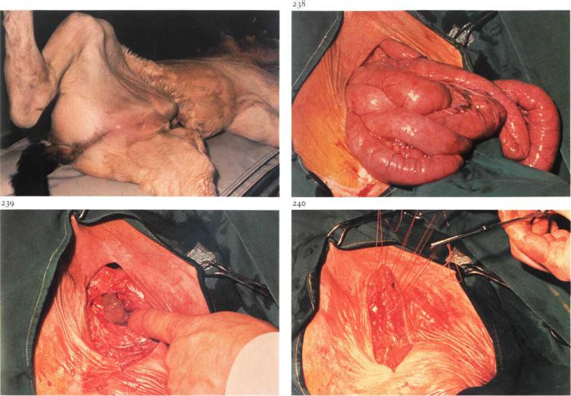
presence of intestines, sometimes called interstitial inguinal hernia or intestinal prolapse [237]. This may be due to a rupture of the vaginal tunic of an indirect inguinal hernia; b a direct inguinal hernia, i.e. herniation through the deep inguinal ring (outside the vaginal ring) into the inguinal canal; c rupture in the abdominal wall, usually cranial to the deep inguinal ring. In contrast to the indirect hernia, reduction of the contents is very difficult and immediate surgical intervention is indicated.
Surgery. Under general anaesthesia the animal is positioned in dorsolateral recumbency with the hindleg on the affected side uppermost and secured in flexion and abduction. After incision of the skin over the external inguinal ring, protruding jejunal loops are usually visible [238]. The intestines, but not the testicle, are returned into the abdominal cavity [239]. If the hernia has occurred through the inguinal canal, unilateral castration is indicated. The hernial ring is closed with interrupted sutures [240], followed by suturing of the subcutis (continuous pattern) and then skin (interrupted). Drainage of the deeper regions of the wound with gauze or latex drains is recommended. Svstemic antibiotics are indicated.