

Атлас анатомии крупных животных
.pdfChapter 7 THE M U S C U L O S K E L E T A L SYSTEM / Carpus 7-9 |
116 |
434
7-9 Arthrotomy in carpal bone fracture
Injuries of the carpal bones are common in horses, especially racing thoroughbreds. Radial carpal bone fractures are usually small chip fractures and third carpal fracture may result in a large slab, usually on the dorsal surface. When dislocation of the fragment is minimal, the fracture may be envisaged clearly only in oblique radiographs [433]. The only effective therapy for displaced fractures is surgical removal of the small fragments or fixation of the larger fragment.
Surgery. The horse is placed in right or left lateral recumbency under general anaesthesia, depending on the fracture site. The carpal joint is slightly flexed, and a vertical (curvilinear) incision is made through skin, subcutaneous structures and joint capsule, avoiding tendons and tendon sheaths. Retraction of the joint capsule is necessary to be able to envisage the exact reposition of the fragment and removal of possible debris [434]. The site and direction of the screws can be established using hypodermic needles
and intra-operative radiography. In this case the fragment of the third carpal bone was fixed with two 3.5 mm navicular lag screws [435]. The joint cavity is flushed with sterile physiological saline. The joint capsule (without penetrating synovial membrane), the dorsal carpal ligament (extensor retinaculum), subcutaneous fascia and the skin are sutured separately with absorbable synthetic material. The limb is firmly bandaged from hoof to mid-radius for about 3 weeks (a plaster cast is used only in exceptional cases). Systemic antibiotics may be given.
Radiographic control is carried out during convalescence; plate 436 shows the situation 6 weeks postoperatively.
Prognosis depends on the possible development of carpal arthrosis.
Chapter 7 THE M U S C U L O S K E L E T A L SYSTEM / Carpus 7-10
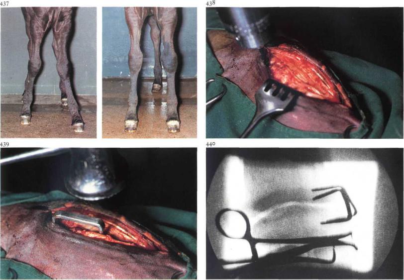
7-10 Correction of valgus deviation
Angular deformity is a common disorder afflicting the distal radius, tibia, and metacarpal / metatarsal bones in the young animal. One of the causes of the deformity is retardation ofenchondral ossification on one side (in most cases the lateral side) ofthe growth plate. In this case the affected foal shows an abnormal curvature of the distal radius, causing the limb distal to that point to deviate laterally [437A]. Ifconservative treatment fails, surgical intervention is necessary. The aim is to reduce growth of the growth plate on the convex side by means of staples or screws and wire.
Surgery. The patient is placed in lateral recumbency under general anaesthesia. The use of an Esmarch's bandage and a pneumatic tourniquet facilitates the surgery. A 5 cm incision is made through skin, subcutaneous tissue and deep fascia from 2 cm proximal to the medial radial epicondyle to almost the level ofthe radiocarpal joint. The growth plate is located by using a hypodermic needle or by radiographic monitoring. The size of the
stainless steel staple depends on the size of the patient. Holes (3 mm diameter, 2.5 cm deep) are drilled through the unincised periosteum proximal and distal to the growth plate to accomodate the staple [438]. The staple is inserted with a hammer and driver [439]. The second staple is placed approximately 20 mm cranial or caudal to the first staple, and at an angle of some 30° to it [440].
The subcutaneous tissue and deep fascia are sutured in a continuous pattern with synthetic absorbable material, and the skin is closed with simple interrupted sutures. A sterile dressing is placed over the wound and an elastic bandage is applied for about i week. Exercise is limited to stall rest until the limb is completely straight [4373]. It is important to remove staples before overcorrection occurs.
Chapter 7 THE M U S C U L O S K E L E T A L SYSTEM / Femur 7-11 |
118 |
441
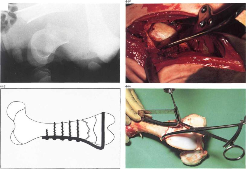
7-11 Plate osteosynthesis of supracondylar fracture
Supracondylar femur fractures occur in foals, ponies and newborn calves [441] (the latter are often delivered in posterior presentation with the help of a calf extractor).
Supracondylar femur fractures are not suitable for the usual osteosynthesis techniques, because of their form and the short, comparatively wide marrow cavity. It is possible, however, to treat supracondylar femur fractures in young animals, especially those with a low bodyweight, with condyle plates fabricated for human use [443].
Surgery. Open reduction is performed in lateral recumbency under general anaesthesia or caudal epidural analgesia (anterior block). After a suitable skin incision the tensor fasciae latae is separated from the biceps femoris. The vastus lateralis is split in the direction of its fibres. If the femoropatellar joint capsule is not already ruptured, arthrotomy exposes the lateral surface of the lateral ridge of the trochlea.
Reposition with the help of reposition forceps [442] is possible only if dislocation of the fracture fragments and contraction of adjacent musculature is not severe. Reposition is more difficult or even impossible in fractures older than 48 hours. If reposition is impossible even after administration of muscle relaxants, the distal end of the proximal fracture fragment is shortened with rongeurs. Reduction of the fragments is maintained with reposition forceps [444].
The distal fracture fragment (condyle) is fixed to the proximal fracture fragment using a condylar plate (DCP) [443]. These plates are available with different angles, and the appropriate plate (usually with an angle of 90°, 95° or 100°) is chosen. With the help of the angle gauge, a Steinman pin or a drill is placed in the distal fracture fragment [444]. The purpose of this pin is to indicate the direction of the two drill holes [445] and to guide the chisel, which has a guide hole [44&A]. The stem of the plate, the length of which depends on diaphyseal length, is contoured to the correct shape indicated by a bending template. The transverse part of the plate (the length

Chapter 7 THE MUSCULOSKELETAL SYSTEM / Femur |
119 |
of which depends on epiphyseal diameter) is inserted using the impactor, which is temporarily attached to the plate [4468]. The stem of the plate is then fixed to the metaphysis and diaphysis using (cancellous and) cortical screws [447].
A vacuum drain is placed in the wound. The femoro-patellar joint capsule, muscle layers, subcutis and skin are closed separately with synthetic absorbable suture material. Systemic antibiotics are administered. Weightbearing on the treated leg after recovery from anaesthesia is permitted. The drain is removed after 3 days.
Radiographic study is conducted immediately postoperatively [448A], and thereafter every 2 weeks. Because growth in the distal growth plate is stunted by the condyle plate, it is important to remove the plate as soon as sufficient callus has formed [4483].

Chapter 7 THE M U S C U L O S K E L E T A L SYSTEM / Tibia 7-12 |
120 |
449
7-12 Plate osteosynthesis
Plate fixation of long bone fractures of large animals is - since the walking cast technique (see 7-17) - used mainly in treating radius-ulna and tibia fractures and seldom in humerus and femur fractures. A tibial diaphyseal fracture in a calf [449] is used to show a plating technique that may be used. Surgery. The patient is positioned in lateral recumbency with the affected limb down. A curvilinear incision through skin and subcutaneous tissue is made on the medial aspect of the tibia. The convexity of the incision is directed caudally. The straight incision through the fascia is made carefully, avoiding the medial saphenous vein, artery and nerve. Deep fascia is dissected from the fractured tibia as far as both epiphyses. Blood clots are removed. After reposition of the fracture fragments and securing with a bone holding forceps, cerclage wiring is employed as a (temporary) fixation method prior to definitive stabilisation with plate and screws [450]. Long bone fractures of large animals require a broad plate. In general, the
longest possible plate is used. Compression plate osteosynthesis is preferred. Compression is achieved with the help of the tension device or a Dynamic Compression Plate. The plate is contoured and then placed over the bone, secured with a bone holding forceps, and fastened with cortical bone screws [451,452] (see also 7-8). Defects at the fracture site should be filled with cancellous bone graft. Before closing, a vacuum drain is put into the wound for 2-3 days.
Fascia, subcutaneous tissue and skin are closed with interrupted sutures using synthetic absorbable material. Systemic antibiotics are administered. In large animals osteosynthesis must in most cases be protected with a plaster cast or Thomas splint.
Chapter 7 THE MUSCULOSKELETAL SYSTEM / Tarsus 7-13 |
121 |
453
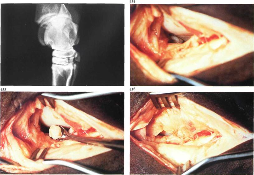
7-13 Arthrotomy in osteochondrosis
Tibio-tarsal joint arthrotomy in horses may be indicated in cases of chip fracture and osteochondrosis. Lameness and/or severe hydrarthrosis may be indications for removal of osteochondrotic fragments and/or curettage of lesions.
Surgery. The tibio-tarsal joint can be approached from the craniolateral or craniomedial side. The craniolateral approach is indicated in case of osteochondrosis at the intermediate ridge of the tibia [453]. The horse is positioned in lateral recumbency under general anaesthesia with the affected leg uppermost.
A 7 cm skin incision is made from the level ofthe lateral malleolus distally and runs lateral to the digital extensor tendon. Superficial and deep fascia are incised avoiding damage to blood vessels. The joint capsule is most easily entered just over the lateral trochlea of the talus. After retracting the wound edges and slight flexion of the joint the intermediate ridge becomes
visible [454]; suction of synovial fluid facilitates the exposure. Removal of osteochondrotic fragments is often accomplished using a Brun curette [455], but occasionally the fragment must be freed with the help of an osteotome. Subsequent curettage of the base of the lesion depends on the findings; flushing of the joint cavity is then obligatory.
Closure is performed in four layers: the fibrous part of the capsule [456], deep fascia, subcutaneous fascia and skin, all in an interrupted pattern using synthetic absorbable material. A firm (possibly elastic) bandage is applied.
After i week the bandage is changed. In cases of severe postoperative joint distension, synovial fluid is aspirated.
Chapter 7 THE MUSCULOSKELETAL SYSTEM / Tarsus 7-14
457
spontaneously occurring ankylosis
• • • • bone spavin predilection sites
122
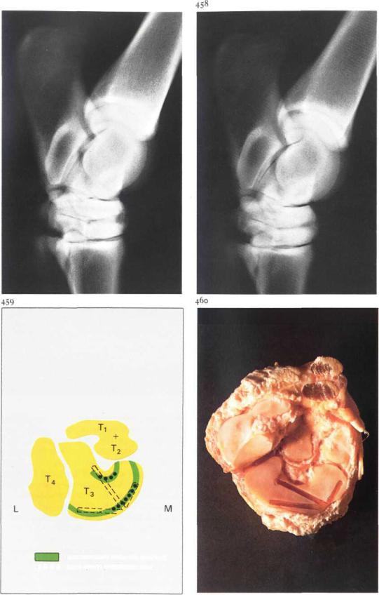
7-14 Arthrodesis of the distal intertarsal joint in the horse
Spavin is an osteoarthrosis of the distal tarsal joints, in which the changes are localised to the central tarsal bone (Tc), third tarsal bone (Ts) and the proximal articular surface of the third metatarsal bone (Mt3). Arthrodesis of the distal intertarsal joint (DIT) is one ofthe possibilities for treatment of bone spavin, and is especially indicated in cases in which the osteoarthritic changes are characterized mainly by osteolysis
[457]-
The principle of arthrodesis is that surgical destruction of parts of the joint surfaces of two apposing bones of a joint with restricted movement induces a rigid ankylosis, as shown in the radiograph ofa DIT six months after arthrodesis [458]. In this operation the drilling of three holes destroys tissue only in the predilection sites of the frequently occurring spontaneous ankylosis [459,460].
Surgery. The horse is anaesthetized in lateral recumbency with the affected limb down. A skin incision is made over the dorso-medial part of the DIT. Care should be taken to avoid the saphenous vein. A cunean tenectomy, in which 2-3 cm of the tendon is excised, is performed [461]. The DIT is identified by inserting four needles of different shape [462].
First needle: dorso-medial in the DIT at the site from which drilling will later begin.
Second needle: medial in the DIT between Tc, T3andTi+2.
Third needle: dorsal in the DIT just lateral to the midline.
Fourth needle: lateral in the tarsal canal, between Tc, T3 and T4. Only by this careful marking ofthe joint space is it possible to drill accurately in the desired directions, especially since attainment of precision is complicated by the curvature of the joint surfaces, and because it is necessary that the drill penetrates the subchondral bone to a uniform depth. Taking intraoperativeradiographs,orpreferably fluoroscopic viewing with an image intensifier, is thus obligatory [464].
After marking is completed, the first needle is removed and a small incision (0.5 cm) is made through the ligaments and joint capsule. All drilling ofthe DIT is carried out through this incision. To reduce the chance of thermal necrosis and prematurely drilling too deeply, it is better
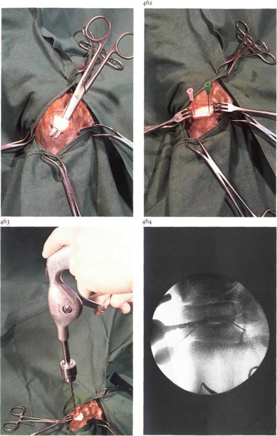
Chapter 7 THE MUSCULOSKELETAL SYSTEM / Tarsus 7-14
461
to use a manually operated drill than an electric or air drill [463]. The dorsal, medial and lateral needles determine the drilling direction of the dorsal, medial and plantar holes respectively. Extreme care should be taken to ensure that the drills do not emerge from the bone and damage the adjacent soft tissue.
When the drill bit (0 4.5 mm) is about i cm into the joint space, radiographic control is used to ensure correct positioning of the drill in the joint space. After drilling further to the desired depth, the drill holes are flushed with sterile physiologic saline. The wound is closed as follows: joint capsule and ligaments with one simple interrupted suture, subcutaneous tissue with continuous, and skin with interrupted sutures, all with synthetic absorbable suture material. Arthrodesis of the tarso-metatarsal joint is necessary only in cases in which this joint is involved. This operation can be performed through one incision, using similar identification and siting procedures. In horses with bilateral spavin both hocks are operated in the same surgery.
Six weeks box rest and four to five months at pasture are recommended.

Chapter 7 THE M U S C U L O S K E L E T A L SYSTEM / Tarsus 7-15 |
124 |
7-15 Arthrotomy and curettage in septic bovine spavin
In cattle serious lameness of the hindleg may be caused by osteomyelitis of the centroquartal bone and the fused second and third tarsal bones. The distal intertarsal joint and/or the tarso-metatarsal joint may be involved in the process, which causes a painful localised swelling at the medial site of the distal tarsal joints [465A]. The osteomyelitis is caused by haematogenous (usually Corynebacterium pyogenes) infection, possibly in combination with local trauma. The result is necrosis and sequestration, and it is thus usually too late to expect antibiotic therapy to be successful. If radiography shows evidence of necrosis and/or sequestration [4653], surgery is indicated.
Surgery. The op'eration is performed with the patient in lateral recumbency, under general anaesthesia or regional intravenous analgesia. The correct site of incision may be ascertained by radiographic control or by aspiration of the process [466].
An incision is made directly over the process and the abscess opened [467], Necrotic tissue (and if present) bone sequesters are removed with a Brun curette [468]. The cavity is packed with gauze soaked in a disinfectant and the hock is bandaged firmly.
Antibiotics are administered systemically for about 10 days. The bandage and gauze drain are changed every second day. If postoperative lameness is severe, analgesics are administered. The prognosis is guarded, since further extension of the osteomyelitis may occur.

Chapter 7 THE M U S C U L O S K E L E T A L SYSTEM / Metacarpus and metatarsus 7-16
470
125
7-16 Resection of fractured splint bone
Fracture occurs anywhere along the length of the splint bone but most often in standardbreds in the distal third of the bone. Trauma is the aetiology in most cases, but some distal fractures have been attributed to stress. This kind of stress is most common in trotters. The constant movement of the fracture fragments prevent healing and may result in non-union with superfluous callus formation [469], which may cause a (peri)- tendinitis of the suspensory ligament. Treatment consists of surgical removal of the distal fragment and the involved part of the proximal fragment.
Surgery. The operation should be performed with the horse in lateral recumbency under general anaesthesia. A skin incision, from the distal end of the bone to 3 cm proximal to the fracture site, is made over the cranial border of the affected splint bone. The periosteum-covered distal fracture fragment and the fracture site are dissected free [470,471]. The periosteum is separated from the distal end of the proximal fragment, which is transected with a chisel just proximal to the hard swelling around the fracture site [472]. The proximal segment should be carefully tapered with a chisel or suitably sized rongeurs to prevent subsequent irritation of the surrounding tissues. If possible, the periosteum is closed with fine synthetic absorbable material over the stump to reduce bone proliferation.
Deep fascia and subcutaneous tissue are sutured separately in a continuous pattern with absorbable material and the skin with simple interrupted sutures. The wound is dressed with a sterile firm (elastic) bandage for at least two weeks. The patient is box rested for 4 weeks. Training
can begin after 6 weeks if undisturbed healing, as evaluated by radiological examination, has taken place.