

G Protein-Coupled Receptors: Conformational ‘‘Gatekeepers’’ |
217 |
involving native tissues that some GPCRs form homodimers and some form heterodimers in their functional forms.
A GPCR class C receptor mGlu5 has been shown to be a homodimer stabilized by a disulfide bridge in the extracellular domain.81 The extracellular VFT domain of mGlu1 receptor has been crystallized as a dimer with a disulfide bridge in di erent relative orientations of the monomers depending on the presence or absence of the agonist glutamate,82,83 suggesting a possible mechanism of activation involving the relative change in the orientation of the monomeric TM bundles in the homodimer.84
Another class C GPCR provided the first conclusive evidence of a functional heterodimer. The g-aminobutyric acid-binding (GABAB) receptor is only functional as a heterodimer between GABAB1 and GABAB2 monomeric subunits, where each subunit plays a distinct role: GABAB1 subunit binds to the agonists and GABAB2 subunit couples to the G protein.85 Sweet and umami taste receptors provide other class C examples of heterodimerization. Of the three genes encoding these receptors (T1R1, T1R2 and T1R3), T1R2-T1R3 heterodimer results in a sweet receptor and the T1R1-T1R3 heterodimer results in an umami receptor.14 The three monomeric receptors don’t display the functional behavior if they are expressed alone.
There is also plenty of evidence now in favor of the presence of functional homodimers and heterodimers in class A GPCRs.80 Here we will mention a more recent example involving the dopamine D2 receptor, which has been shown to form homodimers out of monomers in functionally di erent states; maximal activation was observed when one monomer was bound to the agonist (was active) and the other monomer was bound to an inverse agonist (was inactive).86
Interaction of GPCRs with other membrane and cytoplasmic proteins has been known for a while and has also been reviewed.87 The physiological implications of these interactions are slowly being uncovered as only some of these interactions have been amenable to detailed experimental investigations. A discussion of these interactions is beyond the scope of this chapter.
11.3.3Functional Control of GPCRs by Ligands
As mentioned earlier, GPCRs are pleiotropic in terms of the multiple intracellular signaling cascades they can a ect upon binding to an agonist through both G protein-coupled and b-arrestin coupled pathways. Experimentally, a single functional assay (usually by definition) cannot see all the signaling e ects of a ligand. It is now evident that agonist-bound GPCRs exist in multiple distinct conformations, where each conformation can potentially activate a di erent signaling pathway. From a therapeutic perspective, this may not be desirable if, of the multiple signaling pathways activated by a drug-molecule, one pathway may be mainly responsible for the desirable therapeutic benefit and another may be causing unwanted side-e ects. This opens at least two distinct possibilities of controlling the functional consequences of GPCR activation: biased agonists and allosteric modulators. Each of these possibilities will be
218 |
Chapter 11 |
described below along with a few representative examples. As will be seen, some of these ligands defy the classical definitions of agonists and antagonists.
11.3.3.1Biased Agonism
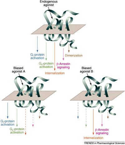
Ligands and especially agonists can induce multiple GPCR conformations upon binding. As has been mentioned before, GPCRs interact with cytosolic G proteins and b-arrestins. Di erent ligands can induce a di erent ensemble of GPCR conformations, which will have a di erent range of interactions with G proteins and/or b-arrestins and hence induce di erent intracellular signaling pathways in a ligand-dependent fashion. As shown in Figure 11.16, agonist
Figure 11.16 Multiple pathways that can be initiated by GPCR activation and biased by ligands. Reprinted with permission from Elsevier from Kenakin, Trends Pharmacol. Sci., 28(8) 407–415. Copyright 2007.
G Protein-Coupled Receptors: Conformational ‘‘Gatekeepers’’ |
219 |
binding to GPCRs can induce a cascade of processes, some through G protein coupling and some through b-arrestin coupling. As shown before, b-arrestin coupling can lead to specific signaling events and receptor endocytosis, which can further lead to either recycling of the receptor or its degradation. Figure 11.16 also shows the e ect of biased agonists A and B that can either exist or be designed to activate a subset of possible signaling pathways.
Classically, the GPCR agonists have been characterized as such by their e ect on G protein-coupled pathways. Experiments measuring b-arrestin signaling are becoming more commonplace so the e ects of classical agonists need to be evaluated and should lead to a detailed characterization of these ligands. The parathyroid hormone (PTH), for example, can activate extracellular signal-related kinase using distinct G protein-dependent and G proteinindependent pathways. The PTH analogs, however, can use the same PTH receptor and separately use either G protein-dependent or -independent pathways. An example is that [Trp1]PTHrp-(1-36) stimulates ERK1/2 via G protein pathway, whereas PTH-1A [[D-Trp12,Tyr34]PTH-(7-34)] stimulates the same via b-arrestin pathway in a G protein-independent manner.78 This can have direct therapeutic consequences because PTH regulates calcium homeostasis as well as bone metabolism and utilization of b-arrestin 2 pathway is critical for this benefit,88 so b-arrestin biased PTH analogs mentioned above provide potentially improved therapy for osteoporosis. Another example is that nicotinic acid is therapeutically very beneficial as an anti-lypolytic agent (via G protein-mediated pathways), but causes cutaneous flushing as a major side-e ect, which has been directly linked to the activation of b-arrestin 1 pathways.89 An analog of this molecule that doesn’t a ect the G protein pathways but blocks the b-arrestin 1 pathways will be highly desirable. This also necessitates new characterization of classical agonists, e.g. a ligand that blocks G protein pathways but uses b-arrestin pathways may have been classified before as an antagonist (or inverse agonist) but now should be more accurately described as a b-arrestin biased agonist.
These studies are also increasing our understanding of the relationship between various signaling pathways and previously unexplained side-e ects of drug molecules. The use of knowledge about biased signaling during the drug design phase has the potential to generate multiple novel ways to control and hopefully cure many ailments with minimal side-e ects.
11.3.3.2Allosteric Ligands and Signal Modulation
Orthosteric ligands bind to GPCRs in regions that fully or partially overlap with that of the endogenous ligand(s), thereby sterically excluding the possibility of both occupying the GPCR at the same time. Allosteric ligands bind to GPCRs in regions that don’t overlap with the endogenous ligand binding site, so both can occupy the receptor at the same time. This can have important signaling consequences because, as mentioned before, agonists induce an ensemble of GPCR conformations with a range of functional implications and allosteric ligands can dramatically modulate those conformations (e.g. by
220 |
Chapter 11 |
stabilizing a subset), which can lead to modulation of signaling and hence function. Allosterism provides a powerful natural tool for modulating signaling cascades, but not many natural modulators are known, probably because of the di culty in identifying these ligands which are structurally dissimilar to endogenous ligands. One of the examples is the unnatural D-amino acid D-serine formed in the brain, which is a strong allosteric modulator of the N-methyl-D-aspartate (NMDA) receptor.90
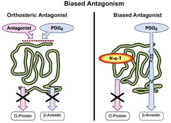
An allosteric modulator can bind to a unique ensemble of GPCR conformations and have three modulatory e ects on GPCR activation: allosteric antagonism (including allosteric inverse agonism), allosteric agonism and allosteric partial antagonism. In allosteric antagonism, the modulator stabilizes more inactive conformations or destabilizes more active conformations, resulting in the net reduction of GPCR activation relating signaling. These are usually referred to as negative allosteric modulators (NAMs). In allosteric agonism, two scenarios arise where the modulator either enhances the e ect of the orthosteric agonist by stabilizing the more active receptor conformations or directly causes the GPCR activation in the absence of the orthosteric agonist. The former kind are referred to as positive allosteric modulators (PAMs), some of which are capable of directly agonizing the receptor in the absence of the orthosteric agonist.91 In allosteric partial antagonism, the modulator (also called biased antagonist) selectively blocks only a subset of the activation related pathways as shown in Figure 11.17. The figure shows that Postaglandin D2 normally activates G protein as well as b-arrestin pathways for its CRTH2 receptors, where both of these pathways can be blocked by an orthosteric
Figure 11.17 Biased antagonism, which can be called allosteric partial antagonism. Reproduced with permission from American Society for Pharmacology and Experimental Therapeutics from Kenakin and Miller, Pharmacol. Rev., 62, 265–304. Copyright 2010.