

G Protein-Coupled Receptors: Conformational ‘‘Gatekeepers’’ |
193 |
11.3.1Structure of GPCRs
GPCRs can be organized into six families (acronymed GRAFTS, a minor variation on the original GRAFS classification proposed13 based on the phylogenetic criteria): glutamate, rhodopsin, adhesion, frizzled, taste2 and secretin.
a)Rhodopsin family (also called Class A or Family 1): This diverse family dominates the human GPCRs with B670 members (out of B800 total). The family is further divided into four subfamilies – a, b, g, d. The a subfamily includes light-sensing rhodopsin receptor, biogenic amine (dopamine, serotonin, histamine, muscarinic) receptors as well as cannabinoid and prostanoid receptors among others. The b subfamily mainly consists of peptide-binding proteins. The g subfamily receptors bind to peptides or lipid-like molecules, some examples being chemokine, angiotensin, somatostatin and opiod receptors. The d subfamily is dominated by olfactory receptors (B388 out of B670 total in rhodopsin family) and also contains purinergic and glycoprotein-binding receptors. The vomeronasal pheromone receptors putatively also belong to the rhodopsin family. Being the largest family, it is not surprising that this family is targeted by the majority of GPCR drugs.
b)Secretin/Adhesion family (also called Class B or Family 2): The secretin receptors of this family bind peptide hormones, whereas Adhesion
receptors bind to extracellular matrix molecules based on the knowledge of receptors de-orphaned so far.13
c)Glutamate family (Class C or Family 3): This family consists of meta-
botropic glutamate receptors, g-aminobutyric acid B (GABAB) receptors, sweet and umami (due to glutamate in monosodium glutamate or MSG, a food additive) taste receptors and calcium-sensing receptor. One of the two taste receptor monomers (T1R1, T1R2) combines with a third monomer
(T1R3) to form functional heterodimers for sweet taste (T1R2 þ T1R3) or umami taste (T1R1 þ T1R3).14
d)Frizzled family: This family consists of B10 frizzled receptors (which bind to Wnt glycoproteins) and a smoothened receptor (which appears to function without binding to any ligand).
e)Taste2 family: This family exclusively consists of B25 bitter taste receptors,15–16 which share the sensing of di erent bitter tastants with a
di erent subset of receptors. These taste receptors have recently been found in the gastrointestinal (GI) tract as well.17 Their function in the gut
is not known but their activation (in mice) has been shown to activate gut hormonal receptors (cholecystokinin or CCK and peptide YY or PYY),18 which are coupled to the glucagon-like peptide 1 (GLP-1) and other glucose metabolism pathways.
The structural topology of the receptors within each of the families mentioned above appears to be similar based on structural and sequence analysis as shown in Box 11.1 (for Family 1, 2 and 3). GPCRs in general are not

194 |
Chapter 11 |
Box 11.1 Reprinted by permission from Macmillan Publishing Ltd.: George et al., Nat. Rev. Drug Discov., 1(10), 808–820. Copyright 2002.
homologous to each other unless they bind to the same ligands and, apart from the seven-TM helix topology, nothing appears to be common across all receptors.
Rhodopsin (Family 1) receptors share some common sequence motifs like D(E)RY at the bottom of TM3, WXPFF motif in TM6, NPXXY motif in TM7 and some conserved prolines usually in the middle of many TMs that produce kinks in their helices. Small molecule ligands typically bind in the extracellular facing half of the TM regions and peptides/proteins bind mainly to the extracellular loops and N-terminus. There is a highly conserved disulfide bridge between cysteines in ECL2 and top of TM3.
Secretin/Adhesion (Family 2) receptors have a long N-terminal ectodomain that binds to ligands and contains many conserved cysteines, which can help the long N-terminus to form a stable tertiary structure (see Box 11.1). These receptors don’t share any sequence motifs with Family 1 receptors even in the TM regions, so it is not obvious if they will have the same TM bundle topology of Family 1 receptors.
Glutamate (Family 3) receptors have a long N-terminus and a long C- terminus as well. Most receptors use their long N-terminus to bind to their
G Protein-Coupled Receptors: Conformational ‘‘Gatekeepers’’ |
195 |
endogenous ligands and the binding pocket is sometimes referred to as the venus fly trap (or VFT; see Box 11.1).
Next we will describe the structure determination e orts aimed at GPCRs and what we have learnt from the available structures generated by these e orts and functional studies of GPCRs.
11.3.1.1Structure Determination
The experimental structure determination of GPCRs had been quite slow until recently relative to other membrane proteins (and obviously soluble proteins) despite intense e orts by many protein crystallography and NMR groups (currently, six GPCR structures present in the PDB out of more than 65,000
structures). Until 2007, crystal structure was available only for bovine rhodopsin.19,20 This lack of structures was due to various factors including poor
protein expression levels, di culties in large-scale receptor purification, the insolubility in media-lacking phospholipids and other di culties in crystallization. Significant technological advances in GPCR crystallization techniques have been made in the last few years that include emergence of lipidic cubic phase crystallization21 and its coupling to the protein fusion methodology22 that replaces a disordered region of protein structure with T4-lysozyme to increase the surface area potential for crystal contacts.
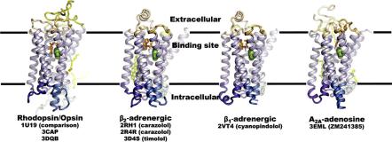
These advances have resulted in the availability of crystal structures23 of two human GPCRs: b2 adrenergic receptor (b2AR) bound to a partial inverse agonist24 and adenosine A2A receptor (A2AR) bound to an antagonist.25 Other advances include increasing GPCR thermal stability by systematic mutagenesis (which led to the structure of turkey b1 adrenergic receptor (b1AR),26 and optimization of receptor purification (which led to the structure of activated ligand-free bovine opsin structure by itself27 and in association with a car- boxyl-terminal peptide fragment of its Ga subunit transducin).28 Figure 11.1
Figure 11.1 Four representative crystallized GPCRs solved to date. Reprinted from structure 17(1), Hanson and Stevens, Discovery of New GPCR Biology: One receptor Structure at a Time, 8–14, Copyright 2009 with permission from Elsevier.
196 |
Chapter 11 |
shows the structures of bovine rhodopsin, human b2 adrenergic, turkey b1 adrenergic and human adenosine A2A receptors.23 They all share the same TM topology (relative positions of TM helices), but can di er (sometimes significantly) in helix tilts and rotations as will be discussed in the next section.
An invertebrate GPCR (squid rhodopsin) has also been crystallized, which showed unusually long TM regions 5 and 6.29 At least two more human GPCR structures are expected this year (dopamine D3 and chemokine CXCR4 receptors) and significantly more in the next decade. Progress is also being made in developing solid-state NMR techniques30 for GPCR structure determination. This rapid growth in GPCR crystal structures since 2007 is beginning to provide insight into the structural biology of these proteins,23,31 however, the progress is expected to remain slow due to intrinsic flexibility of these versatile receptors, which prevents them from packing into ordered crystals.
11.3.1.2Structural Diversity of Current GPCR Structures
The seven-TM helix topology of GPCRs presents unique advantages and challenges for the quantification of sequence-structure relationships. Many comparative modeling programs can predict structures of globular proteins (with 30% or higher sequence identity to a crystallized protein) to a reasonable accuracy, as the belief is that a major fraction of structural folds is now known for globular proteins. The same cannot be said for membrane proteins in general. GPCRs, however, can be thought of as having one structural fold, consisting of seven TM helices interconnected by intracellular and extracellular loops. The TM helices display high sequence conservation as compared to the loop regions as expected (see Table 11.1).
The table shows the sequence identity (Table 11.1A) and sequence similarity (Table 11.1B) (similarity using BLOSUM62,32 where two residues are considered similar if the corresponding substitution element in the BLOSUM62 matrix is 4 0) for the five GPCR sequences that have been crystallized.
To quantify the relationship between sequence and structure for GPCRs, we need to characterize the known structures using some standard geometrical parameters. As crystal structures don’t provide absolute membrane orientation of GPCRs, we use their orientation as predicted by the OPM (Orientation of Proteins in Membrane) database,33 which aligns each newly deposited membrane protein structure to an implicit membrane maximizing the free energy of membrane insertion. The middle of the membrane corresponds to the z ¼ 0 plane or the hydrophobic plane. Each GPCR structure can then be characterized by the six orientation parameters of the seven helices relative to this plane. Figure 11.2A shows how the helix position and tilt are defined. Helix position (R) on the hydrophobic plane is then given by x and y. Value h corresponds to the hydrophobic center residue from the helix that will be

Table 11.1 Sequence comparison across five GPCR sequences that have been crystallized so far, with first number for the whole sequence and the second number for all TMs: A Sequence Identity; B Sequence Similarity (using the Blosum62 matrix).32
‘‘Gatekeepers’’ Conformational Receptors: Coupled-Protein G
197

198 |
Chapter 11 |
A |
B |
C
Figure 11.2 A Definition of the helical axis; B Table showing relative orientation parameters for the GPCRs with crystal structures; C Correlation of deviation in orientation parameters with sequence identity/similarity.
positioned on the hydrophobic plane. Two angles, y and f, specify the tilt angles of the helix and the angle Z corresponds to the helix rotation angle about its axis. The two tilt angles (y,f) and the rotation angle (Z) require a definition of the helical axis, which needs to account for the reality of bent helices as prolines are commonly found in the TM helices. We use a helical axis that corresponds to the lowest moment of inertia vector for the helix obtained by diagonalizing the moment of inertia matrix for the helix using only heavy backbone atoms.
We rotate the membrane-aligned GPCRs from the OPM database in the x–y plane such that the helical axis of TM helix (TMH) 3 goes through the origin, and that of TMH 2 intersects the x-axis. Figure 11.2B shows the relative six orientation parameters for all seven helices for the crystallized GPCRs relative to b2 adrenergic receptor and of bovine rhodopsin (cis-retinal bound form) relative to bovine opsin (the retinal free form) that is considered a conformation along the activation pathway of rhodopsin.
In order to correlate the sequence variability of these GPCRs with their helix geometries, we calculated all-to-all (across these systems) RMS (root-mean- squared) deviations in position R (x,y position in the z ¼ 0 plane), and angles y, f, Z averaged over all helices and plotted them against the corresponding sequence identity and similarity. The equations used for the deviations between