

Полезные материалы за все 6 курсов / Учебники, методички, pdf / Kaplan Pediatrics USMLE 2CK 2021
.pdfUSMLE Step 2 CK λ Pediatrics
Table 13-1. Heart Murmur Gradation
Grade Quality
1Soft, difficult to hear
2Easily heard
3Louder but no thrill
4Associated with thrill
5Thrill; audible with edge of stethoscope
6Thrill; audible with stethoscope just off chest
•Diagnostic tests
−Chest radiograph—evaluate heart size, lung fields, ribs for notching, position of great vessels
−Electrocardiogram
−Echocardiography—definitive diagnosis
−Other—MRI, cardiac catheterization, angiography, exercise testing
•Embryology—knowledge of cardiac embryology is helpful for understanding congenital cardiac lesions, their presentations, symptoms, and treatment.
 Ductus arteriosus
Superior vena cava 
Foramen ovale
Ductus venosus
Inferior vena cava 
Portal vein
Liver
Umbilical vein
O2
|
Umbilical cord |
Shunts in bold |
Right and left |
umbilical arteries |
|
|
O2 |
|
Figure 13-1. Fetal Circulation |
114

Chapter 13 λ Cardiology
PEDIATRIC HEART SOUNDS AND INNOCENT MURMURS
Heart Sounds
First heart sound (S1)
•Closure of mitral and tricuspid valves (MV, TV)
•High pitch, but lower pitch and greater intensity compared to S2
•Usually no discernible splitting of S1 but in completely normal child, a split S1 represents asynchronous closure of the 2 valves (20−30 msec difference); however, what sounds like a split S1 but does not represent pathology:
–Split S1 best heard at apex or right upper sternal border; click (opening of stenotic valve, slightly later than a split S1) may be heard in aortic stenosis
–Apical mid systolic click of mitral valve prolapse
–At upper left sternal border, a click may be heard from pulmonic valve stenosis; compared to aortic stenosis, this changes with respiration (with inspiration, venous return is increased, thus causing the abnormal pulmonary valve to float superiorly after which the click softens or disappears)
–Tricuspid valve abnormalities (e.g., Ebstein anomaly) may cause billowing of the leaflets and result in multiple clicks
•S1 may be inaudible at the lower left sternal border mostly due to sounds that obscure the closure of the MV and TV, e.g., in VSD, PDA, mitral or tricuspid regurgitation and severe right ventricular outflow tract obstruction. Therefore, if the first heart sound is not heard at the lower left sternal border, there is most likely a congenital heart defect, and there will be other clinical and auscultatory findings.
Second heart sound (S2)
•Closure of pulmonary and aortic valves (PV, AV), which close simultaneously on exhalation and a single heart sound is best heard with diaphragm at the upper left sternal border
–Wider splitting of S2 on inspiration is related not only to increased venous return but also to pressures in the aorta and pulmonary artery (PA) (it is significantly higher in the Ao than in the PA, so Ao valve closes first)
•Wider than normal splitting will occur with any lesion that allows more blood to traverse the PV compared to normal
–Increased splitting of S2 may be fixed with respect to respiration if there is increased volume and hence pressure in the right atrium (e.g., ASD); otherwise, it will continue to vary with respiration; may also hear fixed splitting with a right bundle branch block
•Loud single S2: heard with PA hypertension (increased pressure closing the PV causes early closure of the anterior semilunar valve resulting in a loud single S2)
–In D-transposition, the AV is anterior and to the right of the PV, which overwhelms the sound from the PV, so one hears a loud single S2; in truncus arteriosus, there is only 1 valve so there is a single S2
Published by dr-notes.com |
115 |
|
|
|
|

USMLE Step 2 CK λ Pediatrics
Third heart sound (S3)
•Hear early in diastole; creates a gallop rhythm with S1 + S2; very low frequency and is best heard with bell of the stethoscope at cardiac apex; asking patient to lie on left side may increase intensity of S3
•On occasion may be heard normally in children with no pathology: in older people, it represents the presence of CHF or other volume overload and is caused by sudden deceleration of blood flow into LV from the LA
Fourth heart sound (S4)
•Occurs in late diastole, just prior to S1 (presystolic) and is produced by a decrease in compliance (increased stiffness) of the LV
•Low frequency (lower than S3) and best heard with bell of the stethoscope pressed lightly against the skin
•Summation gallop rhythm (S3 + S4) may be found with myocarditis or a cardiomyopathy (combination of volume overload and noncompliant ventricle)
Clinical Recall
A medical student is performing a physical exam on an infant. Cardiac auscultation reveals a loud single S2. What congenital anomaly does the infant likely have?
A.Atrial septal defect
B.Patent ductus arteriosus
C.Ventricular septal defect
D.Ebstein anomaly
E.D-transposition
Answer: E
Innocent Murmurs
Peripheral pulmonic stenosis
•Normal finding age 6 weeks to 1 year
•Generated by blood flowing into the lungs due to (1) pulmonary arteries, which have limited blood flow in utero and are therefore small with significantly increased blood flow after birth (turbulence from RV blood flowing through these arteries) and (2) increasing cardiac output associated with declining [Hgb] over the first weeks of life (physiologic anemia)
•Normal infant with normal S1, then grade 1-2 systolic ejection murmur at the upper sternal border and radiating bilaterally into the axillae; then, normal splitting of S2
Still’s murmur
•Commonly heard first at age 3−5 years
•Represents turbulence or vibrations in either ventricle; child is healthy and asymptomatic
116
Chapter 13 λ Cardiology
•Precordial activity is normal, as are S1 and S2; the murmur is typically low-pitched (bell of stethoscope), musical-quality and often radiates throughout the precordium.
•Murmur is loudest while supine (greater blood flow) and decreases sitting or standing —opposite to the finding of HOCM. Also increases with fever or exercise (hyperdynamic states).
Venous hum
•Only diastolic murmur that is not pathological; represents blood flow returning from the head and flowing from SVC into the RA
•Described as “whooshing” sound (like holding a seashell to your ear at the ocean); is a continuous murmur
–Best heard in sitting position with head in the neutral position
–Murmur becomes softer or disappears while in supine, with slight pressure to the right side of the neck or turning head to opposite side
Aortic outflow murmur
•Heard in adolescents and young adults (especially athletes, due to lower resting heart rate and therefore larger stroke volume
•Best heard in upper right sternal border; represents blood flow in LV outflow tract (without a click, as there is in aortic stenosis)
•Precordial activity is normal, S1 and S2 are normal, the murmur is grade 1-2 ejection
–Going from supine to sitting or standing decreases the murmur (again, opposite to HOCM)
Congenital Heart Disease
In most cases, diagnosis usually made by age 1 month. Murmurs may not be heard in early life because of increased pulmonary vascular resistance (from fetal to neonatal transition physiology).
•Etiology
−Most are unknown
−Associated with teratogens, such as alcohol and rubella
−Genetic predisposition—trisomies; Marfan, Noonan, DiGeorge syndromes (typically non-Mendelian)
•Classification
Table 13-2. Congenital Heart Disease
|
|
Shunting |
|
|
|
|
|
|
|
|
|
|
|
Regurgitant |
|
Stenotic |
Right → Left |
Left → Right |
Mixing |
|
|
|
|
|
|
|
|
MVP |
|
Aortic stenosis |
Tetralogy of Fallot |
Patent ductus |
Truncus arteriosus |
|
|
|
|
|
|
|
|
PI, AI |
|
Pulmonic |
Ebstein anomaly |
Ventricular septal |
TAPVR |
|
|
|
stenosis |
|
defect |
|
|
|
|
|
|
|
|
|
MI, TI |
|
Coarctation of |
Tricuspid atresia |
Atrial septal defect, |
HLH, transposition |
|
|
|
the aorta |
|
endocardial cushion |
of the great vessels |
|
|
|
|
|
defect |
|
|
|
|
|
|
|
|
|
Definition of abbreviations: TAPVR total anomalous pulmonary venous return; HLH hypoplastic left heart; MVP mitral valve prolapse; PI pulmonic insufficiency; AI aortic insufficiency; MI mitral insufficiency; TI tricuspid insufficiency
Published by dr-notes.com |
117 |
|
|
|
|

USMLE Step 2 CK λ Pediatrics
Note
Eisenmenger Syndrome
•Transformation of any untreated left-to-right shunt into a bidirectional or right- to-left shunt
•Characterized by cyanosis
•Results from high pulmonary blood flow, causing medial hypertrophy of pulmonary vessels
and increased pulmonary vascular resistance, resulting in pulmonary arterial hypertension and partial flow reversal
LEFT TO RIGHT SHUNTS
Ventricular Septal Defect (VSD)
A 3-month-old child presents with poor feeding, poor weight gain, and tachypnea. Physical examination reveals a harsh, pansystolic 3/6 murmur at the left lower sternal border, and hepatomegaly.
•Most common congenital heart lesion
•Most are membranous
•Shunt determined by ratio of PVR to SVR
−As PVR falls in first few weeks of life, shunt increases
−When PVR>SVR, Eisenmenger syndrome (must not be allowed to happen)
•Clinical findings
−Asymptomatic if small defect with normal pulmonary artery pressure (most); large defect—dyspnea, feeding difficulties, poor growth, sweating, pulmonary infection, heart failure
−Harsh holosystolic murmur over lower left sternal border ± thrill; S2 widely split and varies with respiration
–With hemodynamically significant lesions, also a low-pitched diastolic rumble across the mitral valve heard best at the apex (increased diastolic flow leads to significant turbulence, which causes the murmur)
Ventricular Septal Defect
(VSD)
|
Left |
|
|
atrium |
|
Right |
|
|
atrium |
Mitral valve |
|
Tricuspid |
|
|
valve |
Left |
|
Right |
||
ventricle |
||
ventricle |
||
|
Figure 13-2. Ventricular Septal Defect
118
Chapter 13 λ Cardiology
•Diagnosis
−Chest x-ray: large heart, pulmonary edema
−EKG: LVH early on, if no signs of dyspnea and increased RV pressure; if left unchecked, PA pressures and RV pressures will increase
−Echocardiogram is definitive
•Treatment
−Small muscular VSD more likely to close in first 1–2 years than membranous
−Less common for moderate to large to close → medical treatment for heart failure
(control failure and prevent pulmonary vascular disease)
−Surgery in first year; indications:
°Failure to thrive or unable to be corrected medically
°Infants at 6–12 months with large defects and pulmonary artery hypertension
°More than 24 months of age with Qp:Qs >2:1 (shunt fraction)
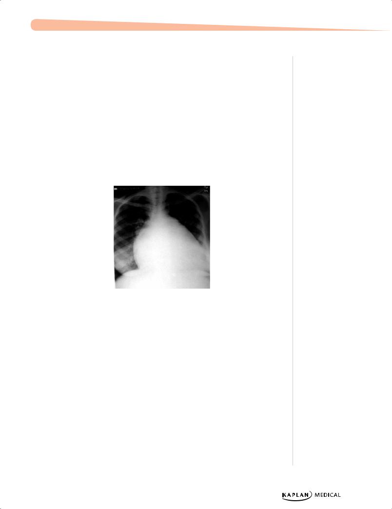
Courtesy of Tom D. Thacher, M.D.
Figure 13-3. Cardiomegaly Due to Ventricular Septal Defect
•Complications
−Large defects lead to heart failure, failure to thrive
−Endocarditis
−Pulmonary hypertension
Atrial Septal Defect (ASD)
•Ostium secundum defect most common (in region of fossa ovalis)
•Clinical
−Few symptoms early in life because of structure of low-flow, left-to-right shunt
−In older children, often with large defects; varying degrees of exercise intolerance
–With hemodynamically significant lesions, also a low-pitched diastolic rumble across the tricuspid valve heard best at the lower sternum
Published by dr-notes.com |
119 |
|
|
|
|
USMLE Step 2 CK λ Pediatrics
Atrial Septal Defect
(ASD)
Right |
Left |
|
atrium |
||
atrium |
||
|
||
|
Mitral |
|
|
valve |
|
Tricuspid |
Left |
|
valve |
ventricle |
|
Right |
|
ventricle
Figure 13-4. Atrial Septal Defect
•Physical examination
−Wide fixed splitting of S2
−Systolic ejection murmur along left mid to upper sternal border (from increased pulmonary flow)
•Diagnosis
−Chest x-ray—varying heart enlargement (right ventricular and right atrial); increased pulmonary vessel markings, edema
−EKG—right-axis deviation and RVH
−Echocardiogram definitive
•Treatment
−Most in term infants close spontaneously; symptoms often do not appear until third decade
−Surgery or transcatheter device closure for all symptomatic patients or 2:1 shunt
•Complications
−Dysrhythmia
−Low-flow lesion; does not require endocarditis prophylaxis
120
Chapter 13 λ Cardiology
Endocardial Cushion Defect
•Pathophysiology
−When both ASDs and VSDs occur, which are contiguous, and the atrioventricular valves are abnormal
−Left-to-right shunt at both atrial and ventricular levels; some right-to-left shunting with desaturation (mild, intermittent cyanosis)
−Atrioventricular valve insufficiency → increase volume load on one or both ventricles; early heart failure, infections, minimal cyanosis, hepatomegaly, and failure to thrive
•Physical examination
−Heart failure early in infancy (hepatomegaly, failure to thrive)
−Eisenmenger physiology occurs earlier (due to increased ejection of blood from the large RV chamber into the narrow infundibulum)
−Moderate-to-severe increase in heart size with hyperdynamic precordium (precordial bulge and lift)
−Widely fixed split S2 (like an isolated ASD)
−Pulmonary systolic ejection murmur, low-pitched diastolic rumble at left sternal border and xiphoid, representing increased diastolic flow across both the MV and TV
•Diagnostic tests
−Chest x-ray—significant cardiomegaly, increased pulmonary artery and pulmonary blood flow and edema
−EKG—signs of biventricular hypertrophy, right atrial enlargement, superior QRS axis
−Echocardiogram (gold standard)
•Treatment—surgery more difficult with heart failure and pulmonary hypertension (increased pulmonary artery pressure by 6−12 months of age); must be performed in infancy
•Complications
−Without surgery—death from heart failure
−With surgery—arrhythmias, congenital heart block
Patent Ductus Arteriosus (PDA)
•Results when the ductus arteriosus fails to close; this leads to blood flow from the aorta to the pulmonary artery
•Risk factors
−More common in girls by 2:1
−Associated with maternal rubella infection
−Common in premature infants (developmental, not heart disease)
•Presentation
−If small—possibly no symptoms
−If large—heart failure, a wide pulse pressure, bounding arterial pulses, characteristic sound of “machinery”
Note
Patients with trisomy 21 are at a higher risk for endocardial cushion defects.
Note
If a PDA persists beyond the first week of life, it is unlikely to close spontaneously.
Published by dr-notes.com |
121 |
|
|
|
|

USMLE Step 2 CK λ Pediatrics
Note
Pulmonic stenosis as a result of valve dysplasia is the common defect in
Noonan syndrome (12q24.1; autosomal dominant;
boys and girls with Turner phenotype).
Pulmonic stenosis (either valve or branched artery) is common in Alagille syndrome (arteriohepatic dysplasia).
122
•Diagnostic tests
−Chest x-ray—increased pulmonary artery with increased pulmonary markings and edema; moderate-to-large heart size
−EKG—left ventricular hypertrophy
−Echocardiogram—increased left atrium to aortic root; ductal flow, especially in diastole
•Treatment
−May close spontaneously
−Indomethacin (preterm infants)
−Surgical closure
•Complications
−Congestive heart failure
−Infective endocarditis
STENOTIC LESIONS
Pulmonic Stenosis
•Pathophysiology
−Deformed cusps → open incompletely during systole; obstruction to right ventricular outflow → increased systemic pressure and wall stress → right ventricular hypertrophy (depends on severity of pulmonary stenosis)
−Arterial saturation normal unless ASD or VSD is present with R → L shunt
−Neonate with severe pulmonary stenosis = critical pulmonary stenosis = R → L shunt via foramen ovale
•Physical examination
−Heart failure only in severe cases, most in first month of life
−Mild cases—normal life, usually no progression
−Moderate to severe—increasing gradient with growth: signs of right ventricular failure (hepatomegaly, peripheral edema, exercise intolerance)
−Pulmonary ejection click after S1 in left upper sternal border and normal S2 (in mild); relatively short, low-to-medium−pitched SEM over pulmonic area radiating to both lung fields
•Diagnosis
−EKG—right ventricular hypertrophy in moderate to severe; tall, spiked P-waves; right atrial enlargement (RAE)
−Chest x-ray—poststenotic dilatation of pulmonary artery; normal-to-increased heart size (right ventricle) and decreasing pulmonary vascularity
−Echocardiogram (gold standard)
•Complications
−Heart failure
−Endocarditis (lower risk)
−Secondary subvalvular muscular and fibrous hypertrophy
•Treatment
−Moderate to severe—balloon valvuloplasty initially; may need surgery
−Neonate with critical pulmonary stenosis—emergent surgery
Chapter 13 λ Cardiology
Aortic Stenosis
•Most are bicuspid aortic valve—usually asymptomatic in children
•Supravalvular stenosis (least common form)—sporadic, familial, or with Williams syndrome (intellectual disability, elfin facies, heart disease, idiopathic hypercalcemia; deletion of elastin gene 7q11.23)
•Clinical presentation—symptoms depend on severity of obstruction
−If severe early in infancy = critical aortic stenosis = left ventricular failure and decreased cardiac output
−If significant decrease in cardiac output—intensity of murmur at right upper sternal border may be minimal
−Mild to moderate—usually asymptomatic with normal growth and development
°Often discovered with murmur on routine physical examination
°Rare—older children present with syncope, fatigue, angina, dizziness
−With increasing severity—decreased pulses, increased heart size, left ventricular apical thrust
−Early systolic ejection click at apex and left sternal border (does not vary with respiration)
°Severe—no click and decreased S1 (decreased left ventricular compliance), decreased S2 (aortic component), and maybe an S4
°SEM upper-right second intercostal space; the louder (harsher) and longer the murmur, the greater the degree of obstruction; radiates to neck and left midsternal border; positive thrill in suprasternal notch
•Diagnosis
−EKG—left ventricular hypertrophy and strain
−Chest x-ray—prominent ascending aorta; may have valve calcification (older children and adults); if severe → increased heart size (left ventricular hypertrophy)
−Echocardiogram (gold standard)
•Treatment
−Balloon valvuloplasty
−Surgery on valves
−Valve replacement
Coarctation of the Aorta
•Narrowing at any point from transverse arch to iliac bifurcation; 90% just below origin of left subclavian artery at origin of ductus arteriosus (juxtaductal coarctation)
Adult versus childhood
•Discrete juxtaductal coarctation (adult type)
−Ascending aortic blood flows normally through narrowed segment to reach descending aorta, but there is left ventricular hypertrophy and hypertension
−If mild, not recognized until later in childhood
−Increased blood pressure in vessels proximal to coarctation and decreased blood pressure and pulses below constriction
°Femoral and other lower pulses weak or absent; bounding in arms and carotids; also delay in femoral pulse compared to radial (femoral normally occurs slightly before radial)
Note
Coarctation of the aorta has a high association with Turner syndrome (70% with bicuspid aortic valve).
Note
Coarctation should be suspected in an asymptomatic child with hypertension.
Published by dr-notes.com |
123 |
|
|
|
|