

Полезные материалы за все 6 курсов / Учебники, методички, pdf / Kaplan Pediatrics USMLE 2CK 2021
.pdfUSMLE Step 2 CK λ Pediatrics
NEWBORN SCREENING
A 1-month-old fair-haired, fair-skinned baby presents with projectile vomiting of 4 days’ duration. Physical exam reveals a baby with eczema and a musty odor. Which screening test would most likely be abnormal?
As per the American College of Medical Genetics, every newborn is screened for a core panel of 29 disorders, with an additional 25 recommended (Expanded Newborn Screening Program; varies per state).
•All states now use tandem mass spectrometry; typically done after 24–48 hrs of feedings prior to baby leaving the birth hospital
•With early discharge, may be performed at first postnatal visit (3–5 days) for improved accuracy
•In addition to a heel stick blood sample, current program also includes a hearing test and pulse oximetry for critical congenital heart disease.
Examples of the more common disorders in the expanded program include:
•Phenylketonuria, tyrosinemia type I, 21-hydroxylase deficiency, classic galactosemia
•HbS/β-thal, Hb SS, HbS/HbC
•Congenital hypothyroidism
•Cystic fibrosis
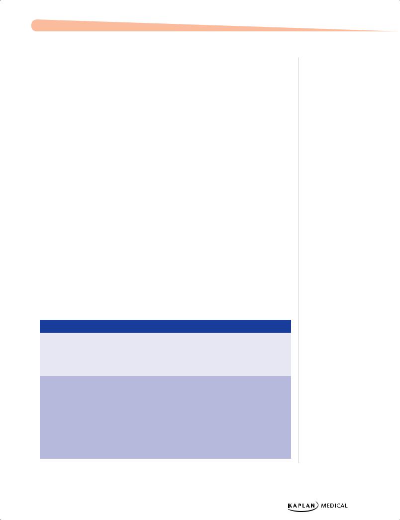
Table 1-4. Two Newborn Screening Diseases*
|
|
Phenylketonuria (PKU) |
|
|
Classic Galactosemia |
|
|
|
|
|
|
|
|
Defect |
Phenylalanine hydroxylase; |
Gal-1-P uridylyltransferase deficiency; |
||||
|
accumulation of PHE in body |
accumulation of gal-1-P with injury to |
||||
|
fluids and central nervous system |
kidney, liver, and brain |
||||
|
|
|
|
|
|
|
Presentation |
Intellectual disability vomiting, |
Jaundice (often direct), hepatomegaly, |
||||
|
growth retardation, purposeless |
vomiting, hypoglycemia, cataracts, |
||||
|
movements, athetosis, seizures |
seizures, poor feeding, poor weight |
||||
|
|
|
|
gain, intellectual disability |
||
|
|
|
|
|
|
|
Associations |
Fair hair, fair skin, blue eyes, |
Predisposition to E. coli sepsis; |
||||
|
tooth abnormalities, |
developmental delay, speech |
||||
|
microcephaly |
disorders, learning disabilities |
||||
|
|
|
|
|
|
|
Other |
Normal at birth; gradual MR over |
May begin prenatally—transplacental |
||||
comments |
first few months |
galactose from mother |
||||
|
|
|
|
|
|
|
Treatment |
Low PHE diet for life |
No lactose—reverses growth failure, |
||||
|
|
|
|
kidney and liver abnormalities and |
||
|
|
|
|
cataracts, but not |
||
|
|
|
|
neurodevelopmental problems |
||
|
|
|
|
|
|
|
G-1-P, galactose-1-phosphate; PHE, phenylalanine
*Items in bold have a greater likelihood of appearing on the exam.
4
Chapter 1 λ The Newborn
Hearing Loss
Pediatric hearing loss is more prevalent than diabetes mellitus and all childhood cancers. A universal newborn hearing screening is recommended prior to newborn discharge, with the goal of evaluating all hearing loss by age 3 months. Usually the otoacoustic emissions test (OAE) is used, where a small earphone/microphone is placed in the ear and sounds are played.
•If hearing is normal, an echo is reflected back into the ear canal and is measured by the microphone.
•If hearing is not normal (patient does not pass), newborns are given the auditory brainstem response test (ABR) (most accurate hearing measure through age 6 months). Sounds are presented through a small earphone, measured with head electrodes, and analyzed by a computer.
•Normal OAE: intact hearing through the cochlea
•Normal ABR: also establishes the integrity of the auditory nerve
As for the causes of hearing loss, up to 60% prelingual is genetic (>60 gene loci, >500 syndromes with hearing loss); 70-80% is autosomal recessive, with 50% having a defect in connexin 26 (a gap junction protein). Examples include Waardenburg syndrome (most common autosomal dominant condition with hearing loss), neurofibromatosis-2 (AD), Alport syndrome (AR).
Up to 25% are nongenetic and up to 25% are idiopathic. Examples include CMV (most common congenital cause; then other congenital infections); otitis media with effusion (OME) (most common childhood cause); bacterial meningitis, especially pneumococcus (occurs early and in >30%); trauma, especially to temporal bone; medication (aminoglycosides, loop diuretics, cisplatin); acoustic (loud music, especially with earbuds/phones; audiograms show high-frequency loss at 4,000 Hz).
FETAL GROWTH AND MATURITY
Table 1-5. Intrauterine Growth Restriction (IUGR)
|
Type |
|
|
Reason |
|
|
Main Etiologies |
|
|
Complications |
|
|
|
|
|
|
|
|
|
|
|
|
|
|
Symmetric |
|
Early, in utero |
|
Genetic syndromes, |
|
Etiology dependent; |
||||
|
|
|
|
insult that |
|
chromosomal |
|
delivery of oxygen and |
|||
|
|
|
|
affects growth |
|
abnormalities, congenital |
|
nutrients to vital organs |
|||
|
|
|
|
of most organs |
|
infections, teratogens, |
|
usually normal |
|||
|
|
|
|
|
|
|
toxins |
|
|
|
|
|
|
|
|
|
|
|
|
|
|
|
|
|
Asymmetric |
|
Relatively late |
Uteroplacental |
|
Neurologic (asphyxia) if |
|||||
|
(head sparing) |
|
onset after fetal |
insufficiency secondary to |
|
significant decreased |
|||||
|
|
|
|
organ |
maternal diseases |
|
delivery of oxygen to |
||||
|
|
|
|
development; |
(malnutrition, cardiac, |
|
brain |
||||
|
|
|
|
abnormal |
renal, anemia) and/or |
|
|
|
|||
|
|
|
|
delivery of |
placental dysfunction |
|
|
|
|||
|
|
|
|
nutritional |
(hypertension, |
|
|
|
|||
|
|
|
|
substances and |
autoimmune disease, |
|
|
|
|||
|
|
|
|
oxygen to the |
abruption) |
|
|
|
|||
|
|
|
|
fetus |
|
|
|
|
|
|
|
|
|
|
|
|
|
|
|
|
|
|
|
Published by dr-notes.com |
5 |
|
|
|
|

USMLE Step 2 CK λ Pediatrics
Gestational Age and Size at Birth
Preterm |
Large for Gestational Age |
Post-term |
|
|
(LGA)—Fetal Macrosomia |
|
|
|
|
|
|
• Premature—liveborn |
• Birth weight >4,500 |
• Infants born after 42 |
|
infants delivered prior to |
grams at term |
weeks’ gestation from last |
|
37 weeks as measured |
• Predisposing factors: |
menstrual period |
|
from the first day of the |
|
||
obesity, diabetes |
• When delivery is delayed |
||
last menstrual period |
|||
• Higher incidence of birth |
≥3 weeks past term, |
||
|
|||
• Low birth weight (<2,500 |
injuries and congenital |
significant increase in |
|
grams), possibly due to |
anomalies |
mortality |
|
prematurity, IUGR, or |
|
• Characteristics |
|
both |
|
–– Increased birth weight |
|
|
|
||
|
|
–– Absence of lanugo |
|
|
|
–– Decreased/absent |
|
|
|
vernix |
|
|
|
–– Desquamating, pale, |
|
|
|
loose skin |
|
|
|
–– Abundant hair, long |
|
|
|
nails |
|
|
|
–– If placental insufficiency, |
|
|
|
may be meconium |
|
|
|
staining |
|
|
|
|
ENDOCRINE DISORDERS
Infants of Diabetic Mothers
You are called to see a 9.5-pound newborn infant who is jittery. Physical exam reveals a large plethoric infant who is tremulous. A murmur is heard. Blood sugar is low.
•Maternal hyperglycemia (types I and II DM) → fetal hyperinsulinemia
•Insulin is the major fetal growth hormone → increase in size of all organs except the brain
•Major metabolic effect is at birth with sudden placental separation → hypoglycemia
•Infants may be large for gestational age and plethoric (ruddy).
•Other metabolic findings: hypocalcemia and hypomagnesemia (felt to be a result of delayed action of parathyroid hormone)
•Common findings
–Birth trauma (macrosomia)
–Tachypnea (transient tachypnea, respiratory distress syndrome, cardiac failure, hypoglycemia)
–Cardiomegaly—asymmetric septal hypertrophy (insulin effect, reversible)
–Polycythemia (and hyperviscosity) → hyperbilirubinemia → jaundice
6
Chapter 1 λ The Newborn
–Renal vein thrombosis (flank mass, hematuria, thrombocytopenia) from polycythemia
–Increased incidence of congenital anomalies
°Cardiac—especially VSD, ASD, transposition
°Small left colon syndrome (transient delay in development of left side of colon; presents with abdominal distention)
°Caudal regression syndrome: spectrum of structural neurologic defects of the caudal region of spinal cord which may result in neurologic impairment (hypo, aplasia of pelvis & LE)
•Prognosis—Infants of diabetic mothers are more predisposed to diabetes and LGA infants are at increased risk of childhood obesity.
•Treatment—careful monitoring and glucose control during pregnancy + close monitoring of infant after delivery; early frequent feeds (oral, NG if hypoglycemia continues) followed by IV dextrose if euglycemia has not resulted
Clinical Recall
Which of the following is commonly seen in infants of diabetic mothers?
A.Microsomia
B.Small heart size
C.Polycythemia
D.Renal artery thrombosis
E.Slow respiratory rate
Answer: C
RESPIRATORY DISORDERS
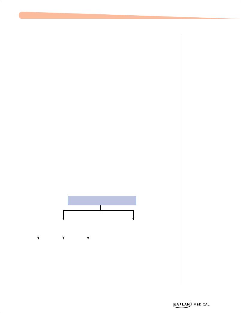
Respiratory Distress
|
|
|
Respiratory |
|
|
|
|
|
Nonrespiratory |
|
|
|
|
|
|
|
|
|
|
|
|
|
|
|
|
|
|
|
|
|
|
|
Cardiac: cyanotic CHD |
||
|
|
|
|
|
|
|
|
|
|||
|
|
|
|
|
|
|
|
|
|||
Respiratory |
|
Transient |
|
Meconium |
Heme: anemia, polycythemia |
||||||
distress |
|
tachypnea |
|
aspiration |
Other: infectious, metabolic, neurologic |
||||||
syndrome |
|
of the |
|
syndrome |
|
|
|
||||
|
|
|
newborn |
|
|
|
|
|
|
|
|
|
|
|
|
|
|
|
|
|
|
||
Pneumonia
Diaphragmatic hernia
Choanal atresia
Figure 1-1. Respiratory Distress
Published by dr-notes.com |
7 |
|
|
|
|

USMLE Step 2 CK λ Pediatrics
Respiratory Distress Syndrome (RDS)
Shortly after birth, a 33-week gestation infant develops tachypnea, nasal flaring, and grunting and requires intubation. Chest radiograph shows a hazy, ground-glass appearance of the lungs.
•Deficiency of mature surfactant (surfactant matures over gestation with the addition of phosphatidyl groups; therefore, the incidence of surfactant deficiency diminishes toward term.)
•Inability to maintain alveolar volume at end expiration → decreased functional residual capacity and atelectasis
•Primary initial pulmonary hallmark is hypoxemia. Then, hypercarbia and respiratory acidosis ensue.
•Diagnosis
–– Best initial diagnostic test—chest radiograph
°° Findings: ground-glass appearance, low lung volume, air bronchograms
–– Most accurate diagnostic test—L/S ratio (part of complete lung profile; lecithin-to- sphingomyelin ratio)
°° Done on amniotic fluid prior to birth
•Best initial treatment—oxygen
•Most effective treatment—intubation and exogenous surfactant administration
•Primary prevention
––Avoid prematurity (tocolytics)
–– Antenatal betamethasone
Transient Tachypnea of the Newborn (TTN)
•Slow absorption of fetal lung fluid → decreased pulmonary compliance and tidal volume with increased dead space
•Tachypnea after birth
•Generally minimal oxygen requirement
•Common in term infant delivered by Cesarean section or rapid second stage of labor
•Chest x-ray (best test)—air-trapping, fluid in fissures, perihilar streaking
•Rapid improvement generally within hours to a few days
Meconium Aspiration
•Meconium passed as a result of hypoxia and fetal distress; may be aspirated in utero or with the first postnatal breath → airway obstruction and pneumonitis → failure and pulmonary hypertension
•Chest x-ray (best test)—patchy infiltrates, increased AP diameter, flattening of diaphragm
•Other complications—air leak (pneumothorax, pneumomediastinum)
8
Chapter 1 λ The Newborn
•Prevention—endotracheal intubation and airway suction of depressed infants with thick meconium
•Treatment—positive pressure ventilation and other complex NICU therapies
Diaphragmatic Hernia
•Failure of the diaphragm to close → abdominal contents enter into chest, causing pulmonary hypoplasia.
•Born with respiratory distress and scaphoid abdomen
•Bowel sounds may be heard in chest
•Diagnosis—prenatal ultrasound; postnatal x-ray (best test) reveals bowel in chest
•Best initial treatment—immediate intubation in delivery room for known or suspected CDH, followed by surgical correction when stable (usually days)
GASTROINTESTINAL AND HEPATOBILIARY DISORDERS
See also GI chapter on this topic.
Umbilical Hernia
•Failure of the umbilical ring closure, weakness of abdominal muscles
•Most are small and resolve in 1-2 years without any treatment
•Surgery if getting larger after 1-2 years, symptoms (strangulation, incarceration), and/or persistent after age 4
Omphalocele
•Failure of intestines to return to abdominal cavity with gut through umbilicus at 11 weeks’ gestation
•Covered in a sac (protection)
•Associated with other major malformations and possible genetic disorders (trisomy)
•Large defects need a staged reduction (use of a surgical Silo), otherwise respiratory failure and ischemia
Gastroschisis
•Defect in abdominal wall lateral to umbilicus (vascular accident; typically not associated with other malformations)
•Any part of the GI tract may protrude
•Not covered by a sac
•Major problem with the intestines: atresia, stenosis, ischemia, short gut
•Surgery based on condition of gut; if no ischemia, large lesions need a staged reduction as with omphalocele
Published by dr-notes.com |
9 |
|
|
|
|

USMLE Step 2 CK λ Pediatrics
Note
Work up for pathologic hyperbilirubinemia when:
•It appears on day 1 of life
•Bilirubin rises >5 mg/dL/day
•Bilirubin >13 mg/dL in term infant
•Direct bilirubin >2 mg/dL at any time
Necrotizing Enterocolitis (NEC)
•Transmural intestinal necrosis
•Greatest risk factor is prematurity; rare in term infants
•Prematurity + immature gut barrier + enteral feeds + possible microorganisms = NEC
•Symptoms usually related to introduction of feeds: bloody stools, apnea, lethargy, and abdominal distention once perforation has occurred
•Pneumatosis intestinalis on plain abdominal film is pathognomonic (air in bowel wall)
•Treatment: cessation of feeds, gut decompression, systemic antibiotics, and supportive care; surgical resection of necrotic bowel may be necessary; early surgical consult is imperative
Imperforate Anus
•Failure to pass stool after birth
•No anal opening visible
•Treatment is surgical correction.
•May be part of VACTERL association.
Jaundice
A 2-day-old infant is noticed to be jaundiced. He is nursing and stooling well. Indirect bilirubin is 11.2 mg/dL; direct is 0.4 mg/dL. Physical exam is unremarkable except for visible jaundice.
•Pathophysiology
––Increased production of bilirubin from breakdown of fetal red blood cells plus immaturity of hepatic conjugation of bilirubin and elimination in first week of life
––Rapidly increasing unconjugated (indirect reacting) bilirubin can cross the bloodbrain barrier and lead to kernicterus (unconjugated bilirubin in the basal ganglia and brain stem nuclei). Hypotonia, seizures, opisthotonos, delayed motor skills, choreoathetosis, and sensorineural hearing loss are features of kernicterus.
Table 1-6. Physiologic Jaundice Versus Pathologic Jaundice
|
Physiologic Jaundice |
|
|
Pathologic Jaundice |
|
|
|
|
|
|
|
|
Appears on second to third day of life (term) |
May appear in first 24 hours of life |
|||
|
|
|
|
|
|
|
Disappears by fifth day of life (term)—7th |
Variable |
|||
|
|
|
|
|
|
|
Peaks at second to third day of life |
Variable |
|||
|
|
|
|
|
|
|
Peak bilirubin <13 mg/dL (term) |
Unlimited |
|||
|
|
|
|
|
|
|
Rate of bilirubin rise <5 mg/dL/d |
Usually >5 mg/dL/d |
|||
|
|
|
|
|
|
10

Chapter 1 λ The Newborn
The causes of hyperbilirubinemia with respect to bilirubin metabolism are as follows:
•RBC metabolism
––Increased RBCs
°° Physiologic jaundice (healthy newborn [normal Hct 42−65]) °° Polycythemia (Hct >65)
i.Increased RBC production: Chronic hypoxia, IUGR, post-mature; IODM, Beckwith-Wiedemann syndrome (insulin effect); trisomies (unknown mechanism)
ii.Extra RBCs entering the circulation: delayed cord clamping, twin-twin transfusion
iii.Treatment: partial exchange transfusion with normal saline (dilutional)
––Increased hemolysis
°° Immune-mediated (labs: high unconjugated bilirubin, may be anemia, increased reticulocyte count, positive direct Coombs test)
i.Rh negative mother/Rh positive baby: classic hemolytic disease of the newborn (erythroblastosis fetalis)
ii.ABO incompatibility (almost all are type O mother and either type A or B baby): most common reason for hemolysis in the newborn
iii.Minor blood group incompatibility (Kell is very antigenic; Kell negative mother), uncommon
°° Non-immune mediated: same as above but Coombs is negative; need to see blood smear
i.Smear shows characteristic-looking RBCs: membrane defect (most are either spherocytosis or elliptocytosis)
ii.Smear shows normal-looking RBCs: enzyme defect (most are G6PD deficiency then pyruvate kinase deficiency)
iii.Extravascular: excessive bruising, cephalohematoma
•Bilirubin is then bound to albumin and carried in the blood; bilirubin may be uncoupled from albumin in the bloodstream to yield free bilirubin, e.g. neonatal sepsis, certain drugs (ceftriaxone), hypoxia, acidosis.
•Bilirubin is transported to the hepatocytes: within the hepatocytes is the conversion of unconjugated (laboratory indirect-acting) fat-soluble bilirubin to conjugated (glucuronide) water-soluble bilirubin (laboratory direct-acting) by the action of hepatic glucuronyl transferase (GT).
––Decreased enzymatic activity of GT
°° Normal newborn first week of life
°° Primary liver disease of systemic disease affecting the liver (sepsis, TORCH, metabolic diseases)
°° No GT activity: Crigler-Najjar syndrome (type I)
Published by dr-notes.com |
11 |
|
|
|
|

USMLE Step 2 CK λ Pediatrics
•Transport through the intrahepatic biliary system to the porta hepatis for excretion into the duodenum; abnormalities of transport and excretion cause a conjugated (direct) hyperbilirubinemia (>2 mg/dL direct-acting bilirubin in the blood in the newborn).
–– Biliary atresia (progressive obliterative cholangiopathy): obstruction at birth due to fibrosis and atresia of the extrahepatic ducts (and so no gall bladder); then variable severity and speed of inflammation and fibrosis of the intrahepatic system which ultimately leads to cirrhosis
°°
°°
Most present in first 2 weeks of life with jaundice (conjugated hyperbilirubinemia), poor feeding, vomiting, lethargy, hepatosplenomegaly, persistent acholic stools and dark urine
Best initial test: U/S (triangular fibrotic cord at porta hepatis; no evidence of normal ductal anatomy; no gallbladder)
°° Most accurate test (next step): percutaneous liver biopsy (is pathognomonic for this process)
°° Best initial treatment (palliative): hepatic portojejunostomy (Kasai procedure)
°° Best long-term management: liver transplant
––Liver disease (primary or secondary to systemic disease): cholestasis (sepsis, perinatal infections, metabolic disease, neonatal hepatitis, severe hypothyroidism and others)
•Intestinal transport and excretion: most bilirubin is eliminated in the stool with final products synthesized with help of colonic bacteria; some bilirubin is eliminated in the urine, some is reprocessed in the liver due to enterohepatic circulation (along with bile acids); intestinal beta-glucuronidase hydrolyzes glucuronide-bilirubin bonds to yield some unconjugated bilirubin, which is absorbed into the portal circulation and transported back to the liver to be acted upon by hepatic glucuronyl transferase
––Increased enterohepatic circulation
°° Intestinal obstruction
°° Decreased colonic bacteria (first week of life, prolonged antibiotics, severe diarrhea)
°° Breastfeeding jaundice (due to decreased intestinal peristalsis)
°° Breast-milk jaundice (due to excessive concentration of glucuronidase in breast milk)
Clinical Recall
Which of the following is not a cause of hyperbilirubinemia?
A.Increased red blood cell production
B.ABO incompatibility
C.Biliary atresia
D.Increased activity of hepatic glucuronyl transferase
E.Decreased enterohepatic circulation
Answer: D
12
Chapter 1 λ The Newborn
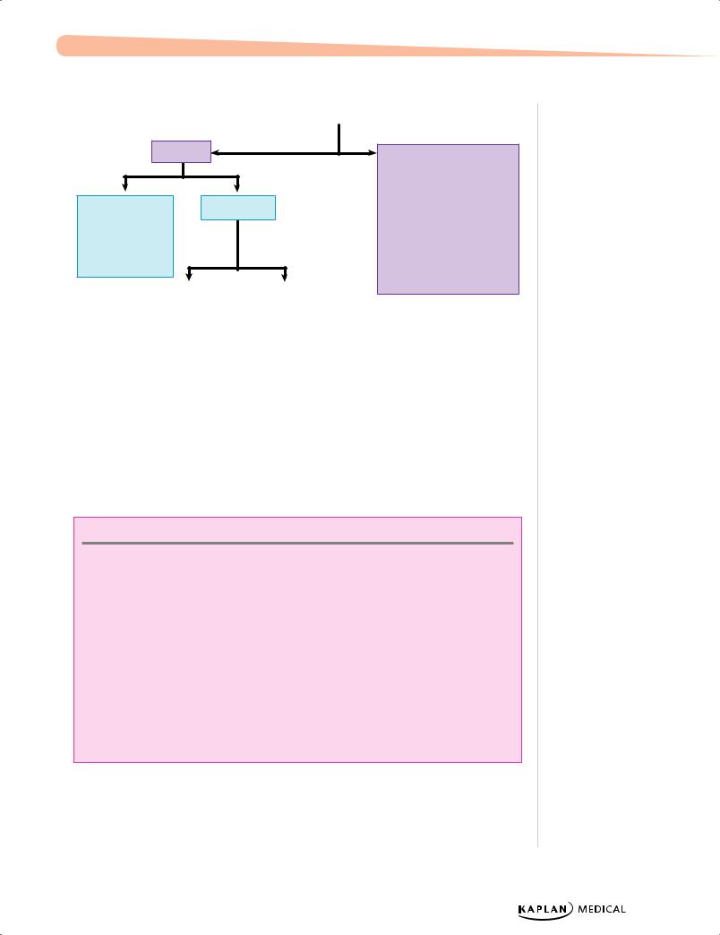
Physiologic |
|
Pathologic |
|
|
|
Indirect |
Coombs (+) |
Coombs (−) |
•Rh/ABO incompatibility
•Minor blood groups
High Hgb |
|
Normal/low Hgb |
|
• Polycythemia |
|
• |
Spherocytosis |
– Twin-twin transfusion |
|
• |
Elliptocytosis |
– Maternal-fetal transfusion |
|
• |
G6PD deficiency |
– Delayed cord |
|
• |
Pyruvate kinase |
– IUGR |
|
• |
Hemorrhage |
– Infant of diabetic mother |
|
• |
Cephalohematoma |
|
|
|
bruising |
|
|
|
|
|
|
• |
Bowel obstruction |
|
|
• |
Breastfeeding |
|
|
• |
Crigler-Najjar |
|
|
• |
Gilbert syndrome |
|
|
|
|
Figure 1-2. Jaundice Workup
Direct
•Sepsis
•TORCH infections
•Hypothyroidism
•Galactosemia
•Cystic fibrosis
•Choledochal cyst
•Biliary atresia
•Dubin-Johnson
•Rotor syndrome
Breastfeeding Jaundice Versus Breast-Milk Jaundice
Breastfeeding jaundice means a baby is not getting enough calories through nursing. It occurs in the first few days of life and is common in first-time breastfeeding mothers. While the infant may become dehydrated, lack of calories is what causes the jaundice. In the absence of food in the
intestines, peristalsis decreases significantly, allowing much more time for intestinal beta-glucuronidase to hydrolyze the glucuronide bonds and produce more unconjugated (indirect-acting) bilirubin. This enters the portal circulation and is transported back to the liver for further action by hepatic glucuronyl transferase. The result is increased unconjugated bilirubin and increased enterohepatic circulation. Treatment is to obtain a lactation consultation and rehydrate the baby.
Breast-milk jaundice (week 2 of life) is caused by increased glucuronidase in breast milk. More glucuronides are hydrolyzed in the intestine, producing more indirect bilirubin and again increasing enterohepatic circulation. The problem is temporary, because the activity of this enzyme decreases steadily over the first 2–3 months of life and then ceases. Treatment is phototherapy if needed. Bilirubin may rise again, but not to the previous level. The baby may then be safely breastfed.
Published by dr-notes.com |
13 |
|
|
|
|