Полезные материалы за все 6 курсов / Учебники, методички, pdf / Kaplan Pediatrics USMLE 2CK 2021
.pdf
USMLE Step 2 CK λ Pediatrics
Note
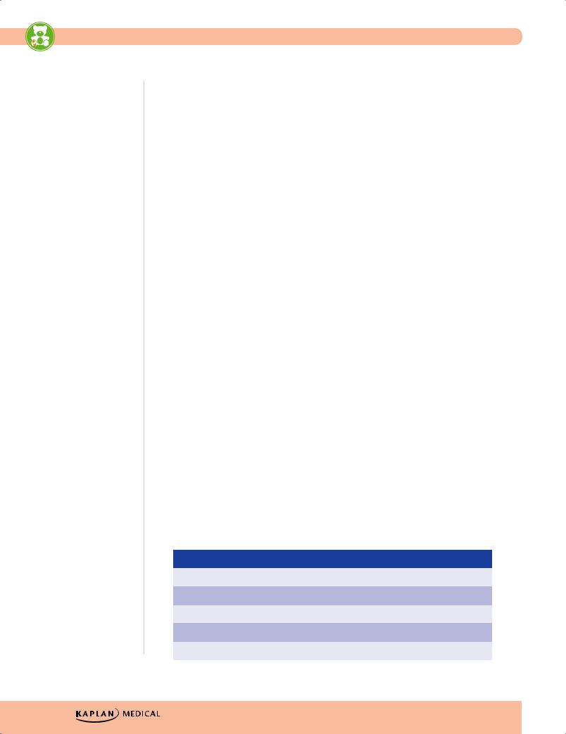
If arthritis is present, arthralgia cannot be used as a minor criterion.
The presence of Sydenham chorea alone is sufficient for diagnosis.
•Prophylaxis (AHA, 2007) for:
−Artificial valves
°Previous history of infective endocarditis
°Unrepaired or incompletely repaired cyanotic disease, including those with palliative shunts and conduits
°A completely repaired defect with prosthetic material or device for first 6 months
°Any residual defect at site of any repair
°Cardiac transplant which develops a problem in a valve
°Given ONLY for dental procedures with manipulation of gingival tissue or periapical area or perforation of oral mucosa; incision or biopsy of respiratory tract mucosa and surgery on infected skin or musculoskeletal structures
°Drug of choice is amoxicillin
Acute Rheumatic Fever
A 6-year-old girl complains of severe joint pain in her elbows and wrists. She has had fever for the past 4 days. Past history reveals a sore throat 1 month ago. Physical examination is remarkable for swollen, painful joints and a heart murmur. Laboratory tests show an elevated erythrocyte sedimentation rate and high antistreptolysin (ASO) titers.
•Etiology/epidemiology
−Related to group A Streptococcus infection within several weeks
−Antibiotics that eliminate Streptococcus from pharynx prevent initial episode of acute rheumatic fever
−Remains most common form of acquired heart disease worldwide (but Kawasaki in United States and Japan)
−Initial attacks and recurrences with peak incidence Streptococcus pharyngitis: age 5–15
−Immune-mediated—antigens shared between certain strep components and mammalian tissues (heart, brain, joint)
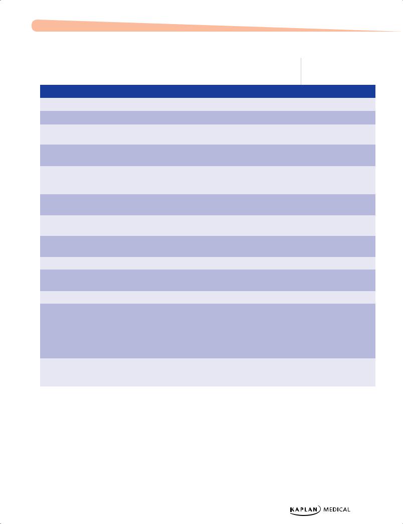
•Clinical presentation and diagnosis—Jones criteria. Absolute requirement: evidence of recent Streptococcus infection (microbiological or serology); then 2 major or 1 major and 2 minor criteria
Table 13-4. Jones Criteria
|
Major Criteria |
|
|
Minor Criteria |
|
|
|
|
|
|
|
Carditis |
|
Fever |
|||
|
|
|
|
|
|
Polyarthritis (migratory) |
|
Arthralgia |
|||
|
|
|
|
|
|
Erythema marginatum |
|
Elevated acute phase reactants (ESR, CRP) |
|||
|
|
|
|
|
|
Chorea |
|
Prolonged PR interval on EKG |
|||
|
|
|
|
|
|
Subcutaneous nodules |
|
Plus evidence of preceding streptococcal infection |
|||
|
|
|
|
|
|
134

Chapter 13 λ Cardiology
•Treatment
−Bed rest and monitor closely
−Oral penicillin or erythromycin (if allergic) for 10 days will eradicate group A strep; then need long-term prophylaxis
−Anti-inflammatory
°Hold if arthritis is only typical manifestation (may interfere with characteristic migratory progression)
°Aspirin in patients with arthritis/carditis without CHF
°If carditis with CHF, prednisone for 2–3 weeks, then taper; start aspirin for 6 weeks
−Digoxin, salt restriction, diuretics as needed
−If chorea is only isolated finding, do not need aspirin; drug of choice is phenobarbital (then haloperidol or chlorpromazine)
•Complications
−Most have no residual heart disease.
−Valvular disease most important complication (mitral, aortic, tricuspid)
•Prevention
−Continuous antibiotic prophylaxis
°If carditis—continue into adulthood, perhaps for life; without carditis—lower risk; can discontinue after patient is in their twenties and at least 5 years since last episode
°Treatment of choice—single intramuscular benzathine penicillin G every 4 weeks
°If compliant—penicillin V PO BID or sulfadiazine PO QD; if allergic to both: erythromycin PO BID
Hypertrophic Obstructive Cardiomyopathy (HOCM)
•Pathophysiology
−Obstructive left-sided congenital heart disease
•Decreased compliance, so increased resistance and decreased left ventricular filling, mitral insufficiency
•Clinical presentation—weakness, fatigue, dyspnea on exertion, palpitations, angina, dizziness, syncope; risk of sudden death
•Cardiovascular examination—left ventricular lift, no systolic ejection click (differentiates from aortic stenosis), SEM at left sternal edge and apex (increased after exercise, during Valsalva, and standing)
•Diagnosis
−EKG—left ventricular hypertrophy ± ST depression and T-wave inversion; may have intracardiac conduction defect
−Chest x-ray—mild cardiomegaly (prominent LV)
−Echocardiogram—left ventricular hypertrophy, mostly septal; Doppler—left ventricular outflow gradient usually mid-to-late systole (maximal muscular outflow obstruction)
•Treatment
−No competitive sports or strenuous exercise (sudden death)
−Digoxin and aggressive diuresis are contraindicated (and infusions of other inotropes)
−Beta blockers (propranolol) and calcium channel blockers (verapamil)
Note
Suspect hypertrophic cardiopathy in an athlete with sudden death.
Published by dr-notes.com |
135 |
|
|
|
|

USMLE Step 2 CK λ Pediatrics
Note
When a child presents with hypertension, think of renal causes.
Clinical Recall
A 16-year-old girl seen in clinic last month for strep throat returns with a few weeks of knee pain that is resolving and 2 days of worsening elbow pain despite no recent trauma. In addition, she has noticed several small ring-like rashes on her arms and abdomen that come and go. What additional finding is needed to diagnose acute rheumatic fever?
A.Cardiac inflammation
B.EKG showing PR interval prolongation
C.Chorea
D.No additional findings are needed
E.Elevated ESR and CRP
Answer: D
HYPERTENSION
A 5-year-old girl is noted to have blood pressure >95th percentile on routine physical examination. The rest of the examination is unremarkable. Her blood pressure remains elevated on repeat measurement over the next few weeks. Past history is remarkable for a treated urinary tract infection 1 year ago. Complete blood cell count is normal; urinalysis is normal. Blood urea nitrogen is 24 mg/dL and creatinine is 1.8 mg/dL.
•Routine blood pressure check beginning at 3 years of age
−If increased blood pressure, check all 4 extremities (coarctation)
−Normal—blood pressure in legs should be 10–20 mm Hg higher than in arms
−If obese, on medications which increase BP, diabetes, or chronic kidney disease, check blood pressure
•Blood pressure increases with age—need standard nomograms
−If mild hypertension, repeat twice over next 6 weeks
−If consistently >95% for age, need further evaluation
−≥95th percentile at 3 different visits
•Etiology—essential (primary) or secondary
−Secondary—most common in infants and younger children
°Newborn—umbilical artery catheters → renal artery thrombosis
°Early childhood—renal disease, coarctation, endocrine, medications
°Adolescent—essential hypertension
136

Chapter 13 λ Cardiology
−Renal and renovascular hypertension—majority of causes may be due to urinary tract infection (secondary to an obstructive lesion), acute glomerulonephritis,
Henoch-Schönlein purpura with nephritis, hemolytic uremic syndrome, acute tubular necrosis, renal trauma, leukemic infiltrates, mass lesions, renal artery stenosis
−Essential hypertension—more common in adults and adolescents
°Positive family history
°Multifactorial—obesity, genetic, and physiologic changes
•Diagnosis
−CBC, blood chemistries, UA, EKG, echo, renal ultrasound, angiogram (less common)
•Treatment
−If obese—weight control, aerobic exercise, no-added-salt diet, monitor blood pressure
−Pharmacologic treatment (secondary hypertension and selective primary)—similar use of drugs as in adults
−No real workup age ≥6 years and family history, obese, with normal history and physical
−DASH diet (Dietary Approaches to Stop Hypertension)
Published by dr-notes.com |
137 |
|
|
|
|


Gastrointestinal Disease 14
Chapter Title
Learning Objective
Diagnose and describe treatments for children who present with gastroenteritis, vomiting, hematochezia, or constipation
GASTROENTERITIS
Acute Diarrhea
A 13-month-old child has had a 3-day history of green watery stools. She has also been vomiting for 1 day. Physical examination reveals a febrile, irritable baby with dry mucous membranes and sunken eyes.
•Etiology
Table 14-1. Causes of Diarrhea (Acute and Chronic)
|
|
Infant |
|
|
Child |
|
|
Adolescent |
|
|||
|
|
|
|
|
|
|
|
|
|
|
|
|
Acute |
|
• |
Gastroenteritis |
• |
Gastroenteritis/ |
• |
Gastroenteritis/ |
|||||
|
|
• |
Systemic infection |
|
|
Food poisoning |
|
|
food poisoning |
|||
|
|
|
|
|
|
|
|
|
|
|||
|
|
• |
Antibiotic |
• |
Systemic infection |
• |
Systemic infection |
|||||
|
|
|
|
|
|
|
|
|
|
|||
|
|
|
|
|
|
|
|
|
|
|
|
|
Chronic |
• |
Postinfectious |
• |
Postinfectious |
• |
Irritable bowel |
||||||
|
|
|
lactase deficiency |
|
|
lactase deficiency |
|
|
syndrome |
|||
|
• |
Milk/soy intolerance |
• |
Irritable bowel |
• |
Inflammatory |
||||||
|
• |
Chronic diarrhea of |
|
|
syndrome |
|
|
bowel disease |
||||
|
|
|
|
|
|
|
|
|
||||
|
|
|
infancy |
• |
Celiac disease |
• |
Lactose |
|||||
|
• |
Celiac disease |
• |
Lactose intolerance |
|
|
intolerance |
|||||
|
|
|
|
|
||||||||
|
• |
Cystic fibrosis |
• |
Giardiasis |
• |
Giardiasis |
||||||
|
|
|
|
|
||||||||
|
|
|
|
|
• |
Inflammatory bowel |
• |
Laxative abuse |
||||
|
|
|
|
|
|
|
|
|
||||
|
|
|
|
|
|
|
disease |
|
|
|
|
|
|
|
|
|
|
|
|
|
|
|
|
|
|
Note
Common Causes of
Bloody Diarrhea
•Campylobacter
•Amoeba (E. histolytica)
•Shigella
•E. coli
•Salmonella
Published by dr-notes.com |
139 |
|
|
|
|

USMLE Step 2 CK λ Pediatrics
•Common organisms
Table 14-2. Common Causes of Acute Diarrhea
|
Bacterial (Inflammatory) |
|
|
Viral |
|
|
Parasitic |
|
|
|
|
|
|
|
|
|
|
|
Campylobacter |
Norovirus |
Giardia lamblia (most common) |
|||||
|
Enteroinvasive E. coli |
Rotavirus |
E. histolytica |
|||||
|
Salmonella |
Enteric adenovirus |
Strongyloides |
|||||
|
Shigella |
Astrovirus |
Balantidium coli |
|||||
|
Yersinia |
Calicivirus |
Cryptosporidium parvum |
|||||
|
Clostridium difficile |
|
|
|
Trichuris trichiura |
|||
|
E. coli 0157:H7 |
|
|
|
|
|
|
|
|
|
|
|
|
|
|
|
|
Note
Antidiarrheal compounds should never be used in children.
•Major transmission is fecal/oral or by ingestion of contaminated food or water
•Clinical presentation
−Diarrhea, vomiting, abdominal cramps, nausea, fever (suggests inflammation and dehydration)
−Can present from an extraintestinal infection, e.g., urinary tract infection, pneumonia, hepatitis
•Management
−Assess hydration and provide fluid and electrolyte replacement
−Prevent spread
−In some cases, determine etiology and provide specific therapy (some are not treated)
−Think about daycare attendance, recent travel, use of antibiotics, exposures, intake of seafood, unwashed vegetables, unpasteurized milk, contaminated water, uncooked meats to isolate differential diagnosis of organisms
•Labs: stool examination (cost-effective, noninvasive)
−Mucus, blood, leukocytes → colitis (invasive or cytotoxic organism)
−Stool cultures—with blood, leukocytes, suspected hemolytic uremic syndrome, immunosuppressed, in outbreaks
−Clostridium difficile toxin—if recent history of antibiotics
−Ova and parasites
−Enzyme immunoassays for viruses or PCR (rarely need to be diagnosed)
140

Chapter 14 λ Gastrointestinal Disease
Chronic Diarrhea |
|
|
|
|
|
|
||
Table 14-3. Organism-Specific Associations and Therapy |
|
|
|
|||||
|
|
|
|
|
|
|
|
|
|
Organism |
|
|
Association |
|
|
Therapy |
|
|
|
|
|
|
|
|
|
|
|
Rotavirus |
|
Watery diarrhea, vomiting, ± fever |
|
Supportive |
|||
|
|
|
|
|
|
|
|
|
|
Enteropathogenic E. coli |
|
Nurseries, daycare |
|
Mostly supportive care |
|||
|
|
|
|
|
|
|
|
|
|
Enterotoxigenic E. coli |
|
Traveler’s diarrhea |
|
Supportive care with trimethoprim sulfamethoxazole in |
|||
|
|
|
|
|
|
|
severe cases |
|
|
|
|
|
|
|
|
|
|
|
Enterohemorrhagic E. coli |
|
Hemorrhagic colitis, HUS |
|
No antibiotic therapy due to ↑ risk of HUS; supportive |
|||
|
|
|
|
|
|
|
care only |
|
|
|
|
|
|
|
|
|
|
|
Salmonella |
|
Infected animals and |
|
Antibiotics indicated only for patients who are |
|||
|
|
|
|
contaminated eggs, milk, poultry |
|
≤3 months of age, toxic, has disseminated disease, or |
||
|
|
|
|
|
|
|
S. typhi |
|
|
|
|
|
|
|
|
|
|
|
Shigella |
|
Person-to-person spread, |
|
Trimethoprim/sulfamethoxazole |
|||
|
|
|
|
contaminated food |
|
|
|
|
|
|
|
|
|
|
|
|
|
|
Campylobacter |
|
Person-to-person spread, |
|
Self-limiting; erythromycin for severe disease |
|||
|
|
|
|
contaminated food |
|
|
|
|
|
|
|
|
|
|
|
|
|
|
Yersinia enterocolitica |
|
Pets, contaminated food |
|
No antibiotics except for infants ≤3 months of age or |
|||
|
|
|
|
|
|
|
culture-proven septicemia |
|
|
|
|
|
|
|
|
|
|
|
Clostridium difficile |
|
History of antibiotic use |
|
Metronidazole or vancomycin |
|||
|
|
|
|
|
|
|
|
|
|
Staphylococcus aureus |
|
Food poisoning (onset within |
|
Supportive care |
|||
|
|
|
|
12 h of ingestion) |
|
|
|
|
|
|
|
|
|
|
|
|
|
|
Entamoeba histolytica |
|
Acute bloody diarrhea |
|
Metronidazole |
|||
|
|
|
|
|
|
|
|
|
|
Giardia |
|
Chronic or intermittent watery |
|
Tinidazole is the FDA-recommended therapy (single |
|||
|
|
|
|
diarrhea, abdominal distension, |
|
dose; has replaced furazolidone) |
||
|
|
|
|
nausea, weight loss, intermittent |
|
|
|
|
|
|
|
|
crampy abdominal pain |
|
|
|
|
|
|
|
|
Contaminated food or water or |
|
|
|
|
|
|
|
|
from infected person |
|
|
|
|
|
|
|
|
|
|
|
|
|
|
Cryptosporidium |
|
Mild diarrhea in |
|
Best treatment is raising CD4 count to normal level + |
|||
|
|
|
|
immunocompromised infants; |
|
supportive care |
||
|
|
|
|
severe diarrhea in AIDS patients |
|
|
|
|
|
|
|
|
|
|
|
|
|
Definition of abbreviations: HUS, hemolytic uremic syndrome
Published by dr-notes.com |
141 |
|
|
|
|

USMLE Step 2 CK λ Pediatrics
Note
Schwachman-Diamond
Syndrome
–Pancreatic insufficiency
–Neutropenia
–Malabsorption
Intestinal lymphangiectasia
–Lymph fluid leaks into bowel lumen
–Steatorrhea
–Protein-losing enteropathy
Disaccharidase Deficiency
–Osmotic diarrhea
–Acidic stools
Abetalipoproteinemia
–Severe fat malabsorption from birth
–Acanthocytes
–Very low to absent plasma cholesterol, triglycerides, etc.
Chronic Diarrhea and Malabsorption
•Patterns
−From birth
−After introduction of a new food
•Clinical presentation
−Chronic nonspecific diarrhea of infancy:
°Weight, height, and nutritional status is normal, and no fat in stool
°Excessive intake of fruit juice, carbonated fluids, low fat intake usually present in history
−Diarrhea with carbohydrates—CHO malabsorption
−Weight loss and stool with high fat—think malabsorption
−Other signs and symptoms suggest other specific diagnosis; see side note
•Workup of chronic diarrhea (simple, noninvasive testing to be done first)
−History and physical, nutritional assessment; stool for pH, reducing substances, fat, blood, leukocytes, culture, C. difficile toxin, ova, and parasites
−Blood studies—complete blood count and differential, ESR, electrolytes, glucose, BUN, and creatinine
−Sweat test, 72-hour fecal fat, breath hydrogen tests
•Initial evaluation
−Fat:
°Most useful screening test is stool for fat (Sudan red stain)
°Confirm with 72-hour stool for fecal fat (gold standard for steatorrhea)
°Steatorrhea is most prominent with pancreatic insufficiency; all require a sweat chloride
°Serum trypsinogen is also a good screen (reflects residual pancreatic function; increased level at birth in CF)
−CHO malabsorption—screen with reducing substances in stool (Clinitest)
°Breath hydrogen test—after a known CHO load, the collected breath hydrogen is analyzed and malabsorption of the specific CHO is identified
−Protein loss—cannot be evaluated directly (large proportion of bacterial protein and dietary protein almost completely absorbed before terminal ileum; amino acids and peptides are reabsorbed)
°Screen—spot stool α1-antitrypsin level
•More common differential diagnosis of malabsorption
−Giardiasis—only common primary infection causing chronic malabsorption; stool test for Giardia antigen duodenal aspirate and biopsy (best test)
−HIV or congenital T- or B-cell defects
−Small-bowel disease—gluten enteropathy, abetalipoproteinemia, lymphangiectasia
−Pancreatic insufficiency—fat malabsorption (cystic fibrosis is most common congenital disorder associated with malabsorption)
−Most common anomaly causing incomplete bowel obstruction with malabsorption is malrotation
−Short bowel—congenital or postnatal loss of >50% of small bowel with or without a portion of the large intestine (presence of ileocecal valve is better)
142

Chapter 14 λ Gastrointestinal Disease
−Celiac disease—associated with exposure to gluten (mostly rye, wheat, barley)
°Patients—mostly 6 months to 2 years; permanent intolerance; genetic predisposition (HLA DQ2)
°Clinical presentation—diarrhea, failure to thrive, growth restriction, vomiting, anorexia, ataxia
°Evaluation—best initial test is blood test for anti-tissue-transglutaminase (IgA) + serum IgA (false if IgA is also deficient); definitive test is small bowel biopsy
°Treatment—lifelong, strict gluten-free diet
Clinical Recall
A 14-year-old boy presents with watery diarrhea and nausea after a hiking trip during which he swam in a small freshwater lake. What is the treatment of choice?
A.Supportive care with rest and fluids
B.Trimethoprim/sulfamethoxazole
C.Metronidazole
D.Neomycin
E.Cefuroxime
Answer: C
VOMITING
Esophageal Atresia (EA) and Tracheoesophageal Fistula (TEF)
•Three basic types:
–Isolated EA
–Isolated (H-type) TEF
–EA and distal TEF
•Most common anatomy is upper esophagus ends in blind pouch and TEF connected to distal esophagus
•H-type—presents chronically and diagnosed later in life with chronic respiratory problems
•Half with associated anomalies—VACTERL association
•Clinical presentation in neonate (EA or EA + TEF)
− At birth—history of polyhydramnios; frothing, bubbling through nose and mouth; suctioning copious amount of fluid; respiratory distress, cyanosis from airway obstruction, amniotic fluid aspiration
− With feedings → immediate regurgitation and aspiration
•Clinical presentation with just TEF—feeding problems and recurrent aspiration
Note
VACTERL Association
Nonrandom association of birth defects:
Vertebral anomalies
Anal atresia
Cardiac defect
TracheoEsophageal fistula
Renal anomalies
Limb abnormalities
Published by dr-notes.com |
143 |
|
|
|
|
