

Полезные материалы за все 6 курсов / Учебники, методички, pdf / Kaplan Pediatrics USMLE 2CK 2021
.pdfUSMLE Step 2 CK λ Pediatrics
Note
The rest of the isolated T-cell defects are extremely rare, known only to immunologists. They are not seen on the exam.
•Diagnosis: very low-to-absent serum IgA with other IGs normal; as with CVID, incidence of autoantibodies, autoimmune disease and malignancy increased; serum antibodies to IgA can cause severe anaphylactic reactions if any blood product with IgA is administered (NOT a transfusion reaction)
•Treatment: IVIG is not indicated (95−99% is IgG) because if usual IVIG (containing IgA) product is given, patients are at risk for severe reaction. Additionally, because it is specifically an IgA deficiency, the IVIG product with the IgA removed cannot be used. Treat the infections (generally milder).
Defects of Cellular Immunity (T-cell Defects)
DiGeorge syndrome (thymic hypoplasia)
DiGeorge syndrome is thymic and parathyroid hypoplasia to aplasia from dysmorphogenesis of the 3rd and 4th pharyngeal pouches. Other structures are also involved: great vessel anomalies (right-sided aortic arch, interrupted aortic arch), esophageal atresia, bifid uvula, congenital heart disease (conotruncal malformations, septal defects), facial dysmorphism (short philtrum, thin upper lip, hypertelorism, mandibular hypoplasia, low-set, often notched ears), and cleft palate.
•Genetics: microdeletions of 22q11.2 (DiGeorge syndrome chromosomal region, DGCR); 22q deletions also seen in velocardiofacial syndrome and conotruncal anomaly face syndrome (CATCH 22 syndromes: Cardiac, Abnormal facies, Thymic hypoplasia, Cleft palate, Hypocalcemia); partial DiGeorge is more common, with variable thymic and parathyroid hypoplasia. About 1/3 with complete DiGeorge have the CHARGE association. Must confirm diagnosis for complete form by molecular genetics (fatal without definitive treatment).
•Clinical findings: from almost no infections with normal growth to severe opportunistic infections and graft-versus-host disease. In most, initial presentation is neonatal hypocalcemic seizures.
•Diagnosis: most with only moderately low absolute lymphocyte counts with variably decreased CD3 T-lymphocytes per the degree of thymic hypoplasia and variable response to mitogen stimulation. Must get a T-cell count on all infants born with primary hypoparathyroidism, CHARGE, truncus arteriosus and interrupted aortic arch
•Treatment: complete form correctable with either culture unrelated thymic tissue transplants or bone marrow or peripheral blood transplantation from HLA-identical sibling
Combined Antibody and Cellular Immunodeficiencies
Severe combined immunodeficiency
Severe Combined Immunodeficiency (SCID) is the absence of all adaptive immune function, and in some, natural killer cells due to diverse mutations. It is the most severe immunodeficiency known.
•Genetics: mutations of any one of 13 genes encoding the components of immune system critical for lymphoid cell development; result in very small thymuses which
94

Chapter 10 λ Immune-Mediated Disease
fail to descend from the neck and a lack of normal components + splenic depletion of lymphocytes and absent (or very undeveloped) remaining lymphatic tissue. X-linked SCID is the most common form in the United States.
•Clinical findings: first 1-3 months of life with recurrent/persistent diarrhea and opportunistic infections that may lead to death; also at risk for graft-versus-host disease from maternal immunocompetent T-cells that crossed the placenta in utero
––If patient continues to live without treatment, typical B-cell related infections will develop
•Diagnosis: all patients have lymphopenia from birth, low-to-absent T-cells and absence of lymphocyte proliferative response to mitogens low-to-absent serum IGs and no antibodies after immunizations. The X-linked form has a low percentage of T and NK cells; autosomal recessive form more common in Europe (mutated forms in 12 genes). ADA deficiency affects primarily T-cell function (most severe lymphopenia from birth; second most common form; deletions of chromosome 20).
•Treatment: stem cell transplantation (HLA-identical or T-cell depleted halfmatched parental); without it, most patients will die in first year but if diagnosed in first 3-4 months and treated, 94% will survive. The ADA form and X-linked have been treated with somatic gene therapy.
Combined immunodeficiency
Combined immunodeficiency is the presence of low but not absent T-cell function and low but not absent antibodies; patients survive longer but have failure-to-thrive and still die relatively early in life which are:
Wiskott-Aldrich syndrome
Wiskott-Aldrich Syndrome is an impaired humoral immune response and highly variable concentrations of the IGs with moderately reduced T-cells and variable mitogen responses.
•Genetics: X-linked recessive (Xp11.22-11.23); encodes a cytoplasmic protein restricted in expression to hematopoietic cell lines (WASP = Wiskott-Aldrich Syndrome Protein)
•Clinical findings: (1) thrombocytopenia presenting in neonatal period or early infancy most commonly with prolonged circumcision bleeding or bloody diarrhea, (2) atopic dermatitis, and (3) recurrent infections in first year of life (early encapsulated bacteria causing otitis, pneumonia, meningitis and sepsis, then later opportunistic infections)
•Diagnosis: clinical and molecular genetics; most common IG pattern is low IgM, high IgA and IgE and normal to slightly low IgG and variably reduced T-cells.
•Treatment: rare survival beyond adolescence (bleeding, infections and EBVassociated malignancies and autoimmune complications) without a bone marrow transplant
Published by dr-notes.com |
95 |
|
|
|
|

USMLE Step 2 CK λ Pediatrics
Ataxia-telangiectasia
Ataxia-telangiectasia is a moderately depressed response to T and B-cell mitogens, moderately reduced CD3 and CD4 T-cells with normal or increased percentages of CD8, T-helper cell and intrinsic B-cell defects, and hypoplastic thymus.
•Genetics: AT mutation (ATM) at 11.22-23
•Clinical findings: (1) ataxia evident with onset of walking and progresses until age 10-12 years when a wheelchair is needed (2) oculocutaneous telangiectasias develop at 3-6 years of age and (3) recurrent sinopulmonary infections most with common viruses and occasional fatal varicella; lymphoreticular malignancies and adenocarcinomas develop later; unaffected relatives also have increased incidence of malignancies
•Treatment: supportive care
Disorders of Phagocytic Function
Leukocyte adhesion deficiency
Leukocyte adhesion deficiency is a rare disorder of leukocyte function causing recurrent bacterial and fungal infections and decreased inflammatory responses in the presence of neutrophilia (increased counts).
•Genetics: autosomal recessive with 3 types; affects neutrophil adhesion; mutation
of 21q22.3 (results in decreased expression of β2-integrin to the endothelial surface, exiting of neutrophils from the circulation and adhesion to microorganisms (which promotes phagocytosis and activation of NADPH oxidase)
•Clinical findings: infant with recurrent, low-grade bacterial infections of the skin, large chronic oral ulcers with polymicrobes and severe gingivitis; respiratory tract and genital mucosa; delayed separation of the umbilical cord with omphalitis; typical signs of inflammation may be absent and there is no pus formation; most common organisms are S. aureus, gram-negatives and
Candida and Aspergillus
•Diagnosis: paucity of neutrophils in affected tissue but circulating neutrophil count is significantly elevated; assessment of neutrophil and monocyte adherence, aggregation, chemotaxis and phagocytosis are all abnormal diagnosis confirmed with flow cytometry
•Treatment: early allogenic stem-cell transplantation for severe forms otherwise supportive care
Chronic granulomatous disease
Chronic granulomatous disease (CGD) is when neutrophils and monocytes phagocytize but cannot kill catalase-positive microorganisms as a result of a defect in production of oxidative metabolites.
•Genetics/pathogenesis: one X-linked and 3 autosomal recessive genes; most are males with X-linked inheritance; neutrophils do not produce hydrogen peroxide, which usually acts as a substrate for myeloperoxidase needed to oxidize halide to hypochlorous acid and chloramines that kill microbes; if organism is catalase positive, the organism’s hydrogen peroxide is metabolized and the organism survives, while catalase-negative organisms are killed
96
Chapter 10 λ Immune-Mediated Disease
•Clinical findings: variable age on onset and severity; recurrent abscesses (skin, lymph nodes, liver), pneumonia, osteomyelitis; most common pathogens are S. aureus and then S. marcescens, B. cepacia, Aspergillus and C. albicans, Nocardia and Salmonella; granuloma formation (due to abnormal accumulation of ingested material) and inflammatory processes are the hallmark (pyloric outlet obstruction, bladder or ureteral obstruction, rectal fistulae or granulomatous colitis
•Diagnosis: flow cytometry using dihydrorhodamine 123 (DHR) to measure oxidant production through increased fluorescence when oxidized by hydrogen peroxide (has taken the place of the NBT); identifying specific genetic subgroup is useful for genetic counseling and prenatal diagnosis
•Treatment: only cure is stem cell transplant; otherwise supportive care including interferon to reduce serious infections
Clinical Recall
Which of the following immune deficiencies is correctly matched to its treatment?
A.X-linked agammaglobulinemia: IVIG
B.DiGeorge syndrome: thyroid transplant
C.CVID: systemic steroids
D.Selective IgA deficiency: bone marrow transplant
E.Wiskott-Aldrich syndrome: treat infections as needed
Answer: A
OTHER IMMUNE DEFICIENCIES
Chédiak-Higashi Syndrome
•Autosomal recessive
•Abnormal secretory/storage granules lead to large and irregular seen in neutrophils
•Oculocutaneous albinism from birth, prolonged bleeding time, peripheral neuropathy, recurrent infections
•Bone marrow transplant or death from infection or lymphoproliferative-like disorder
Complement Deficiencies (Rare)
•Total hemolytic complement screens for most disease of the system; it depends on all 11 components of the classical system; alternative pathway activity (D and B factors) and properdin can be diagnosed with a different assay (AP50)
•All components are autosomal recessive or co-dominant, except for properdin deficiency which is X-linked recessive
Published by dr-notes.com |
97 |
|
|
|
|

USMLE Step 2 CK λ Pediatrics
•Decrease in both C3 and C4 suggests activation of the alternative pathway; this is most useful in distinguishing nephritis secondary to immune complex deposition from that due to nephritic factor
•Defect in complement function: recurrent angioedema, autoimmune disease, chronic nephritis, HUS, recurrent pyogenic infections, disseminated meningococcal or gonococcal infections or a second episode of bacteremia at any age; high incidence of pneumococcal and meningococcal infections
•The only significant one (in terms of numbers of people) is ineffective synthesis of active C1 inhibitor which produces hereditary angioedema.
Graft-Versus-Host Disease (GVHD)
•Major cause of morbidity and mortality after allogenic stem cell transplantation
•Caused by engraftment of immunocompetent donor lymphocytes in an immunocompromised host that shows histocompatibility differences with the donor lead to donor T-cell activation against recipient major or minor MHC antigens
•Acute GVHD: 2-5 weeks post-transplant; erythematous maculopapular rash, persistent anorexia, vomiting and/or diarrhea and abnormal liver enzymes and LFTs; primary prevention is with post-transplant immunosuppressive drugs and corticosteroids
•Chronic GVHD: develops or persists >3 months after transplant; major cause of non-relapse morbidity and mortality in long-term transplant survivors
––Disorder of immune regulation: autoantibody production, increased collagen deposition and fibrosis and signs and symptoms of autoimmune disease
98
Disorders of the Eye |
11 |
Chapter Title |
Learning Objectives
Answer questions about congenital and acquired abnormalities of the eye structuresRecognize and describe treatment approaches to periorbital versus orbital cellulitis
ABNORMALITIES OF THE EYE STRUCTURES
Pupils and Iris
•Coloboma of iris
–Often autosomal dominant
–Defect of lid, iris, lens, retina, or choroid
–Always inferior—keyhole appearance of iris; in lid, manifests as cleft
–Possible CHARGE association
•Leukocoria—white reflex
–Retinoblastoma
–Cataract
–Retinopathy of prematurity
–Retinal detachment
–Larval granulomatosis
Lens
•Cataracts—opacity of the lens; most common etiologies:
–Prematurity
–Inheritance
–Congenital rubella (occasionally other congenital infections)
–Trisomies, other chromosomal defects
–Drugs, trauma, toxins
•Ectopia lentis—instability or displacement of lens; edge of displaced lens may be visible in pupillary aperture
–Differential:
°Trauma—most common
°Uveitis, congenital glaucoma, cataract, aniridia, tumor
°Systemic causes: Marfan syndrome, homocystinuria, Ehlers-Danlos syndrome
Published by dr-notes.com |
99 |
|
|
|
|

USMLE Step 2 CK λ Pediatrics
Note
Chemical: first day
Gonorrhea: first week
Chlamydia: second week (most common)
Note
Congenital nasolacrimal duct obstruction (dacryostenosis)
•Failure of canalization of duct as it enters the nose
•Excessive tears, mucoid material that is produced in the lacrimal sac, erythema
•Treatment—nasolacrimal massage 2–3×/day and warm water cleansing
•Most resolve <1 year of age
Note
Topical erythromycin does not prevent chlamydia conjunctivitis.
Ocular Muscles
•Strabismus
–Definition—Misalignment of the eyes from abnormal innervation of muscles
–Diagnosis—Hirschberg corneal light reflex—most rapid and easily performed; light reflex should be symmetric and slightly nasal to center of each pupil
–Patch the good eye to eliminate amblyopia, then eye muscle surgery
•Pseudostrabismus
–Epicanthal folds and broad nasal bridge
–Caused by unique facial characteristics of infant
–Transient pseudostrabismus; common up to age 4 months
Conjunctiva
A 12-hour-old newborn is noted to have bilateral conjunctival injection, tearing, and some swelling of the left eyelid. Physical examination is otherwise normal.
•Ophthalmia neonatorum
–Redness, chemosis, edema of eyelids, purulent discharge
–Causes:
°Chemical conjunctivitis most common in first 24 hours of life (from silver nitrate and erythromycin)
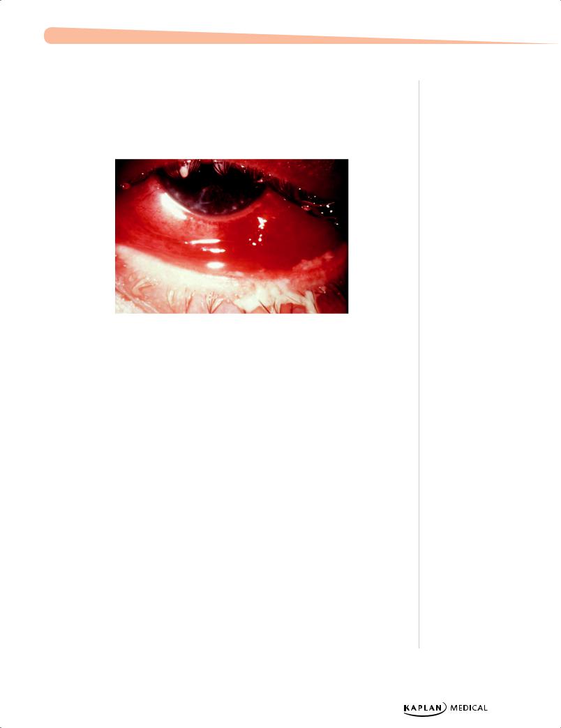
°N. gonorrhea—2–5-day incubation; may be delayed >5 days due to suppression from prophylactic eye treatment; mild inflammatory and serosanguineous discharge, then thick and purulent; complications are corneal ulceration, perforation, iridocyclitis
°C. trachomatis—5–14-day incubation; most common; mild inflammation to severe swelling with purulent discharge; mainly tarsal conjunctivae; cornea rarely affected
–Diagnosis—Gram stain, culture, PCR (polymerase chain reaction) for chlamydia
–Treatment:
°N. gonorrhea: ceftriaxone × 1 dose IM + saline irrigation until clear
°Chlamydia: erythromycin PO × 2 weeks + saline irrigation until clear (may prevent subsequent pneumonia)
•The red eye
–Bacterial conjunctivitis
°General conjunctival hyperemia, edema, mucopurulent exudate (crusting of lids together), and eye discomfort
°Unilateral or bilateral
°S. pneumonia, H. influenza (non-typable), S. aureus, other strep
°Treatment—warm compresses and topical antibiotics
100
Chapter 11 λ Disorders of the Eye
–Viral conjunctivitis
°Watery discharge, bilateral, usually with URI
°Adenovirus, enterovirus
°Epidemic keratoconjunctivitis = adenovirus type 8
°Good hand-washing
phil.cdc.gov.
Figure 11-1. Purulent, Bacterial Conjunctivitis
Secondary to Gonococcal Infection of the Eye
–Allergic
–Chemical
°Household cleaning substances, sprays, smoke, smog
°Extensive tissue damage, loss of sight
–Keratitis—corneal involvement
°H. simplex, adenovirus, S. pneumoniae, S. aureus, Pseudomonas, chemicals
–Foreign bodies → corneal abrasion (pain, photophobia)
–Anterior uveitis = iridocyclitis (from ciliary body to iris)
–Periorbital versus orbital cellulitis
–Dacryocystitis (S. aureus, H. influenza, S. pneumoniae), dacryoadenitis (S. aureus, streptococci, CMV [cytomegalovirus], measles, EBV [Epstein-Barr virus], trauma)
–Treatment—underlying cause and topical steroids
Retina and Vitreous
•Retinopathy of prematurity (ROP)
–Prematurity, hyperoxia, and general illness
–From mild to severe progressive vasoproliferative scarring and blinding retinal detachment
–Treatment—bevacizumab or laser photocoagulation
Published by dr-notes.com |
101 |
|
|
|
|

USMLE Step 2 CK λ Pediatrics
•Retinoblastoma
–Most common primary malignant intraocular tumor
°Recessive-suppressive gene—13q14 → family members need to be screened
–Average age of diagnosis = 15 months for bilateral and 25 months for unilateral
°Rarely discovered at birth
–Initial sign in most = leukocoria
°Appears as white mass
°Second most common—strabismus
–Diagnosis—CT scan to confirm; no biopsy (spreads easily)
–Need to consider enucleation—radiation, chemotherapy, laser therapy, cryotherapy
–Prognosis poor if extends into orbit or optic nerve
EYE INJURIES
Corneal Abrasions
•Symptoms—pain, tearing, photophobia, decreased vision
•Diagnosis—first anesthetize eye, then fluorescein and blue-filtered light (Wood’s lamp)
•Treatment—pain relief and topical antibiotics
Foreign Body
Attempt gentle removal with irrigation or moist cotton-tipped applicator; if embedded body cannot be easily removed, refer immediately to an ophthalmologist.
PERIORBITAL VERSUS ORBITAL CELLULITIS
Periorbital Cellulitis
•Inflammation of lids and periorbital tissue without signs of true orbital involvement; insidious onset; low-grade fever; no toxicity
•Causes—trauma, infected wound, abscess of lid, sinusitis, bacteremia (H. influenza nontypeable, S. pneumoniae, S. aureus)
•May be first sign of sinusitis that may progress to orbital cellulitis
–Physical exam: inflammation with intact eye movements; normal vision; no proptosis
•Diagnosis—clinical (blood culture unlikely to be positive)
•Treatment—oral or IV (depending on severity) antibiotics (cover for S. aureus and gram-positive resistant strains)
102

Chapter 11 λ Disorders of the Eye
Orbital Cellulitis
A 7-year-old boy presents with swelling around the eye 2 days after suffering an insect bite to the eyelid. There is edema, erythema, and proptosis of the eye. Marked limitation of eye movements is noted. He has a low-grade fever.
•Infection of orbital tissue including subperiosteal and retrobulbar abscesses
•Physical examination
–Ophthalmoplegia (eyeball does not move)
–Chemosis
–Inflammation
–Proptosis
•Toxicity, fever, leukocytosis, acute onset
•Causes: paranasal sinusitis, direct infection from wound, bacteremia
•Organisms nontypeable H. influenza, S. aureus, beta hemolytic strep,
S. pneumoniae, anaerobes
•Diagnosis—CT scan with contrast of orbits and surrounding area (best initial test)
•Treatment—Intravenous antibiotics (again, cover for S. aureus) and may require sinus and/or orbital drainage (will give you culture and sensitivities) if no improvement
Clinical Recall
A 5-day-old newborn boy presents with thick, purulent discharge of the right eye and evidence of a corneal ulcer. What is the likely etiology?
A.Syphilis
B.Chlamydia
C.HIV
D.Gonorrhea
E.Silver nitrate
Answer: D
Published by dr-notes.com |
103 |
|
|
|
|