

Полезные материалы за все 6 курсов / Учебники, методички, pdf / Kaplan Pediatrics USMLE 2CK 2021
.pdfUSMLE Step 2 CK λ Pediatrics
–With food allergies, there is an IgE and/or a cell-mediated response.
–Manifestations:
°Skin—urticaria/angioedema and flushing, atopic dermatitis; 1/3 of children with atopic dermatitis have food allergies, but most common is acute urticaria/angioedema
°Gastrointestinal—oral pruritus, nausea, vomiting, diarrhea, abdominal pain, eosinophilic gastroenteritis (often first symptoms to affect infants): predominantly a cell-mediated response, so standard allergy tests are of little value; food protein–induced enterocolitis/proctocolitis presents with bloody stool/diarrhea (most cow milk or soy protein allergies)
°Respiratory—nasal congestion, rhinorrhea, sneezing, laryngeal edema, dyspnea, wheezing, asthma
°Cardiovascular—dysrhythmias, hypotension
•Diagnosis
–Must establish the food and amount eaten, timing, and nature of reaction
–Skin tests, allergen-specific IgE is useful for IgE sensitization: a negative skin test excludes an IgE-mediated form but because of cell-mediated responses, patient may need a food elimination and challenge test in a controlled environment (best test)
•Treatment
–Only validated treatment is elimination
–Epinephrine pens for possible anaphylaxis
Clinical Recall
A 14-year-old-boy has persistent rhinorrhea, itchy eyes and nose, and post-nasal drip. He has no pets, does not smoke, and uses an allergenfree pillowcase. What is the first-line pharmacologic treatment?
A.Continue conservative management
B.Prescribe oral antihistamine
C.Prescribe intranasal corticosteroid
D.Prescribe intramuscular epinephrine
E.Prescribe inhaled steroids
Answer: B
84

Chapter 9 λ Allergy and Asthma
Urticaria and Angioedema
Causes:
•Acute, IgE-mediated (duration <6 weeks)
–Activation of mast cells in skin
–Systemically absorbed allergen: food, drugs, stinging venoms; with allergy, penetrates skin → hives (urticaria)
•Non IgE-mediated, but stimulation of mast cells
–Radiocontrast agents
–Viral agents (especially EBV, hepatitis B)
–Opiates, NSAIDs
•Physical urticarias; environmental factors—temperature, pressure, stroking, vibration, light
•Hereditary angioedema
–Autosomal dominant
–C1 esterase-inhibitor deficiency
–Recurrent episodes of nonpitting edema
•Diagnosis mainly clinical; skin tests, IgE-specific allergens (blood)
•Treatment
–Most respond to avoidance of trigger and oral antihistamine
–Severe—epinephrine, short-burst corticosteroids
–If H1 antagonist alone does not work, H1 plus H2 antagonists are effective; consider steroids
–For chronic refractory angioedema/urticaria → IVIg or plasmapheresis
Anaphylaxis
•Sudden release of active mediators with cutaneous, respiratory, cardiovascular, gastrointestinal symptoms
•Most common reasons
–In hospital—latex, antibiotics, IVIg (intravenous immunoglobulin), radiocontrast agents
–Out of hospital—food (most common is peanuts), insect sting, oral medications, idiopathic
•Presentation—reactions from ingested allergens are delayed (minutes to 2 hours); with injected allergen, reaction is immediate (more gastrointestinal symptoms)
•Treatment
–What the patient should do immediately:
°Injectable epinephrine
°Oral liquid diphenhydramine
°Transport to ER
–Medical:
°Oxygen and airway management
°Epinephrine IM (IV for severe hypotension); intravenous fluid expansion; H1 antagonist; corticosteroids; nebulized, short-acting beta-2 agonist (with respiratory symptoms); H2 antagonist (if oral allergen)
Published by dr-notes.com |
85 |
|
|
|
|
USMLE Step 2 CK λ Pediatrics
Atopic Dermatitis (Eczema)
•Epidemiology/pathophysiology
–Interaction among genetic, environmental, and immunologic factors; familial with strong maternal influence
–Majority develop allergic rhinitis and/or asthma
–Most have increased eosinophils and IgE
•Clinical presentation
–Half start by age 1 year; most by age 1 and 5 years; chronic or relapsing
–Intense cutaneous reactivity and pruritus; worse at night; scratching induces lesions; becomes excoriated
–Exacerbations with foods, inhalants, bacterial infection, decreased humidity, excessive sweating, irritants
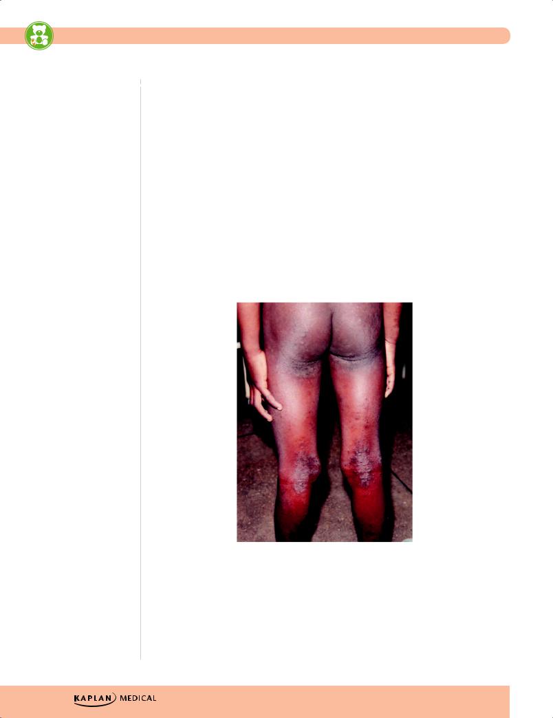
–Patterns for skin reactions:
°Acute: erythematous papules, intensely pruritic, serous exudate and excoriation
°Subacute—erythematous, excoriated, scaling papules
°Chronic—lichenification (thickening, darkening)
Courtesy of Tom D. Thacher, M.D.
Figure 9-1. Subacute and Chronic Atopic Dermatitis Most Commonly
Affects the Flexural Surfaces of Joints
–Distribution pattern:
°Infancy: face, scalp, extensor surfaces of extremities
°Older, long-standing disease: flexural aspects
°Often have remission with age, but skin left prone to itching and inflammation when exposed to irritants
86

Chapter 9 λ Allergy and Asthma
•Treatment
–Identify and eliminate causative factors
–Cutaneous hydration
°Dry skin, especially in winter (xerosis)
°Lukewarm soaking baths followed by application of occlusive emollient (hydrophilic ointments)
–Topical corticosteroids
°Seven classes—the higher potency classes are not to be used on face or intertriginous areas and only for short periods
°Goal—emollients and low-potency steroids for maintenance
–Topical immunomodulators; tacrolimus (calcineurin inhibitor):
°Inhibits activation of key cells
°Ointment safe and effective
°Safe on face
°Can use as young as age 2 years
–Tar preparations
–Phototherapy—UV light
–Systemic: antihistamines (sedating at night; for pruritus); glucocorticoids; cyclosporine (refractory to all other treatment); interferon (if all else fails)
–Treat with antibiotics for bacterial superinfection
•Complications
–Secondary bacterial infection, especially S. aureus; increased incidence of
T. rubrum, M. furfur
–Recurrent viral skin infections—Kaposi varicelliform eruption (eczema herpeticum) most common
–Warts/molluscum contagiosum
ASTHMA
A 6-year-old boy presents to his physician with end-expiratory wheezing scattered throughout the lung fields. He is noted to have nasal flaring, tachypnea, and intercostal retractions. These symptoms are triggered by changes in the weather. He has a family history of asthma and atopic dermatitis. He has never been intubated or admitted to the pediatric ICU. His last hospitalization for asthma was 6 months ago. He takes medication for asthma only when he starts to wheeze.
•Etiology/pathophysiology
–Chronic inflammation of airways with episodic at least partially reversible airflow obstruction
°Genetic and environmental factors: concomitant allergies (perennial in most), induced by common viral agents, tobacco smoke; cold, dry air; strong odors
°Most with onset age <6 years; most resolve by late childhood
°Two main patterns:
Early childhood triggered primarily by common viral infections
Chronic asthma associated with allergies (often into adulthood; atopic)
Published by dr-notes.com |
87 |
|
|
|
|

USMLE Step 2 CK λ Pediatrics
°Some risk factors for persistent asthma: perennial allergies; atopic dermatitis, allergic rhinitis, food allergy; severe lower respiratory tract infections; wheezing other than with URIs (exercise, emotions); environmental tobacco smoke exposure; low birth weight
•Clinical presentation
–Diffuse wheezing, expiratory then inspiratory
–Prolonged expiratory phase
–Decreased breath sounds
–Rales/rhonchi → excess mucus and inflammatory exudate
–Increased work of breathing
–Exercise intolerance
Table 9-1. Bronchiolitis Versus Asthma
|
Feature |
|
|
Bronchiolitis |
|
|
Asthma |
|
|
|
|
|
|
|
|
|
|
|
Etiology |
Most RSV |
Reversible bronchoconstriction with chronic |
|||||
|
|
|
|
|
|
inflammation |
||
|
|
|
|
|
|
|
|
|
Age |
Infants (especially <1 year) |
Most start age <5 years |
||||||
|
|
|
|
|
|
|
|
|
Timing |
• Winter |
• All year |
||||||
|
|
|
|
|
|
• Most with URI in winter |
||
|
|
|
|
|
|
|
|
|
Diagnosis Key Words |
• URI from another household contact |
• Repeated episodes of expiratory wheezing |
||||||
|
|
|
• Getting worse |
• Chronic non-productive cough |
||||
|
|
|
• Fever |
• Chest tightness |
||||
|
|
|
• Tachypnea |
• Respiratory distress |
||||
|
|
|
• Bilateral expiratory wheezing ± |
• May have other atopic disease + family history |
||||
|
|
|
|
respiratory distress |
• May occur primarily with URIs |
|||
|
|
|
|
|
|
|||
|
|
|
• Apnea |
• Cannot make diagnosis of asthma for first-time |
||||
|
|
|
|
|
|
|||
|
|
|
|
|
|
|
wheezing in infant with fever (diagnosis is |
|
|
|
|
|
|
|
|
bronchiolitis) |
|
|
|
|
|
|
|
|
|
|
Best Initial Test |
• Clinical Dx |
Worsening of FEV1/FVC with exercise and |
||||||
|
|
|
• CXR only if severe and therefore |
improvement with beta-agonist |
||||
|
|
|
|
|
|
|||
|
|
|
|
possibility of secondary bacterial |
|
|
|
|
|
|
|
|
pneumonia |
|
|
|
|
|
|
|
|
|
|
|
|
|
Most Accurate Test |
• NP rapid test or PCR for organism |
• Repeated episodes that improve with beta- |
||||||
|
|
|
• ABG only for severe to evaluate |
|
agonist |
|||
|
|
|
|
|
|
|||
|
|
|
|
possible need for ventilation |
|
|
|
|
|
|
|
|
|
|
|
|
|
Treatment |
• Oxygen, if needed |
• Oxygen |
||||||
|
|
|
• Supportive Rx |
• Short-acting beta-agonist |
||||
|
|
|
• May try nebulized hypertonic saline |
• Add oral steroid for acute attack |
||||
|
|
|
• Ribavirin in severe or worsening cases |
• May need chronic maintenance Rx |
||||
|
|
|
|
MAY prevent the need for intubation |
|
|
|
|
|
|
|
|
and ventilation |
|
|
|
|
|
|
|
|
|
|
|
|
|
88

Chapter 9 λ Allergy and Asthma
•Diagnosis
–In children, neither lab tests nor provocation challenge tests are required for diagnosis; they may support the clinical diagnosis or may be used to follow the patient clinically.
–Lung function:
°Gold standard = spirometry during forced expiration. FEV1/FVC <0.8 = airflow obstruction (the forced expiratory volume in 1 second adjusted to the full expiratory lung volume, i.e., the forced vital capacity) in children age ≥ 5 yrs
°Bronchodilator response to inhaled beta-agonist—improvement in FEV1 to >12%
°Exercise challenge—worsening in FEV1 of at least 15%
°Home tool—peak expiratory home monitoring (PEF); a.m. and p.m. PEF for several weeks for practice and to establish personal best and to correlate to symptoms; based on personal best, divide PEFs into zones: green (80–100%), yellow (50–80%), red (<50%)
–Radiology (no routine use):
°Hyperinflation—flattening of the diaphragms
°Peribronchial thickening
°Use to identify other problems that may mimic asthma (e.g., aspiration with severe gastroesophageal reflux) and for complications during severe exacerbations (atelectasis, pneumonia, air leak)
•Treatment—based on asthma severity classification
–Intermittent: symptoms ≤2 days/week and ≤2 nights/mo
°No need for daily controller
–Persistent (mild → moderate → severe) symptoms > intermittent
°Need daily controller
Table 9-2. Asthma Severity Classification and Treatment (simplified from National Asthma Education and Prevention Program)
|
|
|
|
Daytime |
|
|
Nighttime |
|
|
|
|
|
Class |
|
|
Symptoms |
|
|
Symptoms |
|
|
Treatment |
|
|
|
|
|
|
|
|
|
|
|
|
|
|
Intermittent |
|
≤2×/week |
|
≤2×/month |
|
SABA for relief of acute symptoms |
||||
|
|
|
|
|
|
|
|
|
|
|
|
|
Mild |
|
>2×/week |
|
>2×/month |
|
Low-dose ICS |
||||
|
persistent |
|
|
|
|
|
|
|
|
|
|
|
|
|
|
|
|
|
|
|
|
|
|
|
Moderate |
|
Daily |
|
>1×/week |
|
Increased dose ICS or (preferred) |
||||
|
persistent |
|
|
|
|
|
|
|
low-dose ICS + either LABA or LTRA |
||
|
|
|
|
|
|
|
|
|
|
SABA for relief of acute symptoms |
|
|
|
|
|
|
|
|
|
|
|
|
|
|
Severe |
|
Continual; limited |
|
Frequent |
|
Moderateto high-dose ICS + |
||||
|
persistent |
|
activities; frequent |
|
|
|
|
either LABA or LTRA |
|||
|
|
|
|
exacerbations |
|
|
|
|
SABA for relief of acute symptoms |
||
|
|
|
|
|
|
|
|
|
|
||
|
|
|
|
|
|
|
|
|
|
|
|
Note
With all asthma categories, a step-up, step-down dosing is typically used (high at first, then down to minimum necessary to prevent symptoms).
Published by dr-notes.com |
89 |
|
|
|
|

USMLE Step 2 CK λ Pediatrics
Note
Older children can use a metered dose inhaler (MDI); younger children often need to do so with a spacer and face mask. Infants may need to have nebulized medications.
Note
Adjunct Treatment to Prevent Intubation and Ventilation
•IV beta agonist
•IV theophylline
•Heliox (70:30 He:O2); decreased airway resistance and clinical response in
20 min
•IV MgSO4— smooth-muscle relaxant; monitor BP
every 10–15 min (risk of hypotension)
•Asthma medications
–Quick-relief medications
°Short-acting beta-2 agonists: albuterol, levalbuterol (nebulized only), terbutaline, metaproterenol (rapid onset, may last 4–6 hrs; drug of choice for rescue and preventing exercise-induced asthma but inadequate control if need >1 canister/month
°Anticholinergics (much less potent than beta agonists): ipratropium bromide; mostly for added treatment of acute severe asthma in ED and hospital
°Short-course systemic glucocorticoids: outpatient for moderate to severe flare-up, and prednisone 3–7 days; inpatient recommended with IV methylprednisolone IV
•Management of asthma exacerbations
–Emergency department:
°Monitor, oxygen as needed
°Inhaled albuterol q 20 minutes for 1 hour—add ipratropium if no good response for second dose
°Corticosteroids PO or IV
°Can go home if sustained improvement with normal physical findings and
SaO2 >92% after 4 hours in room air; PEF ≥70% of personal best
°Home on q 3–4 hour MDI + 3–7-day oral steroid
–Hospital—for moderate–severe flare-ups without improvement within 1–2 hours of initial acute treatment with PEF <70% of personal best or SaO2 <92% on room air:
°Oxygen
°Nebulized albuterol (very frequently or continuous)
°Add ipratropium q 6 hours
°Intravenous corticosteroids
°May need intravenous fluids
°Mechanical ventilation (rare)
Clinical Recall
A 12-year-old girl is diagnosed with asthma. She has nighttime symptoms twice a week and daily daytime symptoms. Which of the following should NOT be part of her long-term treatment?
A.Inhaled steroids
B.Leukotriene-receptor antagonist
C.Short-acting beta agonist
D.Oral prednisone
E.Long-acting beta agonist
Answer: D
90

Immune-Mediated Disease 10
Chapter Title
Learning Objectives
Explain information related to evaluation of suspected immune deficiency
Categorize specific defects of immune deficiency
Published by dr-notes.com |
91 |
|
|
|
|

USMLE Step 2 CK λ Pediatrics
EVALUATION OF SUSPECTED IMMUNE DEFICIENCY
Table 10-1. Suspecting Immunodeficiency by Major Defect
|
|
B-Cell |
|
|
T-Cell |
|
|
Complement |
|
|
Neutrophil |
|
|
|
|
|
|
|
|
|
|
|
|
|
|
Common organism |
Recurrent bacterial: |
Opportunistic |
Pneumococci, |
Bacteria: |
||||||||
|
streptococci, |
organisms: CMV, EBV, |
Neisseria |
Staphylococci, |
||||||||
|
staphylococci, |
varicella, Candida, |
|
|
|
Pseudomonas, |
||||||
|
Haemophilus, |
Pneumocystis jiroveci, |
|
|
|
Serratia, Klebsiella, |
||||||
|
Campylobacter; Viral: |
mycobacteria |
|
|
|
Salmonella; Fungi: |
||||||
|
enteroviruses; |
|
|
|
|
|
|
Candida, Aspergillus |
||||
|
Uncommon: giardia, |
|
|
|
|
|
|
|
|
|
||
|
cryptosporidia |
|
|
|
|
|
|
|
|
|
||
|
|
|
|
|
|
|
|
|
|
|
|
|
Age onset |
Age 5-7 months or later |
Usually age 2-6 months |
Any age |
Early onset |
||||||||
|
childhood to adult |
|
|
|
|
|
|
|
|
|
||
|
|
|
|
|
|
|
|
|
|
|
|
|
Infections |
Most are recurrent |
Mucocutaneous |
Meningitis, arthritis, |
Skin abscesses, |
||||||||
|
sinopulmonary infections |
candidiasis; pulmonary |
septicemia, recurrent |
impetigo, cellulitis, |
||||||||
|
and recurrent enteroviral |
and GI infections |
sinopulmonary |
suppurative adenitis, |
||||||||
|
meningitis |
|
|
|
infections |
gingivitis, oral ulcers, |
||||||
|
|
|
|
|
|
|
|
|
|
osteomyelitis, internal |
||
|
|
|
|
|
|
|
|
|
|
organ abscesses |
||
|
|
|
|
|
|
|
|
|
|
|
|
|
Other findings |
Autoimmunity, |
Chronic diarrhea and |
Autoimmune |
Prolonged attachment |
||||||||
|
lymphoreticular |
failure-to-thrive; |
disorders, vasculitis, |
of umbilical cord, poor |
||||||||
|
malignancy |
postvaccination |
glomerulonephritis, |
wound healing, |
||||||||
|
|
|
|
dissemination - |
angioedema |
decreased signs of |
||||||
|
|
|
|
varicella, BCG; |
|
|
|
infection |
||||
|
|
|
|
hypocalcemia in infancy; |
|
|
|
|
|
|
||
|
|
|
|
graft-versus-host from |
|
|
|
|
|
|
||
|
|
|
|
transplacental maternal |
|
|
|
|
|
|
||
|
|
|
|
engraftment or |
|
|
|
|
|
|
||
|
|
|
|
nonirradiated blood |
|
|
|
|
|
|
||
|
|
|
|
|
|
|
|
|
|
|
|
|
Best initial test |
Screen with IgA→if low, |
Lymphocyte count (low) |
Screen is total |
Neutrophil count |
||||||||
|
measure IgG and IgM |
|
|
|
hemolytic |
|
|
|
||||
|
(quantitative |
|
|
|
complement |
|
|
|
||||
|
immunoglobulins) |
|
|
|
(CH50)—will be |
|
|
|
||||
|
|
|
|
|
|
|
depressed if any |
|
|
|
||
|
|
|
|
|
|
|
component is |
|
|
|
||
|
|
|
|
|
|
|
consumed |
|
|
|
||
|
|
|
|
|
|
|
|
|
|
|
|
|
Other tests |
Low antibody titers to |
Best cost-effective test |
Identify mode of |
Neutrophil respiratory |
||||||||
|
specific antigens— |
for T-cell function |
inheritance—all are |
burst after phorbol |
||||||||
|
isohemagglutinins, |
– Candida skin test |
autosomal except for |
ester stimulation; most |
||||||||
|
vaccines |
|
|
|
properdin deficiency |
reliable now uses |
||||||
|
|
|
|
|
|
|
(X-linked) |
rhodamine |
||||
|
|
|
|
|
|
|
|
|
|
fluorescence (replaced |
||
|
|
|
|
|
|
|
|
|
|
the NBT test) |
||
|
|
|
|
|
|
|
|
|
|
|
|
|
Specific tests |
Enumerate B-cells with |
Flow cytometry using |
Can easily measure |
Can identify leukocyte |
||||||||
|
flow cytometry |
monoclonal antibodies |
C3 and C4 |
adhesion deficiencies |
||||||||
|
(monoclonal antibodies |
recognizing T-cell CD |
(hereditary |
with flow cytometric |
||||||||
|
to B-cell-specific CD |
antigens |
angioedema); others |
assays of lymphocytes |
||||||||
|
antigens): B cell absent |
(phytohemagglutinin, |
require a research |
and neutrophils (CD18, |
||||||||
|
or present and number |
concanavalin A, |
lab |
CD11, CD15) |
||||||||
|
|
|
|
pokeweed mitogen) |
|
|
|
|
|
|
||
|
|
|
|
|
|
|
|
|
|
|
|
|
Note: For each, the most accurate test is molecular genetic diagnosis.
92

Chapter 10 λ Immune-Mediated Disease
SPECIFIC DEFECTS
Defects of Antibody Production
X-linked (Bruton) agammaglobulinemia
X-linked (Bruton) agammaglobulinemia (XLA) is a profound defect in B-cell development which leads to an absence of circulating B cells and thus leads to severe hypogammaglobulinemia with small-to-absent tonsils and no palpable lymph nodes.
•Genetics: >500 known mutations of the Btk gene (Bruton tyrosine kinase), which is necessary for pre-B-cell expansion and maturation; long arm of X-chromosome
•Clinical findings: boys with pyogenic sinopulmonary infections
•Diagnosis: clinical presentation + lymphoid hypoplasia on exam; all immunoglobulins severely depressed; flow cytometry shows absence of circulating B-cells; gene sequencing for specific mutation
•Treatment: appropriate use of antibiotics + regular monthly IVIG
NOTE: The only 2 B-cell defects for which stem cell transplantation is recommended are CD40 ligand defect (extremely rare; one of the known mutations on the X-chromosome for hyper-IGM syndrome) and X-linked lymphoproliferative disease.
Common variable immunodeficiency
Common Variable Immunodeficiency (CVID) is hypogammaglobulinemia with phenotypically normal B-cells; blood B-lymphocytes do not differentiate into IG-producing cells
•Genetics: majority have no identified molecular diagnosis, so are sporadic; may have a common genetic basis with selective IgA deficiency (occurs in families together and some later with IgA may develop CVID)
•Clinical findings: boy or girl (equal sex distribution) with later onset infections, less severe; clinically similar to XLA, but rare echovirus meningoencephalitis
•Diagnosis: clinical presentation + serum IG and antibody deficiencies as profound or less than in XLA; normal sized lymphoid tissue; later autoimmune disease and malignancy (lymphoma)
•Treatment: need to be screened for anti-IgA antibodies (as in selective IgA deficiency)→ if present, therapy consists of the one IG preparation available that contains no IgA.
Selective IgA deficiency
Selective IgA deficiency is the most common immunodeficiency. It is caused by the absence or near absence of serum and secretory IgA with phenotypically normal B-cells
•Genetics: basic defect is unknown; boys and girls and familial pattern suggests autosomal dominant with variable expression; also seen in families with CVID (as above); both may be triggered by environmental factors
•Clinical findings: same bacteria as others with most infections in respiratory, GI and urogenital tracts; giardiasis is common
Published by dr-notes.com |
93 |
|
|
|
|