

Полезные материалы за все 6 курсов / Учебники, методички, pdf / Kaplan Pediatrics USMLE 2CK 2021
.pdfUSMLE Step 2 CK
Note
Preductal versus postductal is no longer used; it has been found that irrespective of the location, there are only 2 types based on pathology:
•Short, discrete segment of incomplete narrowing which allows for blood flow with left ventricular hypertrophy
•Tubular hypoplasia which does not allow for any hemodynamically significant blood flow; is generally
a longer segment of hypoplasia of arch or even distal to ductus
λPediatrics
°Normally, leg systolic pressure is 10–20 mm Hg higher than in arms; in coarctation, leg systolic pressure is decreased (>5%)
°If pressure is greater in right arm than left arm, suggests coarctation involving left subclavian artery
°Short systolic murmur along left sternal border at third-to-fourth intercostal space → left scapula and neck
−Hypertension due not only to mechanical but also to neurohormonal reasons
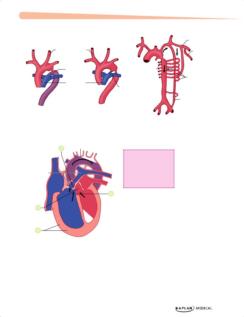
−Over time, patient develops an extensive collateral circulation (systolic or continuous murmurs over left and right sides of chest with thrills), rib notching (dilated intercostal arteries)
•Tubular hypoplasia (preductal, infantile type)
−Severe narrowing starting at one of the head or neck vessels and extending to the ductus
−Right ventricular blood flows across the PDA to supply the descending aorta so the perfusion of the lower part of the body is dependent upon right ventricular output
−Seen as differential cyanosis—upper body is pink, lower is cyanotic; prominent heart failure as ductus closes (if completely atretic = interrupted aortic arch)
−Presents with lower body hypoperfusion, acidosis, and severe heart failure with ductal closure; large heart, systolic murmur along left sternal border
•Diagnostic tests
−Chest x-ray—depends on age and effects of hypertension and collaterals
°Severe (infantile)—increased heart size and pulmonary congestion
°Adult—findings usually occur after first decade:
Increased size of subclavian artery—prominent shadow in left superior mediastinum
Notching of inferior border of ribs from passive erosion of increased collaterals in late childhood
Poststenotic dilatation of ascending aorta
•Diagnosis
−EKG—left ventricular hypertrophy in older children; in neonates, biventricular hypertrophy
−Echocardiogram (gold standard)
•Treatment
−Neonate—PGE1 infusion to maintain patent, ductus, which establishes adequate lower extremity blood flow; surgery after stabilization
−Surgery soon after diagnosis of any significant coarctation
−Adult—treat heart failure and hypertension, then follow with surgery
•Complications
−Associated cerebrovascular disease
−Systemic hypertension
−Endocarditis
−Aortic aneurysms
124

Chapter 13 λ Cardiology
Clinical Recall
A newborn with Noonan syndrome and a cardiac anomaly presents for evaluation. EKG will likely show which of the following?
A.Right ventricular hypertrophy
B.Left ventricular hypertrophy
C.Biventricular hypertrophy
D.Biatrial dilation
E.Left ventricular dilation
Answer: A
RIGHT TO LEFT SHUNTS (CYANOTIC LESIONS)
Cyanotic Lesions Associated with Decreased Pulmonary Blood Flow
Tetralogy of Fallot (TOF)
A 6-month-old infant is prone to episodes of restlessness, cyanosis, and gasping respirations. Symptoms resolve when he is placed in the knee-chest position. Physical examination reveals an underweight infant, with a harsh long systolic ejection murmur and a single second heart sound.
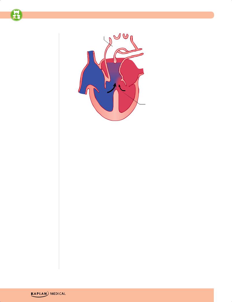
•Components
−Pulmonary stenosis and infundibular stenosis (obstruction to right ventricular outflow)
−VSD
−Overriding aorta (overrides the VSD)
−Right ventricular hypertrophy
•Most common cyanotic lesion
•Pulmonary stenosis plus hypertrophy of subpulmonic muscle (crista supraventricularis) → varying degrees of right ventricular outflow obstruction
−Blood shunted right-to-left across the VSD with varying degrees of arterial desaturation and cyanosis
−If mild, patient may not be visibly cyanotic (pink tetralogy of Fallot)
°With growth and further hypertrophy of infundibulum, cyanosis may be seen later in first year of life
−With severe obstruction, cyanosis in the immediate neonatal period (ductal dependent)
−If not corrected, older children are blue, have marked clubbing, and have dyspnea on exertion (child will squat to increase systemic vascular resistance and to decrease right-to-left shunt)
Note
Common Cyanotic Heart
Disease (5 Ts)
Tetralogy of Fallot
Transposition of great vessels
Truncus arteriosus
Total anomalous pulmonary venous return
Tricuspid atresia
Published by dr-notes.com |
125 |
|
|
|
|

USMLE Step 2 CK
Note
The combination of severe cyanosis in the newborn plus a chest x-ray showing decreased pulmonary blood
flow plus an EKG with left axis deviation and left ventricular hypertrophy is most likely to be tricuspid atresia..
λPediatrics
−Paroxysmal hypercyanotic attacks (tet spells)
°Acute onset of hyperpnea and restlessness → increased cyanosis → gasping → syncope (increased infundibular obstruction with further right-to-left shunting
°Treatment—place in lateral knee-chest position, give oxygen, subcutaneous morphine, give beta-blockers
•Physical examination—substernal right ventricular impulse, systolic thrill along third- to-fourth intercostal space on left sternal border, loud and harsh systolic ejection murmur (upper sternal border), may be preceded by a click; either a single S2 or soft pulmonic component
•Diagnosis
−Chest x-ray—hypertrophied right ventricle causes the apex to be uplifted above the diaphragm → boot-shaped heart plus dark lung fields (decreased pulmonary blood flow)
−EKG—right axis deviation plus right ventricular hypertrophy
−Echocardiogram (gold standard)
•Pre-correction complications—cerebral thromboses, brain abscess, bacterial endocarditis, heart failure, but not common because of early correction
•Treatment
−Depends on degree of obstruction
°PGE1 infusion—prevent ductal closure; given if cyanotic at birth
°Augment pulmonary blood flow with palliative systemic to pulmonary shunt
(modified Blalock-Taussig shunt)
°Corrective surgery (electively at age 4–12 months)—remove obstructive muscle, valvulotomy, and patching of VSD
Tricuspid atresia
•Pathophysiology—no outlet from the right atrium to the right ventricle; entire venous (systemic) return enters the left atrium from a foramen ovale or ASD (there must be an atrial communication); left ventricular blood to right ventricle (atretic) via a VSD and is augmented by PDA; therefore, pulmonary blood flow depends on presence (and size) of VSD
•Clinical presentation
−Will present at birth with severe cyanosis
−Increased left ventricular impulse (contrast to most others with right ventricular impulse), holosystolic murmurs along left sternal border (most have a VSD; though right ventricle is small, it is still a conduit for pulmonary blood flow)
•Diagnosis
−Chest x-ray—pulmonary undercirculation
−EKG—left axis deviation plus left ventricular hypertrophy (distinguishes from most other congenital heart disease)
−Echocardiogram (gold standard)
•Treatment
−PGE1 until aortopulmonary shunt can be performed
−May need an atrial balloon septostomy (to make larger ASD)
−Later, staged surgical correction
126

Chapter 13 λ Cardiology
Ebstein anomaly
•Development associated with periconceptional maternal lithium use in some cases
•Downward displacement of abnormal tricuspid valve into right ventricle; the right ventricle gets divided into 2 parts: an atrialized portion, which is thin-walled, and smaller normal ventricular myocardium
•Right atrium is huge; tricuspid valve regurgitant
•Right ventricular output is decreased because
−Poorly functioning, small right ventricle
−Tricuspid regurgitation
−Variable right ventricular outflow obstruction—abnormal anterior tricuspid valve
leaflet. Therefore, increased right atrial volume shunts blood through foramen ovale or ASD → cyanosis
•Clinical presentation
−Severity and presentation depend upon degree of displacement of valve and degree of right ventricular outflow obstruction
°May not present until adolescence or adulthood
°If severe in newborn → marked cyanosis, huge heart
−Holosystolic murmur of tricuspid insufficiency over most of anterior left chest
(most characteristic finding)
•Diagnosis
−Chest x-ray—heart size varies from normal to massive (increased right atrium); if severe, decreased pulmonary blood flow
−EKG—tall and broad P waves, right bundle branch block
•Treatment
−PGE1
−Systemic-to-pulmonary shunt
−Then staged surgery
Cyanotic Lesions Associated with Increased Pulmonary Blood Flow
Transposition of the great arteries (TGA)
•Pathophysiology
−Aorta arises from the right ventricle, and the pulmonary artery arises from the left ventricle; d = dextroposition of the aorta anterior and the right of the pulmonary artery (normal is posterior and to the right of the pulmonary artery)
−Series circuit changed to 2 parallel circuits; need foramen ovale and PDA for some mixture of desaturated and oxygenated blood; better mixing in half of patients with a VSD
•Clinical presentation
−With intact septum (simple TGA)—as PDA starts to close, severe cyanosis and tachypnea ensue
−S2 usually single and loud (closure of pulmonic valve obscured by closure of aortic valve)
Note
Patients with Ebstein anomaly may have Wolff-Parkinson- White syndrome (delta wave and short PR interval) and present with episodes of supraventricular tachycardia.
Note
TGA is the most common cyanotic lesion presenting in the immediate newborn period. It is seen more often
in infants of diabetic mothers.
Published by dr-notes.com |
127 |
|
|
|
|

USMLE Step 2 CK λ Pediatrics
Note
Truncus arteriosus is one of the major conotruncal lesions associated with the CATCH-22 syndrome, i.e., DiGeorge. Also seen are transposition of the great arteries and aortic arch abnormalities.
−If VSD is present, there is a harsh murmur at the lower left sternal border. If large, then holosystolic murmur, significant mixing of blood lessens cyanosis, but presents as heart failure
•Diagnosis
−Chest x-ray:
°Mild cardiomegaly, narrow mediastinum, and normal-to-increased pulmonary blood flow
°“Egg on a string” appearance—narrow heart base plus absence of main segment of the pulmonary artery
−EKG—normal neonatal right-sided dominance
−Echocardiogram (gold standard)
•Treatment
−PGE1 (keeps PDA patent)
−Balloon atrial septostomy
−Arterial switch surgery in first 2 weeks
Truncus Arteriosus
•Pathophysiology
−Single arterial trunk arises from the heart and supplies all circulations.
−Truncus overlies a ventral septal defect (always present) and receives blood from both ventricles (total mixing).
−Both ventricles are at systemic pressure.
•Clinical presentation
−With dropping pulmonary vascular resistance in first week of life, pulmonary blood flow is greatly increased and results in heart failure.
−Large volume of pulmonary blood flow with total mixing, so minimal cyanosis
−If uncorrected, Eisenmenger physiology
−Single truncal valve, which may be incompetent (high-pitched, early diastolic decrescendo at mid-left sternal border)
−Initially, SEM with loud thrill, single S2, and minimal cyanosis
−With decreasing pulmonary vascular resistance (PVR) → torrential pulmonary blood flow with heart failure; runoff from truncus to pulmonary circulation → wide pulse pressure with bounding pulses and hyperdynamic precordium
−Apical mid-diastolic rumble (increased flow across mitral valve)
•Diagnosis
−Chest x-ray—heart enlargement with increased pulmonary blood flow
−EKG—biventricular hypertrophy
−Echocardiogram (gold standard)
•Treatment
−Treat heart failure
−Then surgery in first few weeks of life
128
Chapter 13 λ Cardiology
Subclavian artery
|
Common carotid |
Subscapular artery |
|
arteries |
|
|
|
|
|
Patent ductus |
Postductal |
|
arteriosus |
coarctation |
|
|
Intercostal |
|
Pulmonary |
arteries |
|
artery |
Ligamentum |
|
|
arteriosum |
|
|
Inferior epigastric artery |
A |
B |
C |
Figure 13-5. Coarctation of the Aorta: (A) Tubular Hypoplasia; (B) Juxtaductal; (C) Collateral Circulation
Aorta
1
1. Pulmonary stenosis
2. Ventricular septal defect
Pulmonary
trunk 3. Hypertrophied right ventricle
RA |
|
LA |
4. Overriding aorta |
|
|
|
2 |
4 |
RV |
LV |
|
|
|
|
3
Figure 13-6. Tetralogy of Fallot
Published by dr-notes.com |
129 |
|
|
|
|
USMLE Step 2 CK λ Pediatrics
Aorta
 Pulmonary artery
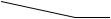 Truncus arteriosus
Interventricular septal defect
Figure 13-7. Truncus Arteriousus
Note
TAPVR always has an atrial connection.
MIXED LESIONS
Total Anomalous Pulmonary Venous Return (TAPVR)
•Pathophysiology
−Complete anomalous drainage of the pulmonary veins into the systemic venous circulation; total mixing of systemic venous and pulmonary venous blood within the heart produces cyanosis
−Right atrial blood → right ventricle and pulmonary artery or to left atrium via foramen ovale or ASD
−Enlarged right atrium, right ventricle, and pulmonary artery; and small left atrium; and left ventricle normal or small
•Clinical manifestations depend on presence or absence of obstruction.
−Obstruction (of pulmonary veins, usually infracardiac):
°Severe pulmonary venous congestion and pulmonary hypertension with decreasing cardiac output and shock
°Cyanosis and severe tachypnea; may not respond to ventilation and PGE1 → need emergent diagnosis and surgery for survival
°Heart failure early with mild-to-moderate obstruction and a large left-to-right shunt; pulmonary hypertension and mild cyanosis
−No obstruction—total mixing with a large left-to-right shunt; mild cyanosis; less likely to be severely symptomatic early
130
Chapter 13 λ Cardiology
•Diagnosis
−Chest x-ray—large supracardiac shadow with an enlarged cardiac shadow forms a “snowman” appearance; pulmonary vascularity is increased
−EKG—RVH and tall, spiked P waves (RAE)
−Echocardiogram (gold standard)
•Treatment: PGE1; surgical correction
Hypoplastic Left Heart Syndrome
•Pathophysiology
−Atresia of mitral or aortic valves, left ventricle, and ascending aorta (or any combination)
−Right ventricle maintains both pulmonary and systemic circulation.
−Pulmonary venous blood passes through foramen ovale or ASD from left atrium
→right atrium and mixes with systemic blood to produce total mixing
−Usually, the ventricular septum is intact and all of the right ventricular blood enters the pulmonary artery.
−Ductus arteriosus supplies the descending aorta, ascending aorta and coronary arteries from retrograde flow.
−Systemic circulation cannot be maintained, and if there is a moderate-to-large ASD
→pulmonary overcirculation
•Clinical presentation
−Cyanosis may not be evident with ductus open, but then gray-blue skin color (combination of hypoperfusion and cyanosis as ductus closes)
−Signs of heart failure, weak or absent pulses, and shock
−Enlarged heart with right parasternal lift; nondescript systolic murmur
•Diagnosis: chest x-ray shows heart enlargement with increased pulmonary blood flow; EKG shows right ventricular hypertrophy and right atrial enlargement with decreased left-sided forces; echocardiogram (gold standard)
Treatment: consider doing nothing if malformations or genotype not compatible with life; best treatment is 3-stage Norwood procedure (better result than cardiac transplantation)
•Other: many patients have a significant abnormality of central nervous system (CNS) and/or kidneys: need careful genetic, neurologic examination and screening tests on any child being considered for surgery
Published by dr-notes.com |
131 |
|
|
|
|

USMLE Step 2 CK λ Pediatrics
Note
Mitral valve prolapse is a common finding in those with Marfan and Ehlers-Danlos syndrome.
Note
Staphylococcal endocarditis is more common in those without underlying heart
disease. Streptococcus viridans is more common in patients with underlying heart disease or after dental procedures.
132
Clinical Recall
Which of the following cardiac anomalies is correctly matched to its classic chest x-ray findings?
A.Hypoplastic left heart syndrome: normal cardiac silhouette with decreased pulmonary vascularity
B.TAPVR: snowman sign with increased pulmonary vascularity
C.Truncus arteriosus: egg on a string sign
D.TGA: massively enlarged right atrium
E.Tricuspid atresia: boot-shaped heart
Answer: B
REGURGITANT LESIONS
Mitral Valve Prolapse
•Abnormal cusps—billowing of one or both leaflets into left atrium from mid to late systole (congenital defect: entire MV complex undergoes myxomatous degeneration, which leads to redundant tissue of the chordae tendineae)
•Usually not recognizable until adolescence or adulthood; girls > boys
−May present with chest pain or palpitations
−Arrhythmias, especially unior multifocal premature ventricular contractions
•Mid-systolic click followed by midto late-blowing decrescendo systolic murmur
•Diagnosis: EKG usually normal; chest x-ray normal; echocardiogram (gold standard)
•No therapy, not progressive; adults (more in men) at risk for cardiovascular complications if have thickened leaflets
OTHER CARDIAC PATHOLOGY
Infective Endocarditis
A 6-year-old boy has had high intermittent fevers for 3 weeks, accompanied by chills. He has a past history of bicuspid aortic valves and recently had dental work.
•Etiology/epidemiology
−Most are Streptococcus viridans (alpha hemolytic) and Staphylococcus aureus
−Organism associations
°S. viridans—after dental procedures
°Group D streptococci—large bowel or genitourinary manipulation
°Pseudomonas aeruginosa and Serratia marcescens—intravenous drug users
°Fungi—after open heart surgery
°Coagulase-negative Staphylococcus—indwelling intravenous catheters

Chapter 13 λ Cardiology
−Highest risk with prosthetic valve and uncorrected cyanotic heart lesions
−Most cases occur after surgical or dental procedures (high risk with poor dental hygiene) are performed.
•Clinical presentation
−Prolonged intermittent fever, weight loss, fatigue, myalgia, arthralgia, headache, nausea, vomiting
−New or changing heart murmur
−Splenomegaly, petechiae, embolic stroke, CNS abscess, CNS hemorrhage, mycotic aneurysm (all more with Staphylococcus)
−Skin findings (rare): late findings (uncommon in treated patients); represent vasculitis from circulating Ag-Ab complexes; if present, are highly suggestive
°Osler nodes—tender, pea-sized, intradermal nodules on pads of fingers and toes
°Janeway lesions—painless, small erythematous or hemorrhagic lesions on palms and soles
°Splinter hemorrhage—linear lesions beneath nail beds
º Roth spots—retinal exudates
•Diagnosis
−Two separate positive blood cultures plus echocardiographic evidence of intracardiac or valve lesion; prosthetic regurgitant flow; abscess; partial dehiscence of prosthetic valve or new valvular regurgitant flow
Table 13-3. Duke Criteria
|
Major Criteria |
|
|
Minor Criteria |
|
|
|
|
|
|
|
|
• Positive blood culture (2 separate |
• Predisposing conditions |
|||
|
for usual pathogens; at least 2 for less |
• Fever |
|||
|
common) |
||||
|
• Emboli or vascular signs |
||||
|
• Evidence on echocardiogram |
||||
|
• Immune complex disease |
||||
|
(intracardiac or valve lesion, prosthetic |
||||
|
|
(glomerulonephritis, arthritis, positive |
|||
|
regurgitant flow, abscess, partial |
|
|||
|
|
rheumatoid factor, Osler node, Roth |
|||
|
dehiscence of prosthetic valve, new |
|
|||
|
|
spots [retinal hemorrhages with white |
|||
|
valvular regurgitant flow) |
|
|||
|
|
centers]) |
|||
|
|
|
|
||
|
|
|
• Single positive blood culture |
||
|
|
|
• Echocardiographic signs not meeting |
||
|
|
|
|
criteria |
|
|
|
|
|
|
|
•Complications
−Most common—heart failure from aortic or mitral lesions
−Others—systemic or pulmonary emboli, myocardial abscess, myocarditis, valve obstruction, heart block, meningitis, osteomyelitis, arthritis, renal abscess, immune complex−mediated glomerulonephritis
•Treatment
−Organism specific for 4−6 weeks (S. viridans, Enterococci, S. aureus, MRSA,
S. epidermidis, HACEK)
−Heart failure—digitalis, diuretic, salt restriction
−Surgery with severe involvement or lack of improvement
Note
Clinical diagnosis of infective endocarditis is made with one of the following:
•2 major
•1 major + 3 minor
•5 minor
Note
HACEK
•Haemophilus spp.
•Actinobacillus actinomycetemcomitans
•Cardiobacterium hominis
•Eikenella corrodens
•Kingella kingae
These are slow-growing gramnegative organisms that are part of normal flora.
Published by dr-notes.com |
133 |
|
|
|
|