

Полезные материалы за все 6 курсов / Учебники, методички, pdf / Kaplan Pediatrics USMLE 2CK 2021
.pdfUSMLE Step 2 CK λ Pediatrics
Hirschsprung Disease
•Etiology—absence of a ganglion cells in bowel wall beginning at internal anal sphincter and extending variably proximally
•Most common reason for bowel obstruction in neonates
•Clinical presentation
−Symptoms usually present at birth
−Suspect in any full-term infant with a delay in passage of meconium (>24 hours)
−May have subsequent history of chronic constipation (if short aganglionic segment)
•Diagnosis
– Rectal suction biopsy is definitive
−Presence of transition zone on barium enema (not necessary to perform)
•Treatment—surgery (most with temporary colostomy) and wait 6−12 months for definitive correction (most achieve continence) or one-stage repair
•Complications—enterocolitis
Table 14-5. Functional Constipation Versus Hirschsprung Disease
|
|
Functional Constipation |
|
|
Hirschsprung Disease |
|
|
|
|
|
|
|
|
Onset constipation |
|
After 2 years of age |
|
At birth |
||
|
|
|
|
|
|
|
Failure to thrive |
|
Uncommon |
|
Possible |
||
|
|
|
|
|
|
|
Enterocolitis |
|
No |
|
Possible |
||
|
|
|
|
|
|
|
Abdominal distention |
|
Usually not |
|
Yes |
||
|
|
|
|
|
|
|
Poor weight gain |
|
Usually not |
|
Common |
||
|
|
|
|
|
|
|
Anal tone |
|
Normal |
|
Normal |
||
|
|
|
|
|
|
|
Rectal |
|
Stool in ampulla |
|
No stool |
||
|
|
|
|
|
|
|
Anorectal manometry |
|
Distention of rectum → |
|
No sphincter relaxation |
||
|
|
relaxation of internal |
|
|
|
|
|
|
sphincter |
|
|
|
|
|
|
|
|
|
|
|
Barium enema |
|
Large amount of stool; no |
|
Transition zone with delayed |
||
|
|
transition zone |
|
evacuation |
||
|
|
|
|
|
|
|
154

Renal and Urologic Disorders |
15 |
Chapter Title |
Learning Objectives
Recognize and describe treatment for urinary tract infection, vesicoureteral reflux, obstructive uropathy, and polycystic kidney disease
Diagnose and describe treatments for disorders presenting with hematuria or proteinuria
URINARY TRACT INFECTION (UTI)
A 12-day-old infant presents with fever of 39 C (102 F), vomiting, and diarrhea. On physical examination the infant appears to be ill and mildly dehydrated.
•Epidemiology and risk factors
−Age <24 months
°7% of febrile infants without a source and T >39 C (>102 F) for 24–48 hrs
°More common in whites than blacks
°Febrile females are more common in first year than age >12 mos
°Male infection correlated to not being circumcised
−Age ≥24 months
°Can describe symptoms and localize
°Most important factors: presence of bowel/bladder withholding behaviors; congenital anomalies; previous history of UTI
•Etiology and pathogenesis
−E. coli is number 1 organism for all ages; then Klebsiella, Proteus Enterococcus, Pseudomonas (these all with later, recurrent infections and immune compromise)
−Most from ascending infection; rare hematogenous spread
•Clinical presentation
−Age <24 mos: fever, irritability, crying, decreased input, less sleep
−Age ≥24 mos: localized symptoms of dysuria, urgency, frequency, suprapubic pain, incontinence for cystitis and abdominal or flank pain, malaise, nausea, vomiting, diarrhea for pyelonephritis
−May also have asymptomatic bacteriuria–positive urine culture without signs or symptoms; may become symptomatic if untreated; almost exclusively in girls
Published by dr-notes.com |
155 |
|
|
|
|

USMLE Step 2 CK λ Pediatrics
•Diagnosis
−Need evidence of inflammation (urinalysis: WBCs and leukocyte esterase) + bacterial growth (UA nitrites, bacteria and positive culture)
−Age <24 mos: may place a urethral bag for UA only and then if positive → need catheterization for culture and sensitivity
−Age ≥24 mos: mid-stream clean catch urine
−Best sensitivity and specificity for positive cultures: leukocyte esterase + nitrites + microscopic WBCs + microscopic bacteria
−Interpretation: ≥50,000 CFU/ml (may be less in neonates, immune deficiency, congenital anomalies or if already on antibiotics)
•Management
−Oral antibiotics are as effective as IV
−Use IV if toxic or cannot tolerate oral
−Choice is tailored to local bacterial susceptibility data, compliance, cost and history of previous treatment/results
−Oral: amoxicillin, trimethoprim-sulfamethoxazole, oral cephalosporins
−Parenteral: best/safest are third-generation cephalosporins
•Imaging
−Age <24 mos: renal and bladder U/S after first febrile UTI; VCUG after first febrile UTI with an abnormal U/S (note: not afebrile)
−All children: VCUG after second febrile UTI (no policy for U/S or nonfebrile UTIs, which generally means that physicians should use their judgement)
−If there is grade 2-5 reflux, obtain a renal radionuclide scan for function, kidney size, and scarring
Clinical Recall
Which of the following children should undergo a voiding cystourethrogram?
A.A 4-month-old uncircumcised male infant with his first positive urine culture
B.A 9-year-old girl with no significant medical history being treated for pyelonephritis
C.A 17-year-old sexually active girl with 2 urinary tract infections in 3 years
D.A 1-year-old boy with hydronephrosis on renal U/S
E.None of the above, as only children with recurrent UTIs should receive a VCUG
Answer: D
156
Chapter 15 λ Renal and Urologic Disorders
VESICOURETERAL REFLUX (VUR)
A 2-year-old girl presents with urinary tract infection. She has had multiple urinary tract infections since birth but has never had any follow-up studies to evaluate these infections. Physical examination is remarkable for an ill-appearing child who has a temperature of 40 C (104 F) and is vomiting.
•Definition—abnormal backflow of urine from bladder to kidney
•Etiology
−Occurs when the submucosal tunnel between the mucosa and detrusor muscle is short or absent.
−Predisposition to pyelonephritis → scarring → reflux nephropathy (hypertension, proteinuria, renal insufficiency to end-stage renal disease [ESRD], impaired kidney growth)
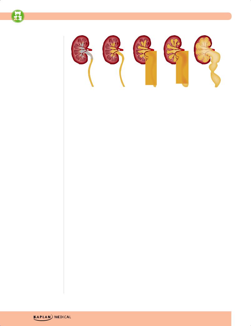
•Grading—see Figure 15-3
•Diagnosis
– VCUG for diagnosis and grading
– Renal scan for renal size, scarring and function; if scarring, follow creatinine
•Natural history
– Increased scarring with grade V (less so with bilateral 4)
– Most below grade V resolve regardless of age at diagnosis or whether it is unilateral or bilateral
– With growth, tendency to resolve (lower > higher grades); resolve by age 6–7 years
•Treatment
– Medical—based on reflux resolving over time; most problems can be taken care of nonsurgically
– Careful ongoing monitoring for and aggressive treatment of all UTIs
– Surgery if medical therapy fails, if grade V reflux, or if any worsening on VCUG or renal scan
– The issue of antibiotic prophylaxis is controversial and thus must be individualized. Studies do show, however, that it would take thousands of doses of antibiotics to prevent a single UTI and that prophylaxis does not prevent scarring. Therefore, it is not currently recommended routinely by the AAP.
Published by dr-notes.com |
157 |
|
|
|
|
USMLE Step 2 CK λ Pediatrics
|
I |
II |
III |
IV |
V |
GRADE |
DESCRIPTION |
|
|
|
|
|
|
|
|
|
|
I |
Reflux into a nondilated ureter |
|
|
|
|
II |
Reflux into the pelvis and calyces without dilation |
|
|
||
IIIReflux with mild to moderate dilation of the ureter, renal pelvis, and calyces, with minimal blunting of the fornices
IV |
Reflux with moderate tortuosity of the ureter and dilation of the |
|
pelvis and calyces |
VReflux causing ureteral tortuosity with severe dilation of ureter, renal pelvis, and calyces and loss of fornices and papillary impressions
Figure 15-1. Vesicoureteral Grading Scale
OBSTRUCTIVE UROPATHY
•Definition—obstruction of urinary outflow tract
•Clinical presentation
–Hydronephrosis
–Upper abdominal or flank pain
–Pyelonephritis, UTI (recurrent)
–Weak, decreased urinary stream
–Failure to thrive, diarrhea (or other nonspecific symptoms)
•Diagnosis
–Palpable abdominal mass in newborn; most common cause is hydronephrosis due to ureteropelvic junction obstruction or multicystic kidney disease
(less so–infantile polycystic disease)
–Most can be diagnosed prenatally with ultrasound.
–Obtain VCUG in all cases of congenital hydronephrosis and in any with ureteral dilatation to rule out posterior urethral valves
•Common etiologies
–Ureteropelvic junction obstruction—most common (unilateral or bilateral hydronephrosis)
–Ectopic ureter—drains outside bladder; causes continual incontinence and UTIs
158

Chapter 15 λ Renal and Urologic Disorders
–Ureterocele—cystic dilatation with obstruction from a pinpoint ureteral orifice; mostly in girls
–Posterior urethral valves:
°Most common cause of severe obstructive uropathy; mostly in boys
°Can lead to end-stage renal disease
°Present with mild hydronephrosis to severe renal dysplasia; suspect in a male with a palpable, distended bladder and weak urinary stream
•Diagnosis—voiding cystourethrogram (VCUG)
•Treatment
–Decompress bladder with catheter
–Antibiotics (intravenously)
–Transurethral ablation or vesicostomy
•Complications
–If lesion is severe, may present with pulmonary hypoplasia (Potter sequence)
–Prognosis dependent on lesion severity and recovery of renal function
DISEASES PRESENTING PRIMARILY WITH HEMATURIA
Acute Poststreptococcal Glomerulonephritis
A 10-year-old boy presents with Coca-Cola–colored urine and edema of his lower extremities. On physical examination, the patient has a blood pressure of 185/100 mm Hg. He does not appear to be in any distress. His lungs are clear to auscultation, and his heart has a regular rate and rhythm without any murmurs, gallops, or rubs. His past medical history is remarkable for a sore throat that was presumed viral by his physician 2 weeks before.
•Etiology
−Follows infection with nephrogenic strains of group A beta-hemolytic streptococci of the throat (mostly in cold weather) or skin (in warm weather)
−Diffuse mesangial cell proliferation with an increase in mesangial matrix; lumpybumpy deposits of immunoglobulin (Ig) and complement on glomerular basement membrane and in mesangium
−Mediated by immune mechanisms but complement activation is mostly through the alternate pathway
•Clinical presentation
−Most 5–12 years old (corresponds with typical age for strep throat)
−1–2 weeks after strep pharyngitis or 3–6 weeks after skin infection (impetigo)
−Ranges from asymptomatic microscopic hematuria to acute renal failure
– Edema, hypertension, hematuria (classic triad)
−Constitutional symptoms—malaise, lethargy, fever, abdominal or flank pain
•Diagnosis
−Urinalysis—RBCs, RBC casts, protein 1–2 +, polymorphonuclear cells
−Mild normochromic anemia (hemodilution and low-grade hemolysis)
Note
For diagnosis of prior Strep infection, use streptozyme (slide agglutination), which detects antibodies to streptolysin O, DNase B, hyaluronidase, streptokinase, and nicotinamide-adenine dinucleotidase.
Published by dr-notes.com |
159 |
|
|
|
|

USMLE Step 2 CK λ Pediatrics
−Low C3 (returns to normal in 6–8 weeks)
−Need positive throat culture or increasing antibody titer to streptococcal antigens; best single test is the anti-DNase antigen
−Consider biopsy only in presence of acute renal failure, nephrotic syndrome, absence of streptococcal or normal complement; or if present >2 months after onset
•Complications
−Hypertension
−Acute renal failure
−Congestive heart failure
−Electrolyte abnormalities
−Acidosis
−Seizures
−Uremia
•Treatment (in-patient, if severe)
−Antibiotics for 10 days (penicillin)
−Sodium restriction, diuresis
−Fluid and electrolyte management
−Control hypertension (calcium channel blocker, vasodilator, or angiotensinconverting enzyme inhibitor)
−Complete recovery in >95%
Other Glomerulonephritides
IgA nephropathy (Berger disease)
•Most common chronic glomerular disease worldwide
•Clinical presentation
–Most commonly presents with gross hematuria in association with upper respiratory infection or gastrointestinal infection
–Then mild proteinuria, mild to moderate hypertension
–Normal C3
•Most important primary treatment is blood pressure control.
Alport syndrome
The school nurse refers a 7-year-old boy because he failed his hearing test at school. The men in this patient’s family have a history of renal problems, and a few of his maternal uncles are deaf. A urinalysis is obtained from the patient, which shows microscopic hematuria.
•Hereditary nephritis (X-linked dominant); renal biopsy shows foam cells
•Asymptomatic hematuria and intermittent gross hematuria 1–2 days after upper respiratory infection
160

Chapter 15 λ Renal and Urologic Disorders
•Hearing deficits (bilateral sensorineural, never congenital); females have subclinical hearing loss
•Ocular abnormalities (pathognomonic is extrusion of central part of lens into anterior chamber
Hemolytic uremic syndrome (HUS)
A 3-year-old child presents to the emergency center with history of bloody diarrhea and decreased urination. The mother states that the child’s symptoms began 5 days ago after the family ate at a fast-food restaurant. At that time the patient developed fever, vomiting, abdominal pain, and diarrhea. On physical examination, the patient appears ill. He is pale and lethargic.
•Most common cause of acute renal failure in young children
•Microangiopathic hemolytic anemia, thrombocytopenia, and uremia
•Most from E. coli O157:H7 (shiga toxin–producing)
−Most from undercooked meat or unpasteurized milk; spinach
−Also from Shigella, Salmonella, Campylobacter, viruses, drugs, idiopathic
•Pathophysiology
−Subendothelial and mesangial deposits of granular, amorphous material—vascular occlusion, glomerular sclerosis, cortical necrosis
−Capillary and arteriolar endothelial injury → localized clotting
−Mechanical damage to RBCs as they pass through vessels
−Intrarenal platelet adhesion and damage (abnormal RBCs and platelets then removed by liver and spleen)
−Hypercoagulable state
•Clinical presentation
−Most common <4 years old
−Bloody diarrhea
−5–10 days after infection, sudden pallor, irritability, weakness, oliguria occur; mild renal insufficiency to acute renal failure (ARF)
•Labs—hemoglobin 5–9 mg/dL, helmet cells, burr cells, fragmented cells, moderate reticulocytosis, white blood cells up to 30,000/mm3, Coombs negative, platelets usually 20,000– 100,000/mm3, low-grade microscopic hematuria and proteinuria
•Many complications, including seizures, infarcts, colitis, intussusception, perforation heart disease, death
Published by dr-notes.com |
161 |
|
|
|
|

USMLE Step 2 CK λ Pediatrics
•Treatment
−Meticulous attention to fluids and electrolytes
−Treat hypertension
−Aggressive nutrition (total parenteral nutrition [TPN])
−Early peritoneal dialysis
−No antibiotics if E. coli O157:H7 is suspected—treatment increases risk of developing HUS
−Plasmapheresis or fresh frozen plasma—may be beneficial in HUS not associated with diarrhea or with severe central nervous system involvement
•Prognosis—more than 90% survive acute stage; small number develop ESRD (end-stage renal disease)
Clinical Recall
A 15-year-old girl recovering from the common cold presents with gross hematuria, causing red blood cell casts and mild proteinuria on urinalysis. There are no hearing difficulties and eye exam is normal. What is the treatment of choice?
A.No treatment beyond control of blood pressure
B.Penicillin
C.Steroids
D.NSAIDs
E.Plasmapheresis
Answer: A
POLYCYSTIC KIDNEY DISEASE
Autosomal-Recessive Type (Infantile)
•Both kidneys greatly enlarged with many cysts through cortex and medulla
•Microcysts → development of progressive interstitial fibrosis and tubular atrophy → renal failure
•Also liver disease—bile duct proliferation and ectasia with hepatic fibrosis
•Clinical presentation
−Bilateral flank masses in neonate or early infancy
−May present with Potter sequence
−Hypertension, oliguria, acute renal failure
−About half have liver disease in newborn period
•Diagnosis
−Bilateral flank masses in infant with pulmonary hypoplasia (if severe)
−Oliguria and hypertension in newborn with absence of renal disease in parents
−Ultrasound–prenatal and postnatal (numerous small cysts throughout)
162
Chapter 15 λ Renal and Urologic Disorders
•Treatment and prognosis
−Symptomatic
−Now more than 80% with 10-year survival
−End-stage renal failure in more than half
−Need dialysis and transplant
Autosomal-Dominant Type (Adults)
•Most common hereditary human kidney disease
•Both kidneys enlarged with cortical and medullary cysts
•Most present in fourth to fifth decade, but may present in children and neonates
•Renal ultrasound shows bilateral macrocysts
•Also systemic cysts—liver, pancreas, spleen, ovaries; intracranial (Berry) aneurysm
(rarely reported in children)
•Diagnosis—presence of enlarged kidneys with bilateral macrocysts with affected first-degree relative
•Treatment—control of blood pressure (disease progression correlates with degree of hypertension); presentation in older children with favorable prognosis
DISEASES PRESENTING WITH PROTEINURIA
Nephrotic Syndrome
A 3-year-old child presents to the physician with a chief complaint of puffy eyes. On physical examination, there is no erythema or evidence of trauma, insect bite, cellulitis conjunctival injection, or discharge.
•Steroid-sensitive minimal change disease is the most common nephrotic syndrome seen in children.
•Features
−Proteinuria (>40 mg/m2/hour)
−Hypoalbuminemia (<2.5 g/dL)
−Edema
−Hyperlipidemia (reactive to loss of protein)
Minimal change disease
•Clinical presentation
−Most common between 2 and 6 years of age
−May follow minor infections
−Edema—localized initially around eyes and lower extremities; anasarca with serosal fluid collections less common
−Common—diarrhea, abdominal pain, anorexia
−Uncommon—hypertension, gross hematuria
Published by dr-notes.com |
163 |
|
|
|
|