°CT scan in term, ultrasound in preterm to diagnose bleed
°Blood and urine culture may be indicated (+CSF)
°Consider newborn screen for inborn errors of metabolism, if abnormal results suggestive or no diagnosis
°Treatment—lorazepam, phenobarbital
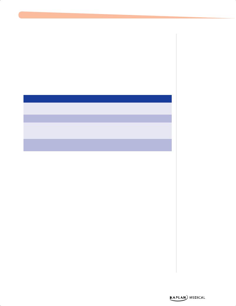
Table 21-1. Neonatal Seizures
Cause
Presentation
Associations
Hypoxic ischemic
12–24 hours
Term; cerebral palsy
encephalopathy
Intraventricular hemorrhage
1–7 days
Preterm
Metabolic
Variable
IODM (infant of diabetic mother), inborn
errors of metabolism, DiGeorge
syndrome
Infection
Variable
TORCH, maternal fever, sepsis/
meningitis
Clinical Recall
A 2-year-old boy with fever, rhinorrhea, and cough is seen in the emergency department after having a first-time generalized tonic-clonic seizure which lasted 6-7 minutes. The exam is notable for a tiredappearing child with no focal neurologic signs or nuchal rigidity. There is no lethargy or irritability. There is no sensitivity to light and no mental status changes or vomiting. What is the next step?
A.Lumbar puncture
B.EEG
C.Brain MRI
D.Prescribe acetaminophen
E.Prescribe ethosuximide
Answer: D
Published by dr-notes.com
225
USMLE Step 2 CK λ Pediatrics
NEUROCUTANEOUS SYNDROMES
A 6-year-old presents to the pediatrician for a routine evaluation. The child is noted to have 10 café-au-lait lesions as well as axillary freckling.
Neurofibromatosis (NF; von Recklinghausen Disease)
NF-1
•Autosomal dominant; but most with new mutation
•Every organ can be affected; features present from birth but complications may be delayed into adulthood
•Diagnosis—a good history and physical examination are needed to make the diagnosis.
−Two of the following are needed:
°At least 5 café-au-lait spots >5 mm prepubertal or at least 6 café-au-lait spots >15 mm postpubertal
°Axillary/inguinal freckling
°>2 iris Lisch nodules (seen on slit lamp only)
°>2 neurofibromas or 1 plexiform neurofibroma
°Osseous lesions, sphenoid dysplasia or cortical thinning of long-bones (LE)
°Optic gliomas
•Complications
−CNS:
°Low-gradegliomas (optic), hamartomas
°Malignant neoplasms (astrocytoma, neurofibrosarcoma, and others)
–Increased incidence of leukemia, rhabdomyosarcoma, Wilms tumor
•Treatment
–Genetic counseling
–Early detection of treatable conditions
–Annual ophthalmologic examination
–Examine family members
NF-2
•Presentation
–Primary feature—bilateral acoustic neuromas
–Hearing loss
–Facial weakness
–Headache
–Unsteady gait
226
Chapter 21 λ Neurology
–Skin findings much less common (glioma, meningioma, schwannoma)
–CNS tumors common
•Treatment
− Developmental and cognitive evaluation and diagnosis
− Prevent pathological fractures if LE cortical thinning present
Tuberous Sclerosis
A 1-month-old infant presents with infantile spasms and has a hypsarrhythmic EEG pattern.
•Autosomal dominant; half with new mutations
•Wide range of manifestations within same family
•The younger the patient, the higher the likelihood of intellectual disability
•Hallmark is CNS tubers found in convolutions of cerebral hemispheres; undergo calcification and project into ventricular cavity, causing obstruction of CSF flow and hydrocephalus.
•Clinical presentation
−Infancy—withinfantile spasms and characteristic skin lesions
°Ash-leafmacule—hypopigmented; increased with Wood UV lamp
°CT scan shows calcified tubers (but may not see till 3–4 years of age)
−Childhood—generalized seizures and skin lesions
°Sebaceous adenoma—red or clear nodules on nose and cheeks
°Shagreen patch—rough, raised lesion with orange-peel consistency; most in lumbosacral area (midline)
•Diagnosis—clinical:characteristic skin lesions and seizure disorder
•Treatment—seizure control
•Complications
−Retinal lesions—either mulberry tumor from optic nerve head or phakomas (round, flat, gray lesions in area of disc)–visual disturbances
−Brain tumors much less common (but may see malignant astrocytoma)
−Half have rhabdomyoma of the heart (can detect in fetus with echocardiogram); most spontaneously regress over first 2 years
−Renal lesion in most—either hamartoma or polycystic kidneys
−Pulmonary—cystic or fibrous changes
Sturge-Weber (SW) syndrome
A newborn is examined in the nursery by the pediatrician. The patient is a product of a term spontaneous vaginal delivery without complications. On physical examination, the patient is noted to have a facial nevus.
•Nevus is always present at birth and always involves at least the upper face and eyelid
Note
Not all babies with a facial nevus have Sturge-Weber syndrome. Obtain a skull x-ray and intraocular pressure.
Published by dr-notes.com
227
USMLE Step 2 CK λ Pediatrics
•Glaucoma in ipsilateral eye
•Presentation
−Seizures in most (focal tonic-clonic,contralateral to the nevus); becomes refractory and slowly develops hemiparesis, intellectual disability
•Diagnosis
−Skull x-ray shows occipital-parietal calcifications (serpentine or railroad-track appearance) and intraocular pressure reading initially (↑)
−CT scan to highlight extent and show unilateral cortical atrophy and hydrocephalus ex vacuo
•Treatment
−Conservative if seizures are well controlled and development is not severely affected
−Hemispherectomy or lobectomy—may prevent intellectual disability and recalcitrant seizures if done in the first year of life
−Regular intraocular pressure evaluation
−Nevus—pulsed laser
−Special education
ENCEPHALOPATHIES
Cerebral Palsy
•Group of motor syndromes from disorders of early brain development
−Neurologic function may change or progress with time
−Some have cognitive dysfunction
−Most born at term with uncomplicated labor and delivery
°Majority have no identifiable antenatal problems
°Only 10% with intrapartum asphyxia
•The most obvious manifestation is impaired ability of voluntary muscles (rigidity and spasticity).
−Other associations—seizures and abnormalities of speech, vision, and intellect
•Other risk factors—increased risk with intrapartum infection, low birth weight, (especially <1,000 g); most of these secondary to intraventricular hemorrhage and periventricular leukomalacia
•Diagnosis
−MRI (location and extent of lesions or abnormalities)
−If spinal involvement, MRI of spine
−Hearing and visual evaluation
−Genetic evaluation
−Complete neurologic and developmental exams
•Treatment
−Multidisciplinary team
−Teach daily activities, exercises, assistance and adaptive equipment, surgical release procedures, communication equipment
The hallmark of neurodegenerative disorders is typically progressive deterioration of neurologic function. This includes loss of speech, vision, hearing, and/or walking; feeding difficulties, cognitive dysfunction, and possible seizures; and regression of developmental milestones.
Friedrich Ataxia
•Abnormal gene encoding for frataxin; autosomal recessive
•Onset of ataxia before <10 years of age
−Slowly progressive
−Loss of DTRs
−Extensor plantar reflex
−Weakness in hands and feet
−Degeneration of posterior columns—loss of position and vibration sense
•Explosive, dysarthric speech
•Skeletal abnormalities, e.g., kyphoscoliosis
•Hypertrophic cardiomyopathy—refractory congestive heart failure, death
Wilson Disease
•Inborn error of copper metabolism; autosomal recessive
•Liver with or without CNS disease (neurologic, psychiatric)
•Liver symptoms first (any liver pathology), neurologic symptoms later (adolescent to adults)
−Dystonia, tremors, basal ganglia problems
−Kayser-Fleischerrings—pathognomonic (all will have with neuropsych symptoms)
−MRI shows dilated ventricles with atrophy of cerebrum and lesions in thalamus and basal ganglia
•Diagnosis—Suspect in any child with acute or chronic liver disease, unexplained neurologic disease, or behavioral or psychiatric changes
−Best screen—serum ceruloplasmin (decreased)
−Confirm with liver biopsy—increased Cu content
−Screen family members
•Treatment
−Chelation with penicillamine (slows progression)
−Definitive treatment with liver transplant
Published by dr-notes.com
229
USMLE Step 2 CK λ Pediatrics
Sphingolipidoses
Tay-Sachs disease
•Deficient β-hexosaminidase-A, accumulate GM2
•Mostly in Ashkenazi Jews (carrier rate 1 in 30)
•Normal developmental until 6 months, then lag and lose milestones
•Seizures, hypotonia, blindness
•Cherry-red macula
Purine Metabolism Disorders
Lesch-Nyhan disease
•X-linked
•Purine metabolism disorder of purine metabolism → excess uric acid
•Delayed motor development after a few months
•Self-mutilation and dystonia, gouty arthritis, tophi, renal calculi
•Choreoathetosis, spasticity
•Diagnosis–Analyze HPRT enzyme
•Treatment
−Manage renal complications, arthritis
−Behavioral modification
−Medication for reduction of anxiety and mood stabilization
Clinical Recall
Which of the following neurodegenerative disorders is correctly matched to a key finding?
A.Lesch-Nyhan disease: cherry red macula
B.Tay-Sachs disease: deficient hexosaminidase-A
C.Wilson disease: error of iron metabolism
D.Friedrich ataxia: dilated cardiomyopathy
E.Niemann-Pick disease: Kayser-Fleischer rings
Answer: B
230
Chapter 21 λ Neurology
NEUROMUSCULAR DISEASE
Spinal Muscle Atrophy (SMA)
A pediatrician examines an infant who is on the examination table in frog-leg position, with subdiaphragmatic retractions and absent tendon reflexes.
•Degenerative disease of motor units beginning in the fetus and progressing into infancy; denervation of muscle and atrophy
•Types
−SMA 1 = severe infantile (Werdnig-Hoffmann disease)
•Clinical presentation—SMA 1 presents in early infancy with
−Progressive hypotonia; generalized weakness; Infant is flaccid, has little movement and poor head control
−Feeding difficulty
−Respiratory insufficiency
−Fasciculations of the tongue and fingers
−Absent DTRs
•Typically appear brighter than others of same age
•Diagnosis
−Simplest, most effective diagnosis is molecular genetic marker in blood for the SMN gene.
−EMG—fibrillation potential and other signs of denervation
−Muscle biopsy shows a characteristic pattern of perinatal denervation.
•Treatment is supportive; there is no cure; most die in first 2 years of life
Myasthenia Gravis
A pediatrician examines an infant with poor sucking and swallowing since birth. The infant is noted to be a floppy baby with poor head control. There is associated ocular ptosis and weak muscles on repeated use.
•Immune-mediated neuronal blockade; motor end plate is less responsive due to, decreased number of available acetylcholine receptors secondary to circulating receptor binding antibodies; generally nonhereditary
•Clinical presentation
−Ptosis and extraocular muscle weakness is the earliest and most consistent finding.
−Dysphagia and facial weakness, and early infant feeding difficulties
−Poor head control
Note
Transient Neonatal
Myasthenia
•Neonates born to mothers with myasthenia;
may have generalized hypotonia and weakness, feeding difficulties, and respiratory insufficiency from days to weeks
•May need ventilation and nasogastric feedings
•After antibodies wane, they are normal and have no risk for disease.
Published by dr-notes.com
231
USMLE Step 2 CK λ Pediatrics
−Limb-girdle weakness and in distal muscles of hands
−Rapid muscle fatigue, especially late in the day
−May have respiratory muscle involvement
•Diagnosis
−EMG more diagnostic than muscle biopsy—decremental response to repetitive
nerve stimulation, reversed after giving cholinesterase inhibitor (edrophonium) → improvement within seconds
−CPK is normal.
−May have anti-acetylcholine (anti-ACh) antibodies (inconsistent)
•Treatment
−Mild—many need no medication
−Cholinesterase-inhibiting drugs—either neostigmine bromide PO or pyridostigmine
−Severe—long-term prednisone; if no response, intravenous immunoglobulin (Ig), then plasmapheresis
−Thymectomy—most effective if patient has high anti-ACh titers and symptoms for <2 years
•Complications—do not tolerate neuromuscular blockade and aminoglycosides potentiate
Hereditary Motor-Sensory Neuropathies (HMSNs)
HMSN I: Marie-Charcot-Tooth disease
•Progressive disease of peripheral nerves; peroneal muscle atrophy; peroneal and tibial nerves
•Autosomal dominant
•Clinical presentation
−Asymptomatic until late childhood or adolescence but may have problem with gait as early as age 2 years
−Clumsy, fall easily; muscles of anterior compartment of lower leg become wasted → stork-like appearance
−Pes cavus, foot drop
−Claw hand (in worse cases)
−Slowly progressive through life, but normal lifespan and remain ambulatory
•Diagnosis
−CPK is normal.
−Decreased nerve conduction velocities (motor and sensory)
−Sural nerve biopsy is diagnostic.
−Blood molecular genetic diagnosis
•Treatment
−Stabilize ankles
−Surgical ankle fusion
−Protection from trauma
−If sensory problems, phenytoin or carbamazepine
232
Chapter 21 λ Neurology
Guillain-Barré syndrome
•Postinfectious polyneuropathy—mostly motor; all ages; most with demyelinating neuropathy
•10 days after a nonspecific viral illness or Campylobacter jejuni or Mycoplasma pneumoniae—Landry ascending paralysis
−Symmetric proximal and distal muscles
−Gradually over days to even weeks
−May have tenderness, pain, paresthesias early
−Bulbar involvement in half—dysphagia, facial weakness, respiratory insufficiency
−May have autonomic involvement—blood pressure lability, bradycardia, asystole
−Spontaneous recovery begins in 2–3 weeks; some have residual weakness; improvement in inverse direction
•Diagnosis
−Significant increase in CSF protein with normal glucose and no cells
−Reduced motor and sensory nerve conductions
•Treatment
−Mostly supportive
−Admit all patients (observe respiratory effort) ° Mild-observation
−Intravenous immunoglobulin 2–5 days
−May need plasmapheresis, steroids, interferon, or other immunosuppressives
Muscular Dystrophy
Duchenne
A 3-year-old boy is brought to the pediatrician because he is very clumsy. According to his parents, he has difficulty climbing stairs and frequently falls. On physical examination hypertrophy of the calves is noted.
•Primary myopathy with genetic basis; is progressive and results in degeneration and death of muscle fibers; most common of the neuromuscular diseases in all races and ethnic groups; X-linked recessive
•Clinical presentation
−First sign may be poor head control in infancy.
−By year 2, may have subtle findings of hip-girdle weakness
−Gower sign as early as age 3 years but fully developed by age 5–6 years; with hipwaddle gait and lordotic posturing
−Calf pseudohypertrophy (fat and collagen) and wasting of thigh muscles
−Most walk without orthotic devices until age 7–10 years, then with devices until 12; once a wheelchair is required, significant acceleration of scoliosis