USMLE Step 2 CK λ Pediatrics
−Then, immediate bone marrow aspirate (>25% homogeneous population of lymphoblasts) and staging lumbar puncture (thus staging at diagnosis is from bone marrow aspirate + lumbar puncture)
−WBC mostly <10,000/mm3 (atypical lymphocytes); poor prognosis if >100,000
−Best test is bone marrow aspirate → lymphoblasts
−If chromosomal abnormalities, poor prognosis
•Treatment
−Remission induction (bone marrow leukemic cell eradication + intrathecal)
−CNS therapy reduces relapse rate to <10%
−Consolidation and intensification
−Maintenance 2–3 years
•Complications
−Majority is bone marrow relapse (15–20%):
°Increased intracranial pressure (ICP) or isolated cranial nerve palsies
°Testicular relapse in 1–2% of boys
−Pneumocystis pneumonia
−Other infections because of immunosuppression
−Tumor lysis syndrome—result of initial chemotherapy (cell lysis): hyperuricemia, hyperkalemia, hyperphosphatemia → hypocalcemia (tetany, arrhythmias, renal calcinosis)
°Treat with hydration and alkalinization of urine; prevent uric acid formation (allopurinol)
•Prognosis: >85% 5-year survival
BRAIN TUMORS
Brain tumors are the second most common tumors in children and have the highest mortality, especially <5 years old. In order of incidence, these are: infratentorial tumors, supratentorial tumors, spinal cord tumors, and tumors of multiple sites.
The predominant type varies with age:
•First year—supratentorial; most common: choroid plexus tumors and teratomas
•Age 1–10—infratentorial; most are juvenile pilocytic astrocytomas (usually cerebellar; low-grade, rarely invasive) and tumors of the medulloblastoma, ependymoma, and brainstem
•Age >10 years—supratentorial; most common: diffuse astrocytoma (also glioblastoma multiforme)
−Craniopharyngioma: histologically benign, slow-growing tumor that predominantly involves the sella and suprasellar space; presenting complaints: headaches, visual symptoms, behavioral changes, growth failure, delayed puberty, amenorrhea, diabetes insipidus, panhypopituitarism; no role for chemotherapy— treated with surgery and radiation
214

−Optic nerve glioma: most frequent optic nerve tumor; benign, slowly progressive; increased incidence in neurofibromatosis; possible symptoms: unilateral visual loss, proptosis, eye deviation, optic atrophy, strabismus, nystagmus; treatment is observation unless symptomatic (chiasm involvement = radiation/chemotherapy, proptosis and visual loss = surgery)
More common in children than adults are tumors of the optic path, hypothalamus, brainstem, and pineal-midbrain.
The clinical presentation of pediatric brain tumors depends on location, type, and age, and symptoms may present as secondary to CSF obstruction (increased ICP, focal brain dysfunction). Common signs and symptoms:
•Supratentorial—subtle changes in personality, mentation, and speech; focal deficits; neuroendocrine if near third ventricle
•Midline or infratentorial—headache, nausea, vomiting, papilledema, blurred vision, diplopia; disturbances in equilibrium, gait, and coordination
•Brainstem—gaze palsy, cranial nerve palsies, upper motor neuron defects, motor weakness
When brain tumor is suspected, perform CT scan (best initial test) followed by MNRI (overall best test). Treatment is specific to the tumor’s type and invasiveness.
OTHER MALIGNANCIES
Wilms Tumor
A mother brings her 3-year-old child to the physician because she found an abdominal mass while bathing the child. The child has been in her usual state of health according to the mother. However, on review of the vital signs, the patient is noted to have an elevated blood pressure.
•Nephroblastoma (Wilms tumor)
•Second most common malignant abdominal tumor
−Usual age 2–5 years
−One or both kidneys (bilateral in 7%)
−Associations:
°Hemihypertrophy
°Aniridia
°Genitourinary anomalies
°WAGR
•Clinical presentation—most are asymptomatic abdominal mass; can have symptoms if encroaching on other organs: hypertension (increased renin), abdominal pain, bowel obstruction (rectal), hematuria
•Diagnosis
−Best initial test–ultrasound
−Abdominal CT scan confirmatory test
Published by dr-notes.com |
215 |
|
|
|
USMLE Step 2 CK λ Pediatrics
•Treatment
−Surgery
−Then chemotherapy and radiation
Note
Patients with neuroblastoma can present with ataxia or opsomyoclonus (“dancing eyes and dancing feet”). These patients may also have Horner syndrome.
− Bilateral renal—unilateral nephrectomy and partial contralateral nephrectomy
•Prognosis—54 to 97% have 4-year survival
Neuroblastoma
A 2-year-old child is brought to the physician because of bluish skin nodules, periorbital proptosis, and periorbital ecchymosis that have developed over the last few days. On physical examination, a hard smooth abdominal mass is palpated.
•From neural crest cells, due to N-myc oncogene; can occur at any site
•8% of childhood malignancies
•Most are
−Adrenal
−Retroperitoneal sympathetic ganglia
Note
Children with pheochromocytoma excrete predominantly
norepinephrine-increased VMA and metanephrine. Children with neuroblastoma usually do not have hypertension, and major metabolites are dopamine and HVA.
−Cervical, thoracic, or pelvic ganglia
•Firm, palpable mass in flank or midline; painful; with calcification and hemorrhage
•Initial presentation often as metastasis—long bones and skull, orbital, bone marrow, lymph nodes, liver, skin
•Diagnosis
−Plain x-ray, CT scan, MRI (overall best)
−Elevated urine homovanillic acid (HVA) and vanillylmandelic acid (VMA) in 95% of cases
−Evaluate for spread—bone scan, bone marrow (neuroblasts) → staging from I (organ of origin) to IV (disseminated)
•Treatment
−Surgery
−Chemotherapy and radiation
−Stem cell transplant (definitive)
Pheochromocytoma
•Catecholamine-secreting tumor from chromaffin cells
•Most common site—adrenal medulla, but can occur anywhere along abdominal sympathetic chain
•Children age 6–14 years; 20% are bilateral, and some with multiple tumors
•Autosomal dominant; associated with neurofibromatosis, MEN-2A and MEN2B, tuberous sclerosis, Sturge-Weber syndrome, and ataxia-telangiectasia
•Clinical presentation
−Episodic severe hypertension, palpitations and diaphoresis, headache, abdominal pain, dizziness, pallor, vomiting, sweating, encephalopathy
−Retinal examination—papilledema, hemorrhages, exudate
•Labs—significant increase in blood or urinary levels of catecholamines and, metabolites

•Diagnosis
−Significant increase in blood/urinary VMA and metanephrine
−CT scan (best initial test), then MRI
−Some adrenal masses are difficult to localize; scan with I-131 metaiodobenzylguanidine (MBIG), which is taken up by chromaffin tissue
•Treatment—surgical removal (high-risk) with preoperative alpha and beta blockade and IV fluids
Rhabdomyosarcoma
A mother brings her 3-year-old daughter to the physician for evaluation because the young girl has “grapes” growing out of her vagina.
•Most common soft-tissue malignancy in children
•Almost any site, which determines presentation; determination of specific histologic type needed for assessment and prognosis. Most are in the head and neck.
•Increased frequency in neurofibromatosis
•Types
−Embryonal—60%
°Intermediate prognosis
−Botryoid (projects; grapelike)—vagina, uterus, bladder, nasopharynx, middle ear
−Alveolar—15%
°Very poor prognosis
°Trunk and extremities
−Pleomorphic—adult form; very rare in children
•Clinical presentation
−Mass that may or may not be painful
−Displacement or destruction of normal tissue
−Easily disseminates to lung and bone
•Diagnosis—depends on site of presentation
−Biopsy, CT, MRI, U/S, bone scan
•Treatment—best prognosis with completely resected tumors (but most are not completely resectable)
−Chemotherapy preand postoperatively; radiation
Published by dr-notes.com |
217 |
|
|
|
USMLE Step 2 CK λ Pediatrics
Clinical Recall
A 7-year-old girl with an abdominal mass diagnosed by MIBG imaging is found to have elevated urinary catecholamines. With which systemic disease is this mass associated?
A.MEN 1
B.von Hippel-Lindau
C.Tuberous sclerosis
D.WAGR
E.Basal cell nevus syndrome
Answer: C
218

Neurology 21
Chapter Title
Learning Objectives
Describe the epidemiology and treatment of febrile and other seizure disorders
Describe CNS anomalies, neurocutaneous syndromes, and neurodegenerative disorders
Recognize and categorize encephalopathies
Categorize and describe the epidemiology and genetics of neuromuscular disease
CENTRAL NERVOUS SYSTEM (CNS) ANOMALIES
Neural Tube Defects
Elevated alpha-fetoprotein is a marker for neural tube defects.
Spina bifida occulta
•Midline defect of vertebral bodies without protrusion of neural tissue; occasionally associated with other anomalies
•Most asymptomatic and of no clinical consequence
•May have overlying midline lumbosacral defect (patch of hair, lipoma, dermal sinus)
Tethered cord
•Ropelike filum terminale persists and anchors the conus below L2
•Abnormal tension—asymmetric lower extremity growth, deformities, bladder dysfunction, progressive scoliosis, diffuse pain, motor delay
•Most associated with a midline skin lesion
•MRI needed for precise anatomy
•Surgical transection
Meningocele
•Meninges herniate through defect in posterior vertebral arches
•Fluctuant midline mass well covered with skin; may transilluminate
•Must determine extent of neural involvement with MRI
−CT scan of head for possible hydrocephalus
−Surgery
Published by dr-notes.com |
219 |
|
|
|
USMLE Step 2 CK λ Pediatrics
Note
Almost every child with a sacral or lower lumbar spine lesion will achieve some form of functional ambulation, and half of those with higher spine defects will have some degree of hip flexor and hip adductor movement.
220
Myelomeningocele
The pediatrician is called to the delivery room because an infant is born with a defect in the lumbosacral area.
•Strong evidence that maternal periconceptional use of folate reduces risk by half
•May occur anywhere along the neuraxis, but most are lumbosacral
•Low sacral lesions—bowel and bladder incontinence and perineal anesthesia without motor impairment
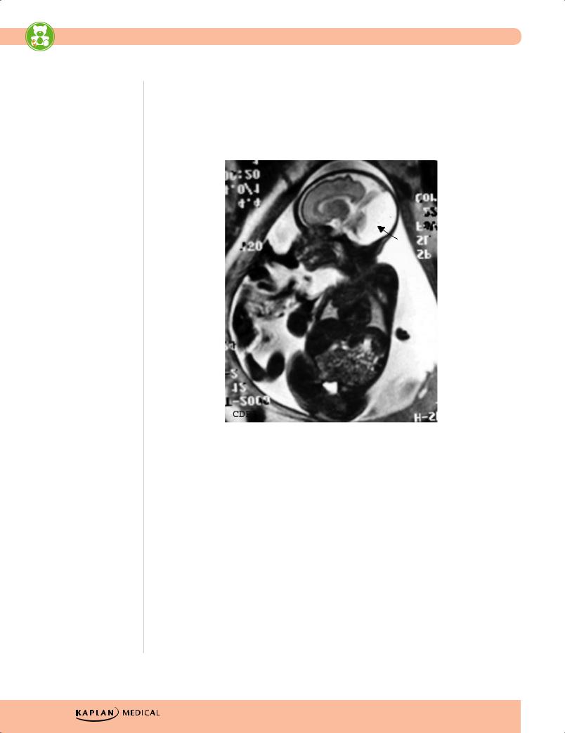
TheFetus.net
Figure 21-1. Arnold-Chiari Malformation, a Defect of the
Hindbrain Usually Accompanied by Myelomeningocele
•Midlumbar lesion—saclike cystic structure covered by thin, partially epithelized tissue
−Flaccid paralysis below the level of the lesion is most common; no deep tendon reflexes (DTRs), no response to touch and pain
−Urinary dribbling, relaxed anal sphincter

•80% associated with hydrocephalus; type II Chiari malformation—may have symptoms of hindbrain dysfunction (feeding difficulty, choking, stridor, apnea, vocal cord paralysis, upper extremity spasticity)
•Evaluation and treatment
−Must evaluate for other anomalies prior to surgery
−Evaluate renal function
−Head CT scan for possible hydrocephalus
−Treatment—ventriculoperitoneal shunt and correction of defect
Hydrocephalus
A 2-month-old infant is noted to have a head circumference >95th percentile.
•Definition—impaired circulation and absorption of CSF or, rarely, from increased CSF production from a choroid plexus papilloma
•Types
−Obstructive (noncommunicative) versus nonobstructive (communicative) from obliteration of subarachnoid cisterns or malfunction of arachnoid villi
°Obstructive—most are abnormalities of the cerebral aqueduct (stenosis or gliosis; congenital, intrauterine infection, mumps, hemorrhage) or lesions near the fourth ventricle (brain tumor, Chiari malformation, Dandy-Walker malformation)
−Nonobstructive—occurs mostly with subarachnoid hemorrhage; also with pneumococcal or TB meningitis or leukemic infiltrates
•Clinical presentation—depends on rate of rise of intracranial pressure
−Infants:
°Increased head circumference
°Bulging anterior fontanel
°Distended scalp veins
°Broad forehead
°“Setting sun” sign
°Increased DTRs
°Spasticity, clonus
−Older child (subtler symptoms)
°Irritability
°Lethargy
°Poor appetite
°Vomiting
°Headache
°Papilledema
°Sixth-nerve palsy
•Treatment for all types of hydrocephalus—shunting
Published by dr-notes.com |
221 |
|
|
|
USMLE Step 2 CK λ Pediatrics
Dandy-Walker malformation
•Cystic expansion of fourth ventricle due to absence of roof
•Associated agenesis of posterior cerebellar vermis and corpus callosum
•Presents with increasing head size and prominent occiput, long-tract signs, cerebellar ataxia, and delayed motor development, positive transillumination
TheFetus.net
Figure 21-2. Dandy Walker Malformation, the Result of Agenesis or Hypoplasia of the Cerebellar Vermis, Cystic Dilatation of the Fourth Ventricle, and Enlargement of the Posterior Fossa
SEIZURES
Seizures are triggered recurrently from within the brain versus somatic disorders that may trigger a seizure from outside the brain. Epilepsy is present when at least 2 unprovoked seizures occur >24 hours apart.
Febrile Seizures
An 18-month-old child is brought to the emergency center after having a generalized tonic-clonic seizure that lasted approximately 5 min. The parents say that the child had been previously well but developed cold symptoms earlier today with a temperature of 39 C (102 F).
222
•Occurs between age 6 months to 5 years; incidence peaks at age 14–18 months and may reoccur with fever
•Usually positive family history
•Temperature usually increases rapidly to >39 C (102 F)
•Typical: generalized tonic-clonic seizures, <10–15 minutes; brief postictal period
•Atypical: >15 minutes, more than 1 in a day, and focal findings
•Simple febrile seizure has no increased risk of epilepsy—risk for febrile seizures is increased with atypical seizure, family history of epilepsy, initial seizure before age 6 months, abnormal development, or preexisting neurologic disorder
−Workup/Evaluation
°Must determine cause of fever, must not look like meningitis
°No routine labs, no EEG, no neuroimaging
−Treatment—control fever
Partial Seizures
Simple seizures
•Asynchronous tonic or clonic movements; most of the face, neck, and extremities; average duration 10–20 seconds
•Some have an aura and may verbalize during the attack; no postictal period
•EEG —spike and sharp waves or multifocal spikes
•Treatment—phenytoin and other anticonvulsants
Complex seizures
•Impaired consciousness at some point, may be very brief; one-third with aura (always indicates focal onset)
•Automatisms common after loss of consciousness (lip-smacking, chewing, swallowing, increased salivation)
•Interictal EEG—anterior temporal lobe shows sharp waves or focal spikes
•MRI—many will show abnormalities in temporal lobe (sclerosis, hamartoma, cyst, infarction, arteriovenous malformation [AVM], glioma)
•Treatment—carbamazepine (drug of choice) and other add-ons
Generalized Seizures
Absence (petit mal) seizures
•Sudden cessation of motor activity or speech with blank stare and flickering eyes
•More in girls; uncommon <5 years of age
•No aura; usually <30 seconds; no postictal period
•EEG —3/second spike and generalized wave discharge
•Treatment—ethosuximide (drug of choice), valproic acid (second line)
Published by dr-notes.com |
223 |
|
|
|