

Полезные материалы за все 6 курсов / Учебники, методички, pdf / Kaplan Pediatrics USMLE 2CK 2021
.pdfUSMLE Step 2 CK λ Pediatrics
Fragile X Syndrome
•Genetics
–Fragile site on long arm of X in affected males and some carrier females— Molecular diagnosis—variable number of repeat CGG (preferred diagnosis = DNA-based molecular analysis)
–With the genetic mutation, can get trinucleotide expansion during meiosis to a premutation state (50-200 repeat CGG); this is passed on to progeny and may
then further expand to the full mutation (>200 CGG); then, epigenetic methylation occurs → gene silencing → protein inactivation = full syndrome. More likely meiotic expansion in future generations = genetic anticipation
–X-linked dominant—males (most common cause of inherited intellectual disability); due to lyonization (random inactivation of one X), there are generally fewer abnormalities seen in girls but they may present with decreased IQ
–There is no meiotic expansion in males; can only pass premutation to daughters
–Males with the full syndrome are infertile
•Findings
–Mild to profound intellectual disability; learning problems; anxiety, depression, and autistic-like behaviors
–Large ears, dysmorphic facial features, large jaw, long face
–Large testes—mostly in puberty (macroorchidism)(fertile)
•Natural history—normal lifespan
EARLY OVERGROWTH WITH ASSOCIATED DEFECTS
Beckwith-Wiedemann Syndrome
•Genetics
–Usually sporadic
–IGF-2 disrupted at 11p15.5 (imprinted segment); therefore, both copies of the gene are expressed (normally one is silenced), leading to overgrowth
•Findings
–Macrosomia
–Macroglossia—may need partial glossectomy
–Pancreatic beta cell hyperplasia—excess islets → hypoglycemia; hypoglycemia may be refractory; glucose control most important initial management
–Umbilical abnormalities, diastasis recti, omphalocele
–Hemihypertrophy → increased risk of abdominal tumors (Wilms)
•Management—obtain ultrasounds and serum AFP every 6 months through 6 years of age to look for Wilms tumor and hepatoblastoma
24

Chapter 2 λ Genetics/Dysmorphology
UNUSUAL BRAIN AND/OR NEUROMUSCULAR FINDINGS WITH ASSOCIATED DEFECTS
Prader-Willi Syndrome
•Genetics
–Most with deletion at 15q11-q13–imprinted segment
–Paternal chromosome responsible
–The same deletion causes both Prader-Willi and Angelman syndromes. This may be due to the normal process of imprinting, which is epigenetic (change in the chromatin and not the gene sequence) silencing (due to hypermethylation) of certain genes in either the male or female germ cells. The alleles in the opposite germ line are expressed and therefore in the zygote this results in monoallelic gene expression so that for any imprinted segment there is a functional haploid state. It is established in the germ line and maintained in all somatic cells.
°If the deletion occurs in the male germ cell, then the inheritance is from the only expressed genes, which are maternal. This is Prader-Willi syndrome.
°If the deletion occurs in the female germ cell, then the inheritance is from the only expressed genes, which are paternal. This is Angelman syndrome.
–Negligible recurrence risk
•Findings
–First year, difficulty feeding with poor growth; then, increased feeding and weight gain plus slow height attainment (short stature)
–Obesity—onset from 6 months to 6 years
–Mild to severe intellectual disability
–Food-related behavioral problems (binge eating)
–Small hands and feet, puffy; small genitalia
–Hypothalamic-pituitary dysfunction (growth, thyroid, adrenal) hypogonadotropic-hypogonadism; hypothalamic-pituitary dysfunction other than GH deficiency and hypogonadism is variable
•Natural history—decreased life expectancy relative to morbid obesity
Angelman Syndrome (Happy Puppet Syndrome)
•Genetics—also deletion of 15q11-q13, but maternally derived (imprinted segment)
•Findings
–Severe MR
–Paroxysms of inappropriate laughter
–Absent speech or <6 words (100%); most can communicate with sign language
–Ataxia and jerky arm movements resembling a puppet’s movements (100%)
–Seizures—most at age 4 years, may stop by age 10
Published by dr-notes.com |
25 |
|
|
|
|

USMLE Step 2 CK λ Pediatrics
OSTEOCHONDRODYSPLASIAS
Achondroplasia/Hypochondroplasia
•Genetics: autosomal dominant; most common short-limb dwarfism; 90% from new gene mutation; older paternal age; mutations in gene for fibroblast growth factor receptor 3 at 4p16.3 (FGFR3)
•Findings
–Short stature (increased upper-to-lower segment ratio; short-limbed dwarfism)
–Proximal femur shortening
–Megalocephaly, small foramen magnum (may have hydrocephalus), small cranial base, prominent forehead
–Lumbar lordosis
•Natural history
–Normal intelligence
–Spinal cord compression is rare (cervicomedullary junction); usually occurs in first year of life
–Tendency of late childhood obesity
–Small eustachian tube—otitis media and hearing loss
–Early cervical compression, respiratory problems, obstructive and central apnea, later cardiovascular disease
CONNECTIVE TISSUE DISORDERS
Marfan Syndrome
•Genetics: autosomal dominant with wide variability; mutation in fibrillin gene (FBN1)—15q21.1
•Findings
–Early rapid growth of the appendicular skeleton and anterior ribs
–Major findings are skeletal, cardiovascular, and ocular
–Tall stature with long, slim limbs and little fat
–Arm span > height
–Arachnodactyly
–Decreased U:L segment ratio (as with XXY)
–Joint laxity with kyphoscoliosis
–Pectus excavatum or carinatum
–Lens subluxation (upward; defect in suspensory ligament); secondary glaucoma, myopia, retinal detachment
–Ascending aortic dilatation with or without dissecting aneurysm (uncommon in children and adolescents unless case is severe) with secondary aortic regurgitation. Mitral valve disease (MVP and regurgitation) is the most common in children.
•Natural history
–Prevent scoliosis
–Vascular complications chief cause of death
–Evaluate heart and aorta
26
Chapter 2 λ Genetics/Dysmorphology
Ehlers-Danlos Syndrome
•Genetics: type I most common (now 6 types); autosomal dominant with wide variability
•Findings
–Droopy ears
–Hyperextensible skin, fragile, easy bruisability, poor wound healing
–Joint hyperlaxity; tendency toward hip, shoulder, knee, and clavicular dislocation
–MVP, tricuspid valve prolapse, aortic root dilatation; dissecting aneurysm, ASD
–Blue sclera, myopia, glaucoma, ectopia lentis, retinal detachment
–Intracranial aneurysm
Osteoporosis in Children
Osteomalacia is undermineralization of normal bone volume, while osteoporosis is normal mineralization but a decrease in bone volume, especially trabecular (vertebral). By definition, with osteoporosis there is also osteopenia, a decreased amount of total bone tissue). It is associated with pathological (atraumatic) fractures.
•Primary osteoporosis: heritable connective tissue disorders
•Secondary osteoporosis: neuromuscular disorders, chronic illness, endocrine disorders, drug-induced, inborn errors of metabolism
Note
Ehlers Danlos patients tend to have thin sclerae, allowing the darker underlying choroid to shine through with a blue-gray tinge.
Table 2-1. Primary Osteoporosis in Children
|
Disease |
|
|
Defect |
|
|
Genetics |
|
|
Comment |
|
|
|
|
|
|
|
|
|
|
|
|
|
|
Osteogenesis |
Structural or quantitative defect of type I |
• Autosomal dominant: |
|
Most common genetic |
||||||
|
imperfecta |
collagen, the primary component of the |
|
all racial and ethnic |
|
cause of osteoporosis |
|||||
|
|
|
extracellular matrix of bone and skin |
|
groups |
|
|
|
|||
|
|
|
|
|
|
• Autosomal recessive: |
|
|
|
||
|
|
|
|
|
|
|
ethnic groups with |
|
|
|
|
|
|
|
|
|
|
|
consanguinity |
|
|
|
|
|
|
|
|
|
|
|
|
|
|
|
|
|
Ehlers-Danlos |
Quantitative deficiency of fibrillar collagen |
Four autosomal |
One AD type, vascular |
|||||||
|
syndrome |
(collagen molecules packed together to |
dominant types and |
has decreased longevity |
|||||||
|
|
|
form long, thin fibrils) |
2 autosomal recessive |
|
|
|
||||
|
|
|
|
|
|
|
|
|
|
|
|
Marfan syndrome |
Mutations in the gene (FBN1) encoding for |
Autosomal dominant |
Mostly skeletal, ocular |
||||||||
|
|
|
the extracellular matrix protein fibrillin-1, |
|
|
|
and cardiovascular |
||||
|
|
|
the major constituent of microfibrils |
|
|
|
findings |
||||
|
|
|
|
|
|
|
|
|
|
|
|
Homocystinuria |
Classic form: cystathionine-β-synthase |
Autosomal recessive |
Phenotype similar to |
||||||||
|
|
|
deficiency: increase of both methionine |
|
|
|
Marfan syndrome but |
||||
|
|
|
and homocysteine in body fluids and |
|
|
|
some differences |
||||
|
|
|
decrease to absence of plasma cystine |
|
|
|
|
|
|
||
|
|
|
|
|
|
|
|
|
|
|
|
Polyostotic fibrous |
Postzygotic activating mutation causing |
Noninherited; 2x more |
Other endocrinopathies |
||||||||
dysplasia (McCune- |
overproduction of endocrine protein |
in girls |
due to overproduction |
||||||||
Albright syndrome) |
products independent from normal |
|
|
|
(pituitary, thyroid, |
||||||
|
|
|
feedback control; precocious puberty with |
|
|
|
adrenal) |
||||
|
|
|
polyostotic fibrous dysplasia and café-au- |
|
|
|
|
|
|
||
|
|
|
lait spots |
|
|
|
|
|
|
||
|
|
|
|
|
|
|
|
|
|
|
|
Published by dr-notes.com |
27 |
|
|
|
|

USMLE Step 2 CK λ Pediatrics
ENVIRONMENTAL AGENTS
Table 2-2. Environmental Agents
|
Embryopathy |
|
|
Major Findings |
|
|
Comments |
|
|
|
|
|
|
|
|
|
|
|
Fetal alcohol |
|
• Neurobehavioral and developmental abnormalities |
|
Most common teratogen; may not have a |
|||
|
|
|
|
(in worst cases, intellectual disability) |
|
maternal history, so must make diagnosis by |
||
|
|
|
|
• Mid-face dysmorphism (from abnormal frontal |
|
first 3 listed findings |
||
|
|
|
|
|
|
|
||
|
|
|
|
lobe development): short palpebral fissures, |
|
|
|
|
|
|
|
|
maxillary hypoplasia, short and smooth philtrum |
|
|
|
|
|
|
|
|
and indistinct philtrum-vermillion border |
|
|
|
|
|
|
|
|
• Pre and postnatal growth deficiency: symmetric |
|
|
|
|
|
|
|
|
IUGR then short stature, slow growth, and |
|
|
|
|
|
|
|
|
acquired microcephaly |
|
|
|
|
|
|
|
|
• PLUS in worse cases: cardiac and joint anomalies |
|
|
|
|
|
|
|
|
|
|
|
|
|
|
Fetal hydantoin |
|
IUGR, hypertelorism; flat, broad nasal bridge and |
|
Similar features with carbamazepine, |
|||
|
|
|
|
hypertelorism, short nose, cleft lip and palate, |
|
primidone and phenobarbital; no dose- |
||
|
|
|
|
malformed ears, web neck, hirsutism, congenital |
|
response relationship |
||
|
|
|
|
heart disease |
|
|
|
|
|
|
|
|
|
|
|
|
|
|
Fetal valproate |
|
Neural tube defects, prominent metopic ridge, cleft |
|
|
|
||
|
|
|
|
lip and palate, radial defects, hypospadias, |
|
|
|
|
|
|
|
|
congenital heart disease, absence of first rib |
|
|
|
|
|
|
|
|
|
|
|
|
|
|
Fetal warfarin |
|
Nasal hypoplasia, microphthalmia, microcephaly, |
|
|
|
||
|
|
|
|
Dandy-Walker malformation, intellectual disability, |
|
|
|
|
|
|
|
|
scoliosis, congenital heart disease |
|
|
|
|
|
|
|
|
|
|
|
|
|
|
Retinoic acid |
|
Affects neural crest and branchial arch development: |
|
• All treated females must take a pregnancy |
|||
|
|
|
|
microtia, anotia; hypertelorism, flat, depressed |
|
test, use definitive method of birth control |
||
|
|
|
|
nasal bridge, intellectual disability, learning |
|
plus 1 back-up method, receive counseling |
||
|
|
|
|
problems, conotruncal anomalies |
|
about teratogenicity; no problems if stopped |
||
|
|
|
|
|
|
|
prior to 15th postmenstrual day |
|
|
|
|
|
|
|
|
• Also obtain baseline liver tests and lipid |
|
|
|
|
|
|
|
|
panel |
|
|
|
|
|
|
|
|
|
|
Clinical Recall
A newborn girl found to be small for gestational age has wide-spaced eyes, increased body hair, and a ventricular septal defect on echocardiography. What was she most likely exposed to in utero?
A.Valproic acid
B.Phenytoin
C.Warfarin
D.Retinoic acid
E.Alcohol
Answer: B
28
Chapter 2 λ Genetics/Dysmorphology
MISCELLANEOUS CONDITIONS
Potter Sequence
•Etiology
–Renal agenesis/dysgenesis or other type of urinary tract defect must occur prior to 31 days’ gestation → oligohydramnios (also from chronic leakage)
–Leads to fetal compression (mid-face, ears)
–Lack of alveolar sac development → pulmonary hypoplasia
•Findings
–Pulmonary hypoplasia
–Potter facies—hypertelorism, epicanthal folds, low-set flattened ears, micrognathia, compressed flat nose
–Breech presentation
–Abnormal positioning of hands and feet; deformations, limb anomalies
–Death from respiratory insufficiency (hypoplasia)
VACTERL Association
• Nonrandom association of V = Vertebral defects
A = Anal atresia (imperforate anus) C = Cardiac defects (VSD and others) T = TE fistula
E = Esophageal atresia R = Renal defects
L = Limb defects (radial)
CHARGE Syndrome
Most cases now known to be caused by a mutation in CHD7 gene (8q12.2), which provides instructions for making a protein that regulates chromatin remodeling. When this is the cause, it follows autosomal dominant inheritance; a small number have no known cause or inheritance pattern.
• Nonrandom association of
C = Coloboma (from isolated iris to anophthalmos; retinal most common) H = Heart defects (TOF, PDA, and others)
A = Atresia choanae
R = Retardation of growth and/or development G = Genital hypoplasia (in males)
E = Ear anomalies and/or deafness
Note
U/S is necessary for the parents/siblings of patients with oligohydramnios secondary to agenesis and/or dysgenesis of both kidneys. This is because 9% of first-degree relatives have asymptomatic malformations.
Published by dr-notes.com |
29 |
|
|
|
|

Growth and Nutrition |
3 |
Chapter Title |
Learning Objectives
Demonstrate steps in evaluation of growth
Solve problems related to breastfeeding, feeding of solids, and other feeding issues
Answer questions related to growth disorders
CHILDHOOD GROWTH
Basic Principles of Growth
In the first week of life, a newborn typically loses up to 10% of birth weight (BW) due to the elimination of large amounts of extravascular fluid. By 2 weeks, BW should be regained or surpassed. In the first month of life, a neonate should gain ~30 grams (1 oz) per day, which slows to ~20 grams/day at 3–4 months.
•By 6 months, an infant typically doubles BW, and by 1 year, triples BW.
•Growth rate slows further between 6 and 12 months and then appetite begins to decline through 18 months of age.
•Then height and weight increase at a steady rate, but head-circumference rate of growth decreases somewhat (2–5 years).
•Between age 6 and 12 years: 3–6 growth spurts each year for 8-week periods each; slower brain growth; myelination complete by age 7
•Between age 10 and 20 years: acceleration in early adolescence. Boys’ highest growth stops at age 18. Their average peak is 13.5 years (2–3 years later than girls, and continues 2–3 years after girls have stopped). Girls’ average peak is 11.5 years and it stops at age 16.
Assessment of Growth
•A child is genetically programmed to stay on 1–2 growth curves after age 2 years.
•The height percentile at age 2 years correlates with final adult height percentile.
•Low-birth-weight and very-low-birth-weight infants may continue to show catch-up growth through early school age.
•Weight/height <5th percentile is the single best growth curve indicator for acute malnutrition. In nutritional insufficiency, weight decreases before length, and weight/height
Published by dr-notes.com |
31 |
|
|
|
|

USMLE Step 2 CK λ Pediatrics
is low. For causes of decreased linear growth, length decreases first or at the same time as weight (e.g., GH deficiency).
•Body mass index (BMI) is accepted as best clinical indicator for measure of underand overweight.
•For bone age-reference standards, use radiographs of left hand and wrist. Skeletal maturity is linked more to sexual maturity than chronologic age.
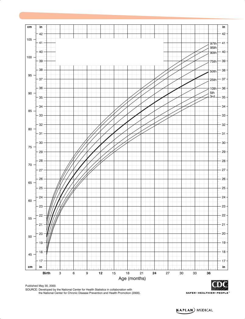
Growth Patterns
The growth chart is the best tool to determine patterns of growth, with separate charts for boys and girls. The charts measure weight for age, height for age, head circumference for age, weight for height, and BMI. Each chart has multiple curves (either 5–95% or 3–97%).
Evaluation of Growth
•Growth velocity: yearly increments of growth; should follow a growth curve
slope = change in height change in age
•Chronologic age (CA): actual age
•Bone age (BA): x-ray of left hand and wrist (non-dominant hand)
32
Chapter 3 λ Growth and Nutrition
Length-for-age percentiles: Boys, birth to 36 months
Published by dr-notes.com |
33 |
|
|
|
|