- •Preface
- •Acknowledgments
- •Introduction
- •Cardiac Tissue Engineering
- •Objectives and Scopes
- •Organization of the Monograph
- •Bibliography
- •Introduction
- •The Heart and Cardiac Muscle Structure
- •Myocardial Infarction and Heart Failure
- •Congenital Heart Defects
- •Endogenous Myocardial Regeneration
- •Potential Therapeutic Targets and Strategies to Induce Myocardial Regeneration
- •Bibliography
- •Introduction
- •Human Embryonic Stem Cells
- •Induced Pluripotent Stem Cells
- •Direct Reprogramming of Differentiated Somatic Cells
- •Cardiac Stem/Progenitor Cells
- •Summary and Conclusions
- •Bibliography
- •Introduction
- •Basic Biomaterial Design Criteria
- •Biomaterial Classification
- •Natural Proteins
- •Natural Polysaccharides
- •Synthetic Peptides and Polymers
- •Basic Scaffold Fabrication Forms
- •Hydrogels
- •Macroporous Scaffolds
- •Summary and Conclusions
- •Bibliography
- •Biomaterials as Vehicles for Stem Cell Delivery and Retention in the Infarct
- •Introduction
- •Stem Cell Delivery by Biomaterials
- •Cardiac Stem/Progenitor Cells
- •Clinical Trials
- •Summary and Conclusions
- •Bibliography
- •Introduction
- •Myocardial Tissue Grafts Created in Preformed Implantable Scaffolds
- •Summary and Conclusions
- •Bibliography
- •Introduction
- •Bioreactor Cultivation of Engineered Cardiac Tissue
- •Mass Transfer in 3D Cultures
- •Bioreactor as a Solution for Mass Transfer Challenge
- •Perfusion Bioreactors
- •Inductive Stimulation Patterns in Cardiac Tissue Engineering
- •Mechanotransduction and Physical/Mechanical Stimuli
- •Mechanical Stimulation Induced by Magnetic Field
- •Electrical Stimulation
- •Summary and Conclusions
- •Bibliography
- •Introduction
- •Prevascularization of the Patch by Incorporating Endothelial Cells (ECs)
- •The Body as a Bioreactor for Patch Vascularization
- •Summary and Conclusions
- •Bibliography
- •Introduction
- •Decellularized ECM
- •Injectable Biomaterials
- •Injectable hydrogels based on natural or synthetic polymers
- •Injectable Decellularized ECM Matrices
- •Mechanism of Biomaterial Effects on Cardiac Repair
- •Immunomodulation of the Macrophages by Liposomes for Infarct Repair
- •Inflammation, Apoptosis, and Macrophage Response after MI
- •Summary and Conclusions
- •Bibliography
- •Introduction
- •Evolution of Bioactive Material Approach for Myocardial Regeneration
- •Bioactive Molecules for Myocardial Regeneration and Repair
- •Injectable Systems
- •Sulfation of Alginate Hydrogels and Analysis of Binding
- •Injectable Affinity-Binding Alginate Biomaterial
- •Summary and Conclusions
- •Bibliography

122. THE HEART—STRUCTURE, CARDIOVASCULAR DISEASES, AND REGENERATION
2.3MYOCARDIAL INFARCTION AND HEART FAILURE
Coronary heart disease (CHD) is now the leading cause of death worldwide [4]. In 2008, CHD caused 1 of every 6 deaths in the United States. Recent data show that death rates from CHD have decreased in North America and in many countries in Western Europe [5]. This decline has been due to improved prevention, diagnosis, and treatment (pharmacological or interventional), in particular reduced cigarette smoking among adults, and lower average levels of blood pressure and blood cholesterol. However, the burden of CHD is increasing in developing and transitional countries, partly as a result of increasing longevity, urbanization, and lifestyle changes. It is expected that 82% of the future increase in coronary heart disease mortality will occur in developing countries.
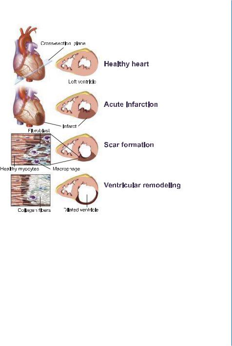
Myocardial infarction (MI) is the most common manifestation of CHD, accounting for 50% of all CHD cases in the US. MI results from temporary or permanent occlusion of the main coronary arteries (Fig. 2.2), causing significant blood supply reduction to the beating heart muscle (mainly left ventricle).
All the myocardium that is supplied by the occluded artery becomes ischemic, resulting in chest pain and electrocardiographic evidence of transmural (full-thickness) ischemia (ST-segment elevation) in the leads reflective of that region of the heart. Because of its high metabolic rate, the myocardium (cardiac muscle) (Fig. 2.3a) begins to undergo irreversible injury within 20 minutes of ischemia, and a wavefront of cell death subsequently sweeps from the inner layers toward the outer layers of myocardium over a threeto six-hour period. Although cardiomyocytes are the most vulnerable population, ischemia also kills vascular cells, fibroblasts, and nerves in the tissue. Myocardial cell necrosis (Fig. 2.3b) elicits a vigorous inflammatory response. Hundreds of millions of marrow-derived leukocytes, initially composed of neutrophils and later of macrophages, enter the infarct. The macrophages phagocytose the necrotic cell debris and likely direct the subsequent phases of wound healing. Concomitant with removal of the dead tissue, a hydrophilic provisional wound repair tissue rich in proliferating fibroblasts and endothelial cells – termed granulation tissue (Fig. 2.3c) – invades the infarct zone from the surrounding tissue. Over time, granulation tissue remodels to form a densely collagenous scar tissue (Fig. 2.3d). In most human infarcts, this repair process requires two months to complete. Infarcts in smaller experimental animals such as mice or rats heal substantially faster [7].
The sudden loss of a significant portion of myocardium leads to a decrease in contractile function of the heart. These changes are compensated by an increase in left-ventricular volume, which augments contractility in the non-infarcted myocardium via the Frank-Starling mechanism (Fig. 2.2). A negative consequence of the enhanced left-ventricular volume, however, is the intensified stress on the ventricular wall. This is partially counteracted by scar formation in the infarct zone and cardiomyocyte hypertrophy in the non-infarcted myocardium (Fig. 2.2). These mechanisms provide temporary compensation for the loss of myocardium contractility. However, in large infarcts, these mechanisms fail, and further deterioration in cardiac function occurs. In these patients, MI results in thinning of the injured wall and dilation of the ventricular cavity, a process termed ventricular remodeling (Fig. 2.2 and Fig. 2.3e). These structural changes markedly increase the mechanical

2.3. MYOCARDIAL INFARCTION AND HEART FAILURE 13
Figure 2.2: Heart failure: from acute crisis to chronic disease. In a healthy heart, the heart’s left ventricle pumps newly oxygenated blood to the rest of the body, and its walls are normally thick with cardiac muscle fibers. When blood supply to the beating muscle is reduced as a result of coronary artery occlusion, myocytes die from oxygen deprivation, and the infarct develops. Within hours and days, existing extracellular matrix degradation takes place. The infarct is infiltrated by macrophages and collagen-producing myofibroblasts. The infarcted wall of the ventricle becomes thin and rigid. As healthy myocytes die at the border of the scarred area, the infarct expansion continues. Developed pressure overload is initially compensated by the hypertrophy of the healthy myocardium. Ultimately, however, this compensating mechanism fails, and infarct wall thinning and ventricle dilatation continues. As a result, the heart is unable to pump effectively, leading to life-threatening condition of heart failure. Reprinted with permission from [6].

14 2. THE HEART—STRUCTURE, CARDIOVASCULAR DISEASES, AND REGENERATION
D E
9LDEOH 1HFURWLF
F G
*UDQXODWLRQ WLVVXH 6FDU
3RVW LQIDUFW YHQWULFXODU UHPRGHOLQJ
H
&RQWUDFWLRQ IRUFH /9 YROXPH (MHFWLRQ IUDFWLRQ
  /9 ZDOO VWUHVV
Figure 2.3: Histological stages of myocardial infarction. a-d: changes in infarct area after ischemic event. e. ventricular remodeling. See text for details. Reprinted with permission from [7].
stress on the ventricular wall and promote progressive contractile dysfunction, eventually leading to chronic and congestive heart failure (CHF) [7, 8, 9, 10].
Over recent decades, major improvements have been realized in the management of patients with MI [11, 12]. These include in-hospital treatments (e.g., pharmacological lysis and anti-platelet and anti-thrombin therapies), interventional therapies and surgery (e.g., cardiac catheterization, percutaneous coronary intervention, coronary artery bypass surgery, and heart transplantation), as well as drug regimens for prevention and long-term treatment (e.g., aspirin, ACE inhibitors, β- blockers, and statins). The improved management of acute coronary events, however, has led to a significant increase in the number of patients who suffer from chronic conditions, namely CHF.

2.4. CARDIAC EXTRACELLULAR MATRIX (ECM) 15
A major disadvantage of the above therapies is their inability to replace, at least partially, cardiac muscle loss after infarction. Thus, there is a need for alternative approaches able to overcome the limitations of standard therapies. The ultimate goal of such novel therapies is the induction of myocardial tissue regeneration (therapeutic, endogenous, or combined) in situ or ex vivo.
2.4CARDIAC EXTRACELLULAR MATRIX (ECM)—ITS FUNCTION AND PATHOLOGICAL CHANGES AFTER MI
The architectural complexity of the myocardium and the potential role of ECM in maintaining the unique myocyte orientations throughout the LV free wall were described by Streeter and Basset [13]. Using a structural engineering approach, these authors demonstrated that myocyte orientation and myocardial fiber angles are highly organized and move in a continuous fashion from the endocardium to the epicardium. It is the structural network of matrix proteins composed of proteins of highly organized structure and architecture, such as type I and type III collagen, that provide structural integrity to adjoining myocytes and contribute to overall LV pump function through the coordination of myocyte shortening. Scanning electron microscopy studies demonstrated the three-dimensional structure of the myocardial ECM and how the fibrillar weave surrounded and supported individual myocytes as well as fascicles of myocytes [14]. Moreover, these initial studies demonstrated the complexity of the ECM and the structural interaction with the vascular compartment. Further research demonstrated that the myocardial ECM maintains alignment of myofibrils within the myocyte through a collagen-integrin-cytoskeleton-myofibril relation (Fig. 2.4).The main components of myocardial ECM are listed in Table 2.1.
In addition to a fibrillar collagen network, a basement membrane, proteoglycans, and glycosaminoglycans, the myocardial ECM contains a large reservoir of bioactive molecules [14]. For example, it has been demonstrated that the concentration of bioactive signaling molecules such as angiotensin II (ANG II) and endothelin (ET)-1 are over 100-fold higher within the myocardial interstitium than in plasma [18, 19]. Moreover, cytokine activation and signaling such as that for tumor necrosis factor-α (TNF-α) is highly compartmentalized within the myocardial interstitium [20]. Growth factors such as transforming growth factor-β (TGF-β) are stored in a latent form within the myocardial interstitium and thereby form a reservoir of signaling molecules that directly influence myocardial ECM synthesis and degradation [14]. Moreover, mechanical stimuli such as stress or strain are likely transduced through the myocardial ECM to the cardiac myocyte, which in turn would directly affect myocyte growth. Thus, structural changes that would occur within the myocardial ECM would in turn affect myocyte biology and the overall structure and function of the myocardium.
Significant alterations in the structure and composition of the myocardial ECM occur following MI. Cardiac wound repair after MI involves temporarily overlapping phases, which include an inflammatory phase and tissue remodeling phase [14, 21]. The first phase starts after coronary artery occlusion with or without reperfusion and involves degradation of normal ECM, invasion of inflammatory cells at the site of initial injury, and the induction of bioactive peptides and cytokines.

16 2. THE HEART—STRUCTURE, CARDIOVASCULAR DISEASES, AND REGENERATION
Figure 2.4: A schematic presentation of myocardial environment and major ECM components. Insert: Collagen weave surrounding individual myocytes and collagen struts tethering adjacent myocytes comprise the endomysium. Groups of myocytes are bundled within the perimysium. Capillaries and coronary microvessels have free diffusion access to cardiac myocytes throughout the ECM. In the scheme: major ECM components in the myocardium include fibrillar collagen, fibronectin, elastin, proteoglycans, and glycosaminoglycans. ECM also serves as controlled reservoir of growth factors. The interaction of ECM with cells is mediated by integrins on cell surface. Cadherins and gap junction proteins comprise the cell-cell interaction complex. Insert: reprinted with permission from [15].
Degradation of the ECM during the acute phase is considered to be an essential event that allows for the ingress of inflammatory cells as well as proliferation and maturation of macrophages and fibroblasts, and provides the necessary substructure for scar formation. Very early post-MI, there is a disappearance of the normal collagen matrix, increased release of hydroxyproline (an amino acid primarily found in collagen), and reduced collagen cross-linking within the ischemic region, all indicating that excessive ECM degradation takes place. These early ECM events occurred prior to the egress of inflammatory cells into the MI region. In this early time period, LV myocardial ECM

|
|
2.4. CARDIAC EXTRACELLULAR MATRIX (ECM) 17 |
||
|
|
|
||
|
Table 2.1: The main components and function of myocardial extracellular |
|
||
|
|
matrix [16, 17] |
|
|
|
&RPSRQHQW |
|
0DLQ )XQFWLRQ |
|
|
|
|
6WUXFWXUDO VXSSRUW PDLQWDLQ VKDSH |
|
|
|
|
|
|
|
&ROODJHQ ILEULOV W\SHV , DQG ,,, |
|
7UDQVPLVVLRQ RI IRUFH |
|
|
|
|
|
|
|
|
|
7HQVLOH VWUHQJWK W\SH , UHVLOLHQFH W\SH ,,, |
|
|
(ODVWLQ |
|
5HVLOLHQFH YHVVHO ZDOO VWUHWFK FDUGLDF ZDOO VWUHWFK |
|
|
|
DQG UHOD[DWLRQ |
|
|
|
|
|
|
|
|
+\GURSKLOLF JO\FRVDPLQRJO\FDQV |
|
'LIIXVLRQ RI QXWULHQWV PHWDEROLWHV JURZWK IDFWRUV |
|
|
|
F\WRNLQHV HWF |
|
|
|
|
|
|
|
|
|
|
|
|
|
3URWHRJO\FDQV |
|
0HFKDQLFV IOXLG G\QDPLFV" |
|
|
,QWHJULQV PDWUL[ UHFHSWRUV |
|
0\RF\WH ILEUREODVW (&0 LQWHUDFWLRQV PDWUL[ |
|
|
|
UHPRGHOLQJ |
|
|
|
|
|
|
|
|
|
|
|
|
|
)LEURQHFWLQ DQG ODPLQLQ |
|
$GKHVLYH ILEURXV SURWHLQV |
|
|
|
|
|
|
|
&HOOV |
|
||
|
|
3URGXFH ILEULOODU FROODJHQ |
|
|
|
|
|
|
|
|
)LEUREODVWV |
|
|
|
|
|
&RQYHUW WR P\RILEUREODVWV DIWHU LQMXU\ |
|
|
|
|
|
|
|
|
|
|
|
|
|
0DFURSKDJHV |
|
3KDJRF\WRVLV LQIODPPDWRU\ UHVSRQVH |
|
|
|
|
(QGRWKHOLDO FHOOV VPRRWK PXVFOH FHOOV SHULF\WHV |
|
|
2WKHU FHOOV |
|
QHXURQV |
|
|
|
|
|
|
degradation and remodeling were associated with an increased probability of rupture [22]. Thus, dynamic changes occur within the myocardial ECM in the initial and early phases of the post-MI period that directly affect the mechanical properties of the LV myocardium.
As the MI period progresses over the next several days, an influx of inflammatory cells into the injured myocardium occurs, which results in further proteolysis of cellular and ECM proteins. In addition, this inflammatory response causes proliferation and differentiation of fibroblasts and other interstitial cells, and the elaboration of bioactive molecules which contribute to a robust synthesis of ECM for the purposes of scar formation [14]. These changes within the MI region yield distinctive cellular and extracellular phenotypic changes. For example, the differentiation and proliferation of fibroblasts within the MI region demonstrate a unique protein signature and function to not only synthesize ECM proteins critical for scar formation, but also contribute to the biophysical properties of the scar itself and have been termed myofibroblasts.The later phase of post-MI remodeling results in ECM changes within all regions of the LV: the MI region, the viable myocardium within the border zone, and the remote region. Within the MI region, the newly formed ECM provides a
